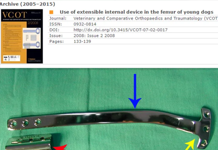
Use of extensible internal device in the femur of young dogs. 1,2Departments of Veterinary Surgery and Anesthesiology, and 5Animal Reproduction and Radiology, School of Veterinary Medicine and Animal Science, São Paulo State University (UNESP), Botucatu Campus, São Paulo, Brazil 3,4Department of Orthopedics and Traumatology, Santa Casa Medical School, São Paulo, Brazil 6Department of Basic Sciences, School of Animal Science, Pirassununga Campus, São Paulo University (USP), São Paulo, Brazil
L. T. Justolin1, S. C. Rahal2, P. P. R. Baptista3, E. S. Yoname4, M. J. Mamprim5, J. C. C. Balieiro6
Use of extensible internal device in the femur of young dogs
Summary
An extensible internal device (EID) was developed to preserve growth plate during the treatment of fracture complications or segmental bone loss from tumour resection in children. Since this type of extensible, transphyseal, internal fixation device has only been used in a few paediatric cases; the aim of this study was to evaluate an in vivo canine study, a surgical application of this device, and its interference with longitudinal growth of the non-fractured distal femur. Ten clinically healthy two- to three-month-old poodles weighing 1.5–2.3 kg were used. Following a medial approach to the right distal femur, one extremity of the EID, similar to a T-plate, was fixed in the femoral condyle with two cortical screws placed below the growth plate. The other extremity, consisting of an adaptable brim with two screw holes and a plate guide, was fixed in the third distal of the femoral diaphysis with two cortical screws. The EID was removed 180 days after application. All of the dogs demonstrated full weight-bearing after surgery. The values of thigh and stifle circumferences, and stifle joint motion range did not show any difference between operated and control hindlimbs. The plate slid in the device according to longitudinal bone growth, in all but one dog. In this dog, a 10.5% shortening of the femoral shaft was observed due to a lack of EID sliding. The other dogs had the same longitudinal lengths in both femurs. The EID permits longitudinal bone growth without blocking the distal femur
Autor : Prof. Dr. Pedro Péricles Ribeiro Baptista
Oncocirurgia Ortopédica do Instituto do Câncer Dr. Arnaldo Vieira de Carvalho
Consultório: Rua General Jardim, 846 – Cj 41 – Cep: 01223-010 Higienópolis São Paulo – S.P.
Fone:+55 11 3231-4638 Cel:+55 11 99863-5577 Email: drpprb@gmail.com

