Technical Aspects of Laparoscopic . Situs inversus is a rare anomaly characterized by transposition of organs to the opposite side of the body. We report a 16-
year-old woman with known situs inversus totalis and gallstone disease who underwent a successful laparoscopic
cholecystectomy. Diagnostic and technical challenges of the operation are discussed.
Technical Aspects of Laparoscopic Cholecystectomy in a Patient with Situs Inversus Totalis – Case Report
INTRODUCTION
Situs inversus is a autosomal recessive morphogenetic abnormality, characterized by the transposition of the abdominal viscera to the opposite side.1 This inversion of the topography can occur in the abdominal cavity and the chest or, more rarely, in one of the two. Its incidence is estimated at 1:5,000 to 1:20,000 live births.2 The clinical diagnosis of gallstones in these patients is more difficult because the clinical presentation is confusing, especially because of the pain localized to the left hypochondrium. There is no evidence showing a higher incidence of gallstones in people with situs inversus than in those with the orthotopic topography of the abdominal viscera.2 Several studies have shown that laparoscopic cholecystectomy is safe in these patients, however, due to the rarity of this condition, there is no standardization of procedure’s technique.1-5 Our objective is to present the case of a women with situs inversus and cholelithiasis who underwent laparoscopic cholecystectomy and discuss the technique used.
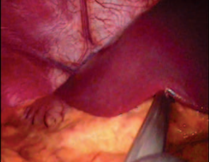
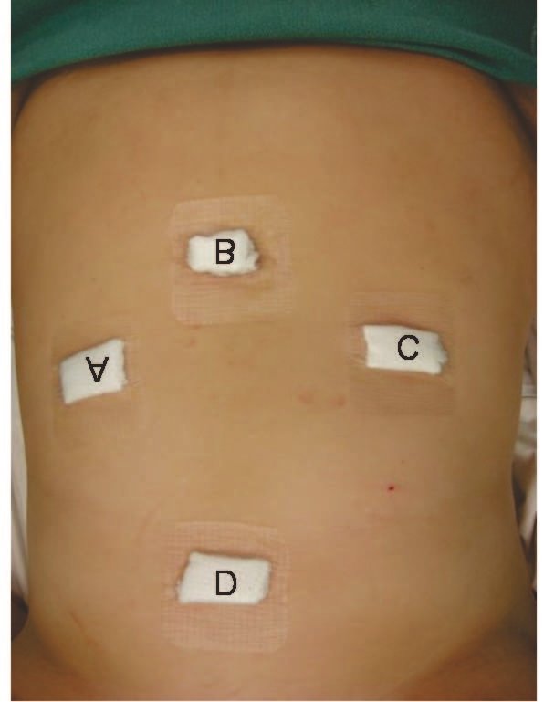
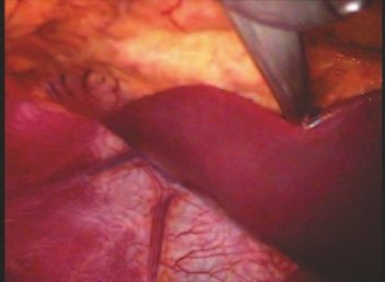
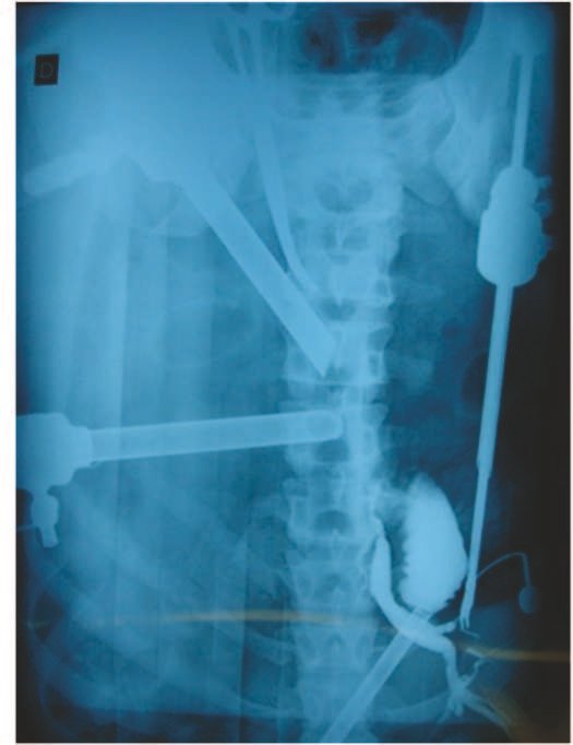
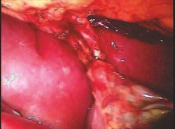
The patient was an overweight (BMI = 26.9) 16 year old adolescent female with an established diagnosis of situs inversus totalis, who presented with a four month history of biliary colic, that localized to the left hypochondrium. Chest radiograph, electrocardiogram, and ultrasound revealed dextrocardia, sinus rhythm and situs inversus totalis with the presence of multiple gallstones with an average diameter of 6 mm. The laparoscopic cholecystectomy was performed with the patient in the semi-lithotomy position with the surgeon between patient’s legs. The trocars were positioned as shown in Figure 1. After the optic was introduced, the mirrored anatomy of the abdominal organs was noted (Figure 2). The surgeon maneuvers his instruments through the pararectus trocars and performs the dissection of the infundibulum with the right-hand forceps while both the Assistant surgeon on the right and the Assistant surgeon on the left of the patient pull the bottom of the gall bladder postero-superiorly performed intraoperatively (Figure 3) to identify anatomical variations of the biliary tree; none was noted. After 90 minutes of surgery the gallbladder was removed through the umbilicus. The patient was discharged the next day.
In 1600, Fabricius described the transposition of the abdominal organs in a man.5 The first report of a laparoscopic cholecystectomy in a patient with situs inversus was published in 1991.5 Although it is a condition in which there is an alteration of the anatomy, there is no predisposition to gallbladder disease. The technical challenge performing a laparoscopic cholecystectomy in a patient with inversion of the abdominal organs – when confronted with the mirror image – consists in adapting the position of the surgeon, the Assistants, and the trocars for the dissection of the gallbladder hilum and the exposure of the gallbladder. Most reports in the literature describe the mirrored arrangement of both the trocars and the surgical team1,3,5 corresponding to the inversion of the abdominal organs. This positioning, which at first seems more logical, accentuate the cognitive bias and hampers the dissection of the Calot’s triangle. The surgeon is not accustomed to seeing the falciform ligament crossing superiorly and to the left across the video screen. There is constant crossing of the instruments as the base of gallbladder is brought forward, a frequent need for dissection with the left hand,4 and even placement of an extra trocar. 2 In this context it was suggested that laparoscopic cholecystectomy would be more easily performed by a left-handed surgeon.4 When operating between the legs of the patient, the adaption to the inversion of the position of the intracavitary organs seems faster. The surgeon performed the dissection of the gallbladder hilum with his right hand in the region anterior and posterior to Cabot’s triangle (Figure 4) and there were no crossing of the instruments. The camera and the forceps adjacent to the xiphoid were handled by both the first and second Assistant surgeons as needed during the surgeon’s dissection. The placement of clips and the sectioning of the cystic duct were performed with the surgeon’s left hand, while the catheter for cholangiography was inserted with the right hand. If a 10mm trocar is placed in the left flank, the surgeon
Autor : Prof. Dr. Pedro Péricles Ribeiro Baptista
Oncocirurgia Ortopédica do Instituto do Câncer Dr. Arnaldo Vieira de Carvalho
Consultório: Rua General Jardim, 846 – Cj 41 – Cep: 01223-010 Higienópolis São Paulo – S.P.
Fone:+55 11 3231-4638 Cel:+55 11 99863-5577 Email: drpprb@gmail.com

