This digital library houses the book on Oncology and Orthopedic Oncosurgery.

It includes academic lectures, presentations from national and international congresses, published papers, case discussions, performed surgical procedures, and proprietary techniques developed.

The digital format was chosen because the web allows the inclusion of texts with numerous visual resources, such as images and videos, which would not be possible in a printed book.

The content is intended for students, healthcare professionals, and the general public interested in the field.

Skull Osteoma Resection Technique

86654aa91382ffa290d510f1d2dd3353 3
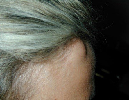
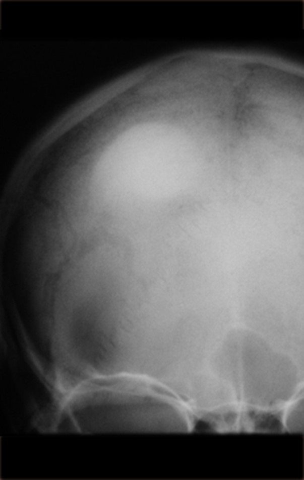
Skull Osteoma Resection Technique. Female patient, 48 years old, with a tumor on her forehead for three years. She reports a slow and progressive, painless appearance, which only makes aesthetics more difficult. She has not seen growth in the last year. Nodular, hard lesion, attached to deep planes, approximately three centimeters in diameter. Figures 1, 2 and 3 illustrate the clinical aspect of the lesion and figures 4 and 5 show the radiographic aspect of the image.

Skull osteoma resection technique

Figura 1: Nódulo firme, saliente, na base de implantação do cabelo.
Figura 2: Visão de perfil.
Figure 1: Firm, prominent nodule at the base of hair implantation. Figure 2: Profile view.
Figura 3: Paciente com o penteado encobrindo a lesão.
Figure 3: Patient with hairstyle covering the lesion.
Figura 4: Radiografia do crânio com lesão nodular, densa, homogênea, na calota.
Figure 4: X-ray of the skull with a nodular, dense, homogeneous lesion in the cap.
Figura 5: Detalhe da lesão de condensação óssea.
Figure 5: Detail of the bone condensation lesion.
To better document these images, we performed a computed tomography (Figures 6,7,8 and 9).
Figura 6: Tomografia axial do crânio com lesão acometendo as duas tábuas, com abaulamento maior da cortical externa.
Figure 6: Axial tomography of the skull with lesion affecting both tables, with greater bulging of the external cortex.
Figura 8 Tomografia com reconstrução em corte coronal, confirmando a íntima relação com a cortical externa.
Figure 7 : Coronal tomography showing that the lesion only affects the outer cortex.
Figure 8 : Tomography with reconstruction in coronal section, confirming the intimate relationship with the external cortex.
Figura 9: Reconstrução tomográfica em 3 dimensões da lesão do crânio.
Figure 9: 3-dimensional tomographic reconstruction of the skull lesion.
Figura 10: Tricotomia e planejamento da incisão cirúrgica.
Figure 10: Trichotomy and surgical incision planning.
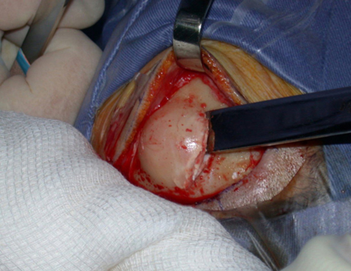
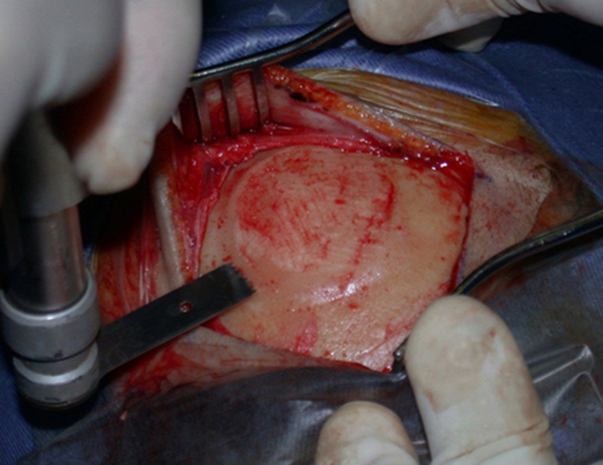
Analysis of the history, clinical picture and images of a homogeneous, compact lesion, with precise limits, producing mature bone, allowed the diagnosis of osteoma, with resection of this lesion for aesthetic reasons. The surgery was performed under general anesthesia and local infiltration to reduce bleeding (figures 10 to 20).
Figura 11: Assepsia e antissepsia, com colocação de campo plástico.
Figure 11: Asepsis and antisepsis, with plastic field placement.
Figura 12: Infiltração local com anestésico com vaso constritor.
Figure 12: Local infiltration with anesthetic with a constrictor vessel.
Figura 13: Incisão, hemostasia e descolamento do periósteo.
Figure 13: Incision, hemostasis and detachment of the periosteum.
Figura 14: Ostectomia com formão.
Figure 14: Ostectomy with chisel.
Figura 15: Superfície cruenta do leito operatório.
Figure 15: Bloody surface of the operating bed.
Figura 16: Regularização com serra elétrica.
Figure 16: Regularization with an electric saw.
Figura 17: Aplanamento com formão.
Figure 17: Flattening with a chisel.
Figura 18: Leito cirúrgico regularizado, sem saliências.
Figure 18: Regularized surgical bed, without protrusions.

Ostectomy with electric saw

The electric saw did not prove to be the most suitable instrument for performing ostectomy and regularization, as we can see. This was best done with the chisel (figure 17).
Figura 19: Fragmentos do osteoma ressecado.
Figure 19: Fragments of the resected osteoma.
Figura 20: Pós-operatório imediato.
Figure 20: Immediate postoperative period.

Author: Prof. Dr. Pedro Péricles Ribeiro Baptista

 Orthopedic Oncosurgery at the Dr. Arnaldo Vieira de Carvalho Cancer Institute

Leave a Reply

Hello! How can we assist you?