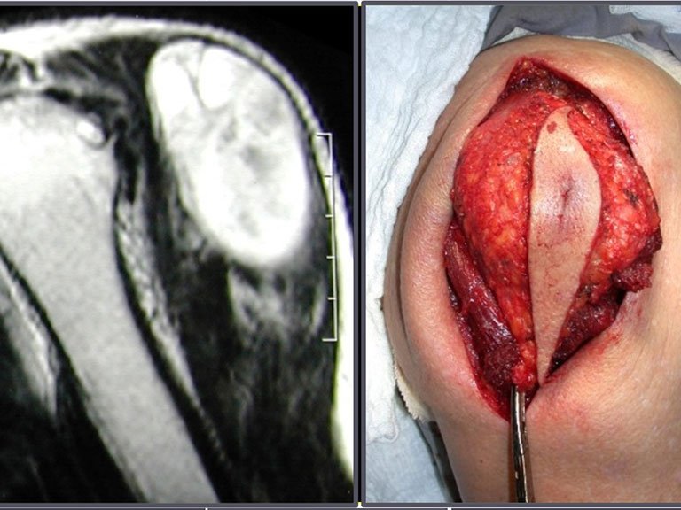
Sarcomas in Soft Tissues
Introduction:
Orthopedic oncological surgery encompasses the treatment of musculoskeletal lesions, including benign and malignant bone neoplasms, pseudo-tumor lesions and benign and malignant soft tissue neoplasms. Soft tissue sarcoma is a malignant neoplasm derived from the mesoderm, occurring in soft tissues, such as muscles, fascia, tendons, etc., differentiating itself from carcinomas, which have embryonic origin in the ectoderm.
Etiology:
Most soft tissue sarcomas do not have a defined cause, but some risk factors are well described, such as previous radiotherapy, lymphedema, Li-Fraumeni syndrome, type I neurofibromatosis, individual genetic propensity and HIV infection.
Incidence:
It is a rare tumor, representing approximately 12% of pediatric neoplasms and only 1% of all malignant tumors in adults. In the USA, there are an estimated 12,000 new cases per year, resulting in around 4,700 deaths. Around 60% of soft tissue sarcomas arise in the limbs, predominantly in the thigh, followed by the chest wall and retroperitoneum.
Classification:
The classification of soft tissue sarcoma is based on histological subtype such as liposarcoma, synovial sarcoma, rhabdomyosarcoma etc. Histological grade is also used, being divided into Grade 1 (well differentiated), Grade 2 (moderately differentiated) and Grade 3 (poorly differentiated).
Clinical condition:
The initial clinical picture is characterized by a palpable tumor bulge, often painless, with progressive growth, mainly in the thigh. Some patients may experience pain and paresthesia due to the compressive effect of the tumor. Fever or weight loss are exceptional symptoms.
Staging:
At diagnosis, soft tissue sarcoma rarely metastasizes, occurring more frequently as large-volume, deep-to-muscular, high-grade tumors. The pattern of dissemination is mainly hematogenous, with predominant lung metastases.
Imaging exams:
Exams include radiography, magnetic resonance imaging, tomography, PET-CT and scintigraphy. Biopsy is indicated for histological diagnosis, and can be performed in several ways, including percutaneous needle biopsy, incisional (surgical), ultrasound or tomography-guided biopsy.
Pathology:
The pathologist plays a crucial role in the diagnosis, carrying out examinations such as freezing, to ensure the representativeness of the sample, and subsequently microscopic study in paraffin, for histopathological diagnosis as well as determining the histological grade of the tumor. Immunohistochemistry is an important resource to complement the study of the sample.
Author: Prof. Dr. Pedro Péricles Ribeiro Baptista
Orthopedic Oncosurgery at the Dr. Arnaldo Vieira de Carvalho Cancer Institute
Office : Rua General Jardim, 846 – Cj 41 – Cep: 01223-010 Higienópolis São Paulo – SP
Phone: +55 11 3231-4638 Cell:+55 11 99863-5577 Email: drpprb@gmail.com

