This digital library houses the book on Oncology and Orthopedic Oncosurgery.

It includes academic lectures, presentations from national and international congresses, published papers, case discussions, performed surgical procedures, and proprietary techniques developed.

The digital format was chosen because the web allows the inclusion of texts with numerous visual resources, such as images and videos, which would not be possible in a printed book.

The content is intended for students, healthcare professionals, and the general public interested in the field.

Simple Bone Cyst of the Tibia

5ca1b0a18c411c3ebfc35c9dad7da921 3

Simple bone cyst of the tibia

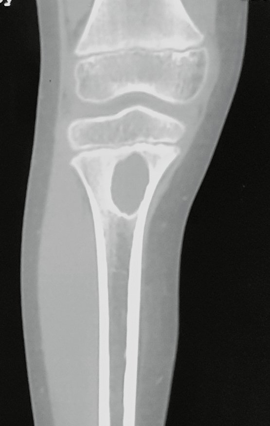
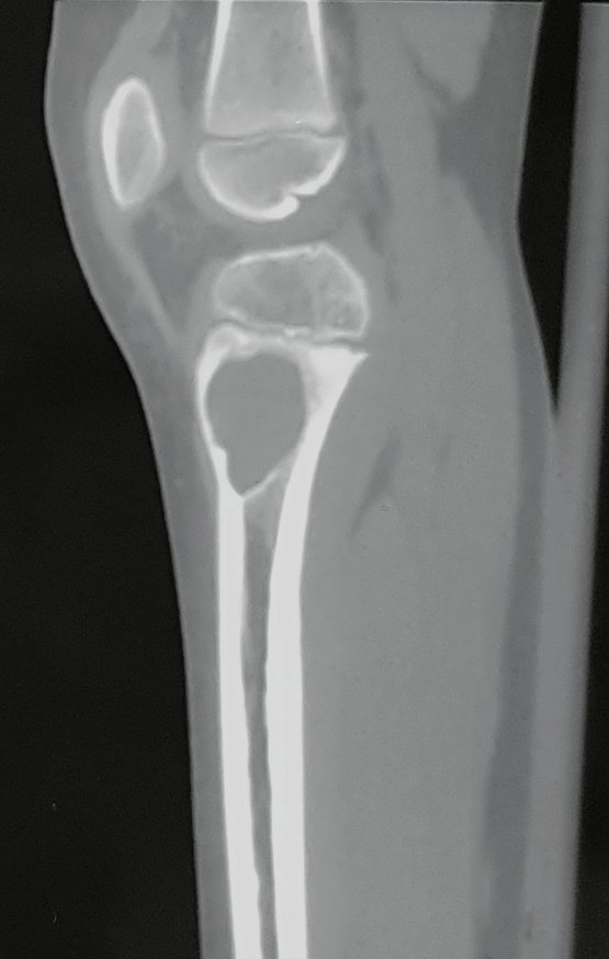
Simple Tibial Bone Cyst. An eight-year-old male patient suffered a fall with an abrasion on his right knee. He underwent an x-ray in the emergency room which revealed a bone rarefaction lesion in the proximal metaphysis of the right tibia, figures below.
Figura 1: Radiografia do 1/3 proximal da tíbia direita, frente, com lesão metafisária de rarefação óssea, de limites precisos.
Figure 1: Radiograph of the proximal 1/3 of the right tibia, front, with metaphyseal lesion of bone rarefaction, with precise limits.
Figura 2: Radiografia do 1/3 proximal da tíbia direita, perfil, com lesão metafisária de rarefação óssea, de limites precisos
Figure 2: Radiograph of the proximal 1/3 of the right tibia, profile, with metaphyseal lesion of bone rarefaction, with precise limits
Figura 3: Tomografia coronal com lesão metafisária cística.
Figure 3: Coronal tomography with cystic metaphyseal lesion.
Figura 4: Tomografia sagital com lesão metafisária cística.
Figure 4: Sagittal tomography with cystic metaphyseal lesion.
Figura 5: Tomografia axial, densidade de tecidos moles, com lesão metafisária cística.
Figure 5: Axial tomography, soft tissue density, with cystic metaphyseal lesion.
Figura 6: Tomografia axial, densidade de tecido ósseo, com lesão metafisária cística.
Figure 6: Axial tomography, bone tissue density, with cystic metaphyseal lesion.
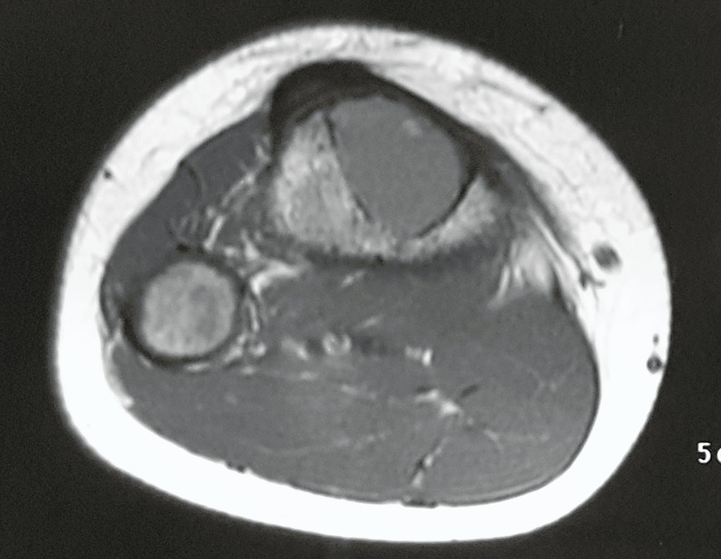
Figura 7: RM axial T1, com lesão de baixo sinal.
Figure 7: Axial T1 MRI, with low signal intensity lesion.
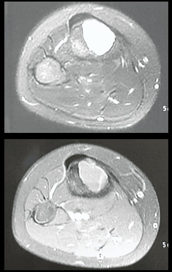
Figura 8: RM axial T1, com supressão de gordura e abaixo com contraste, apenas na periferia da lesão
Figure 8: Axial T1 MRI, with fat suppression and below with contrast, only at the periphery of the lesion
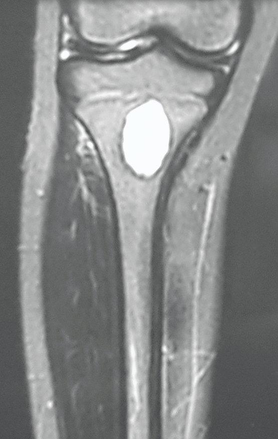
Figura 9: RM coronal T1, com supressão de gordura.
Figure 9: Coronal T1 MRI, with fat suppression.
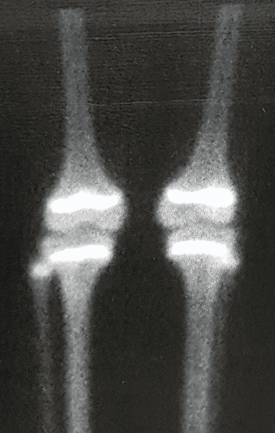
Figura 10: cintilografia pré-operatória, com captação aparentemente simétrica.
Figure 10: Preoperative scintigraphy, with apparently symmetric uptake.
Figura 11: cintilografia detalhada, com maior aumento, notando lesão cística em metáfise de tíba direita.
Figure 11: Detailed scintigraphy, with higher magnification, noting a cystic lesion in the metaphysis of the right tiba.
Figura 12: laudo de cintilografia óssea.
Figure 12: Bone scintigraphy report.

Author: Prof. Dr. Pedro Péricles Ribeiro Baptista

 Orthopedic Oncosurgery at the Dr. Arnaldo Vieira de Carvalho Cancer Institute

Hello! How can we assist you?