This digital library houses the book on Oncology and Orthopedic Oncosurgery.

It includes academic lectures, presentations from national and international congresses, published papers, case discussions, performed surgical procedures, and proprietary techniques developed.

The digital format was chosen because the web allows the inclusion of texts with numerous visual resources, such as images and videos, which would not be possible in a printed book.

The content is intended for students, healthcare professionals, and the general public interested in the field.

Simple Bone Cyst of the Femur

Bf13848f7f02f488b2e12e009a8b0df3 3

Simple Bone Cyst of the Femur

Simple Femoral Bone Cyst. A 12-year-old female patient, evaluating her dentition with a panoramic x-ray, was referred to a physiatrist due to the anatomical variation of the spine.

The physiatrist requested a scanogram of the lower limbs and recommended Pilates, without diagnosing the injury to the left femur. He later went to see a pediatrician who diagnosed the injury.

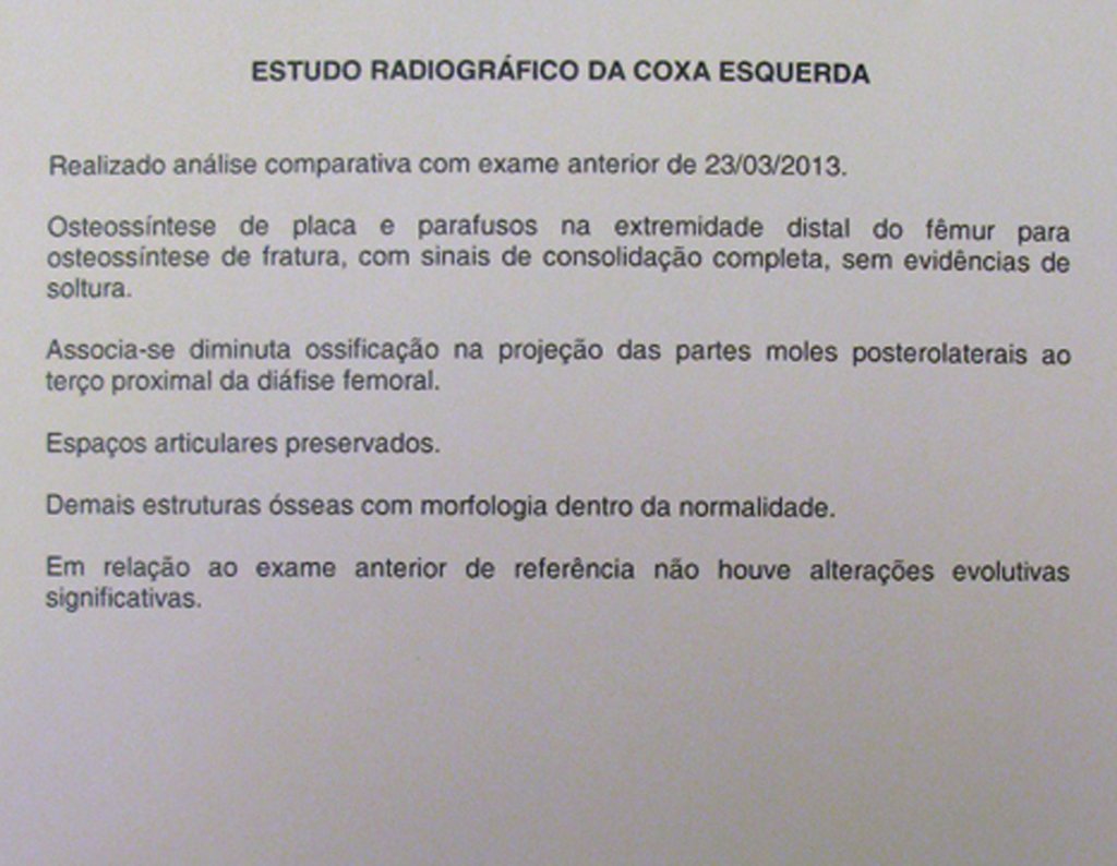
Radiografias do quadril frente e perfil do lado esquerdo Estudo radiográfico da coxa esquerda.
Front and lateral hip radiographs on the left side Radiographic study of the left thigh.
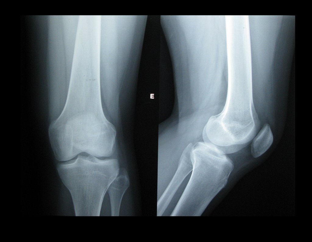
Radiografias do joelho frente e perfil
Front and lateral knee radiographs
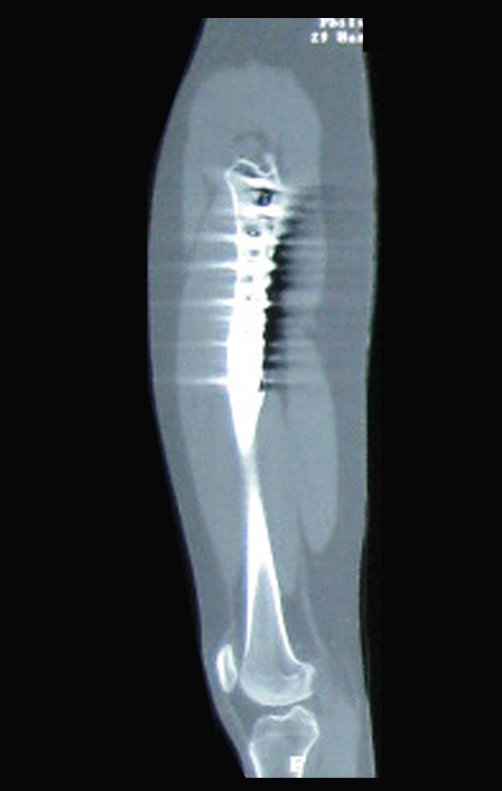
Tomografia computadorizada do quadril esquerdo/perfil.
Computed tomography of the left/front hip.
Tomografia computadorizada do quadril esquerdo/perfil.
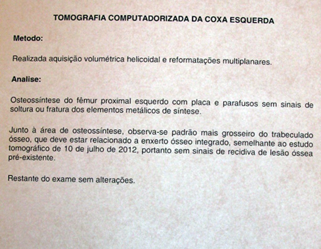
Computed tomography report of the left thigh. Computed tomography of the left hip/profile.
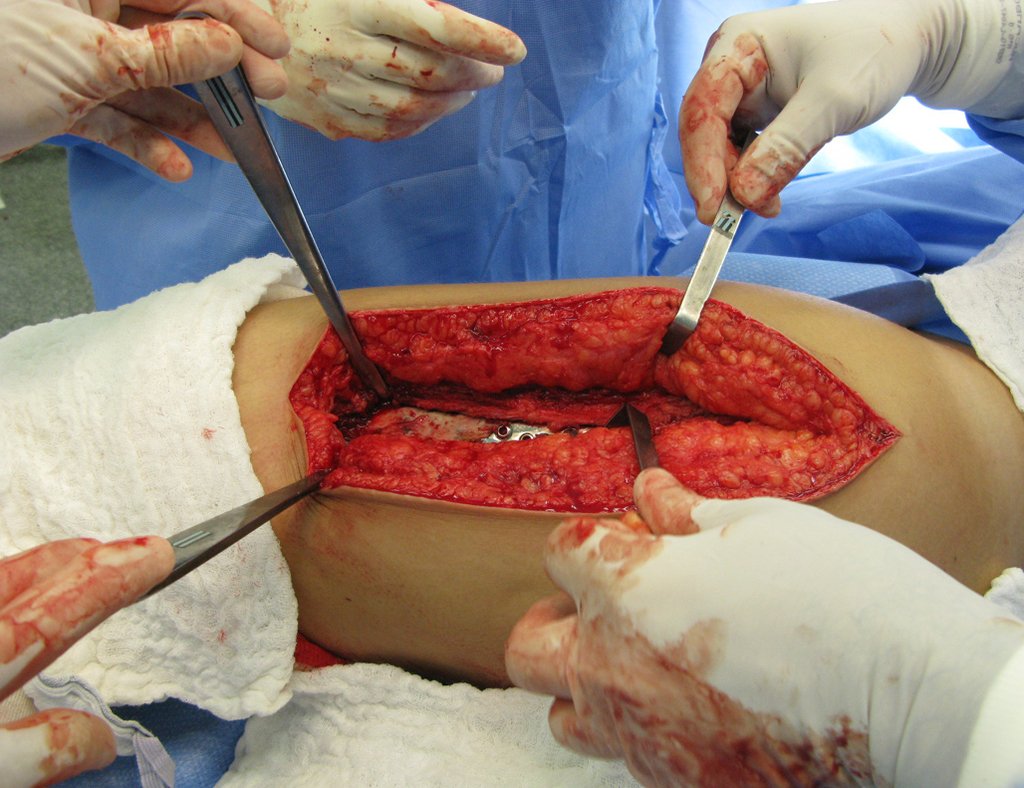
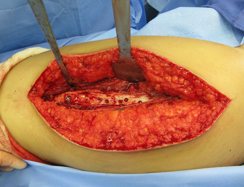
Incisão cirúrgica da coxa esquerda, onde se visualiza a placa.
Surgical incision on the left thigh, where the plaque is visible.
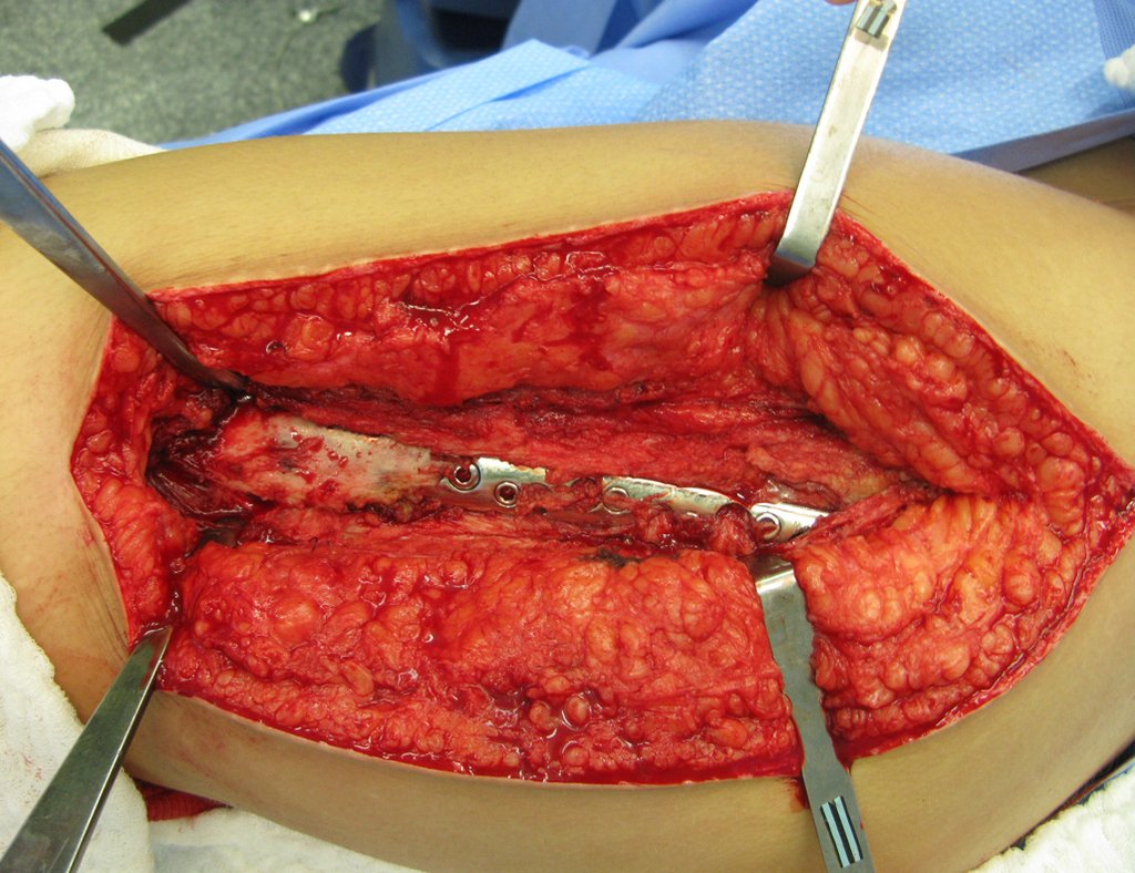
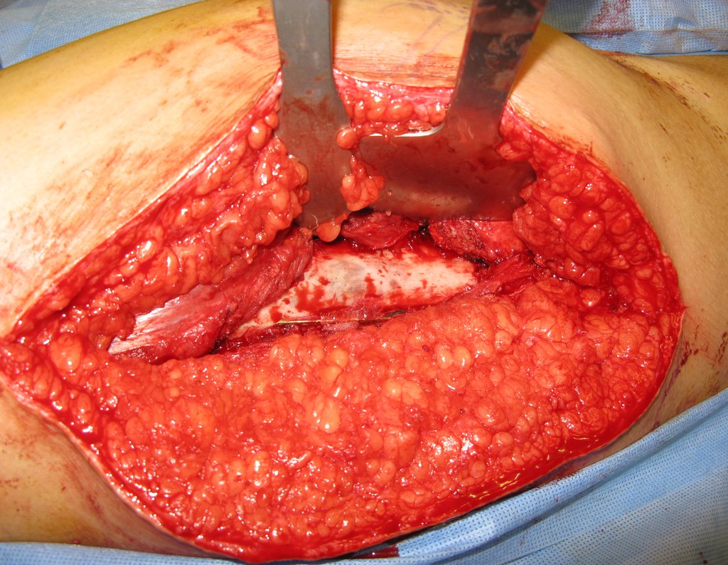
Nesta imagem podemos ver a placa sendo revestida por osso.
In this image we can see the plate being covered by bone.
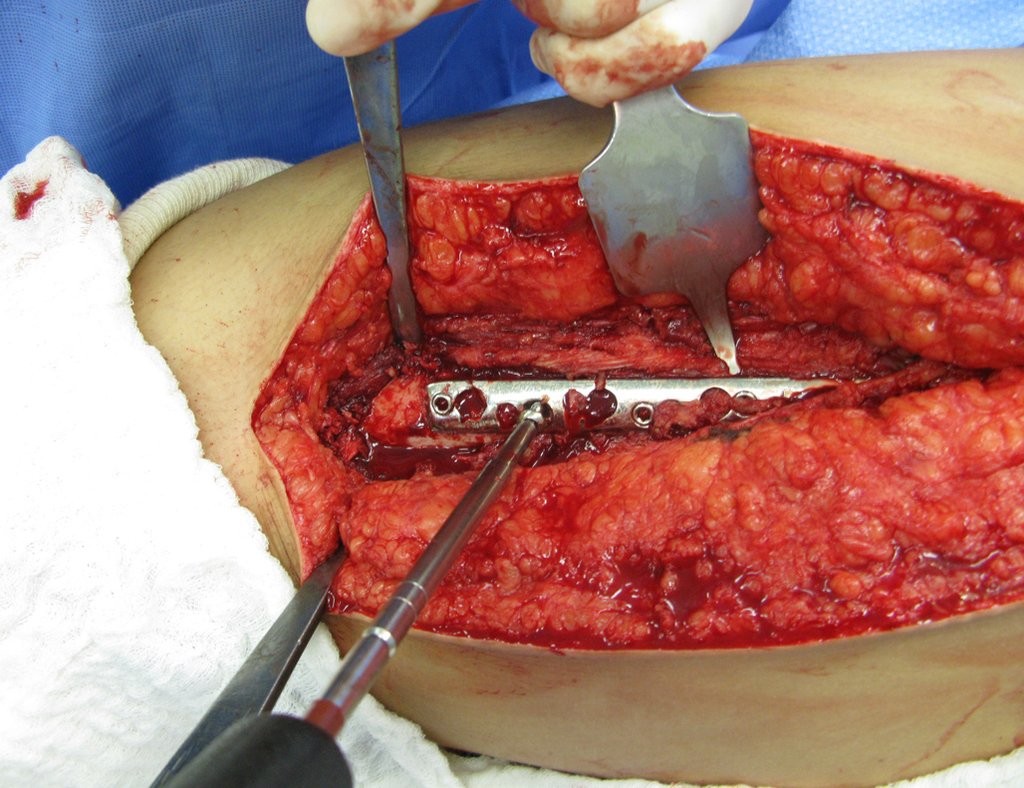
Início da retirada dos parafusos
Start of screw removal
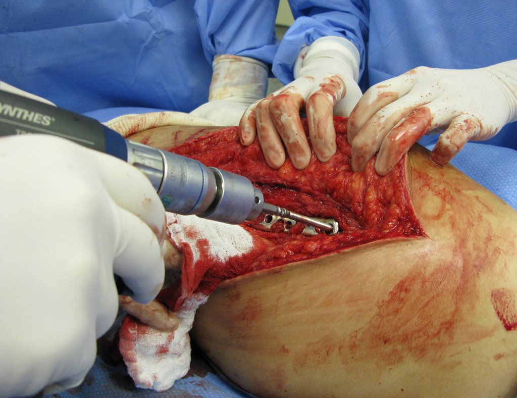
Foi usado também uma desparafusadeira para retirar demais parafusos.
A screwdriver was also used to remove other screws.
Após a retirada de todos os parafusos.
After removing all screws.
intra op
intra op
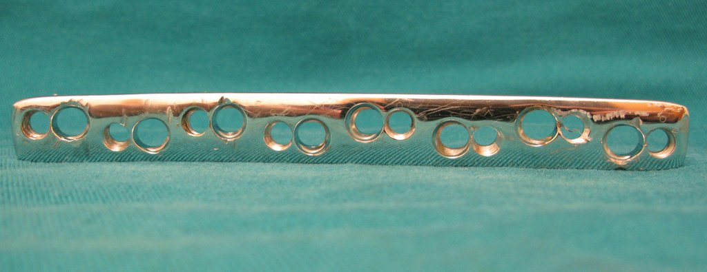
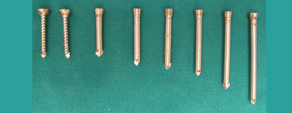
Os 8 parafusos retirados.
Plate removed. The 8 screws removed.
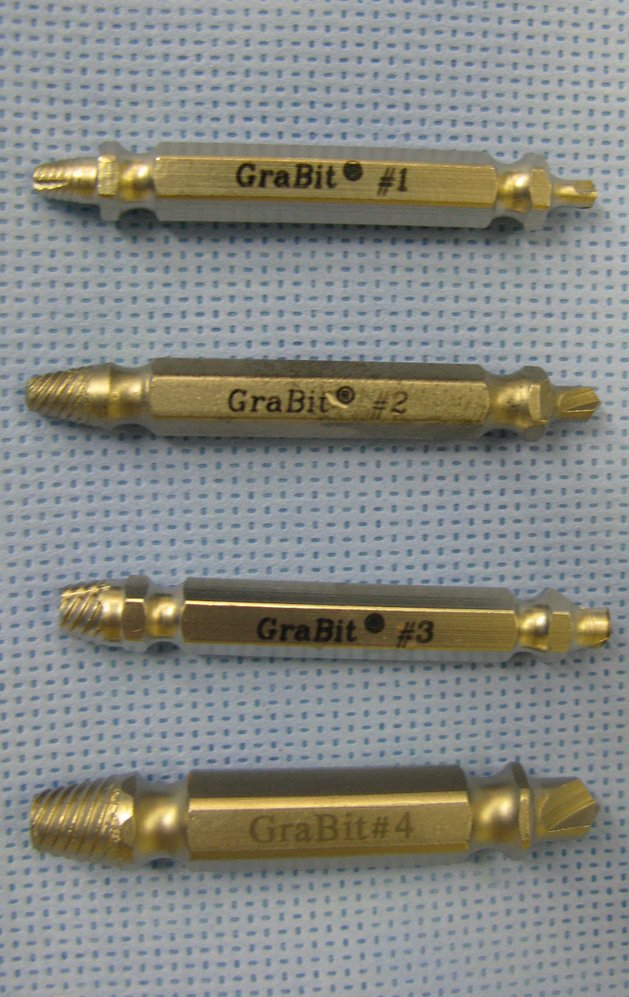
Nesta imagem podemos a chave abaixo, que foi usada para a retirada do parafuso.
In this image we can see the key below, which was used to remove the screw.
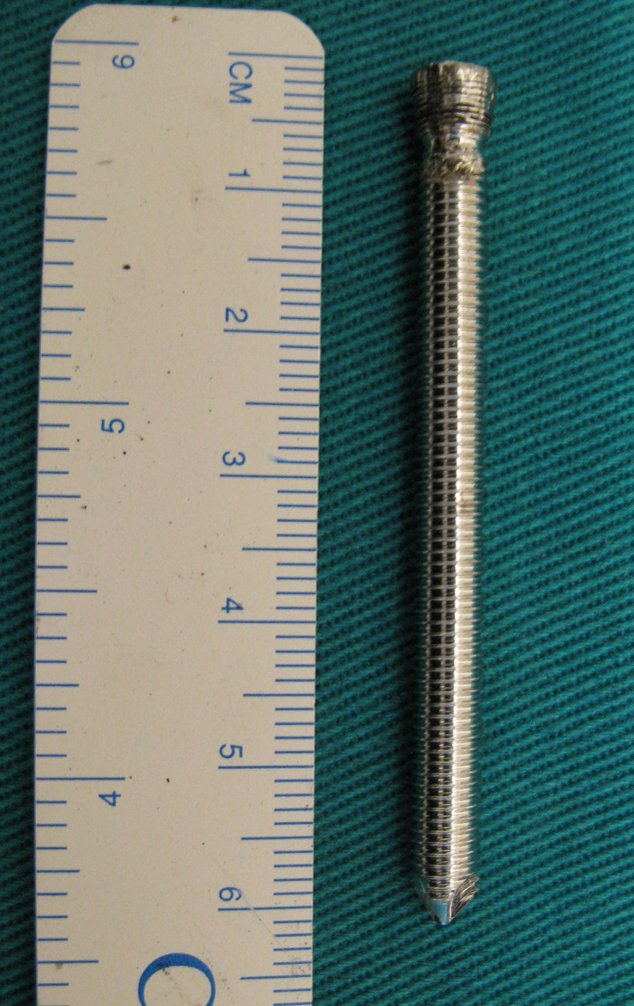
O tamanho de um dos parafusos retirados.
The size of one of the screws removed.
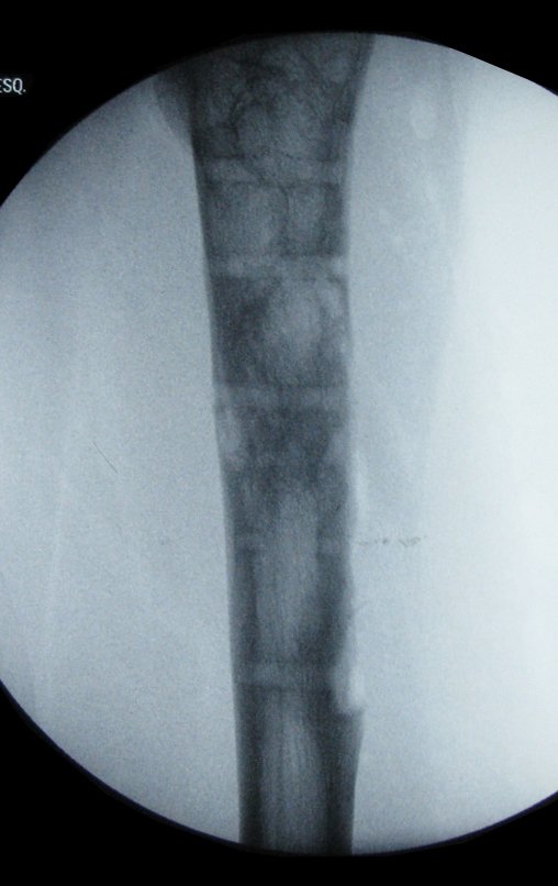
Nesta radiografia do quadril frente podemos visualizar a marca da retirada dos parafusos.
In this x-ray of the front hip we can see the mark of the screws being removed.
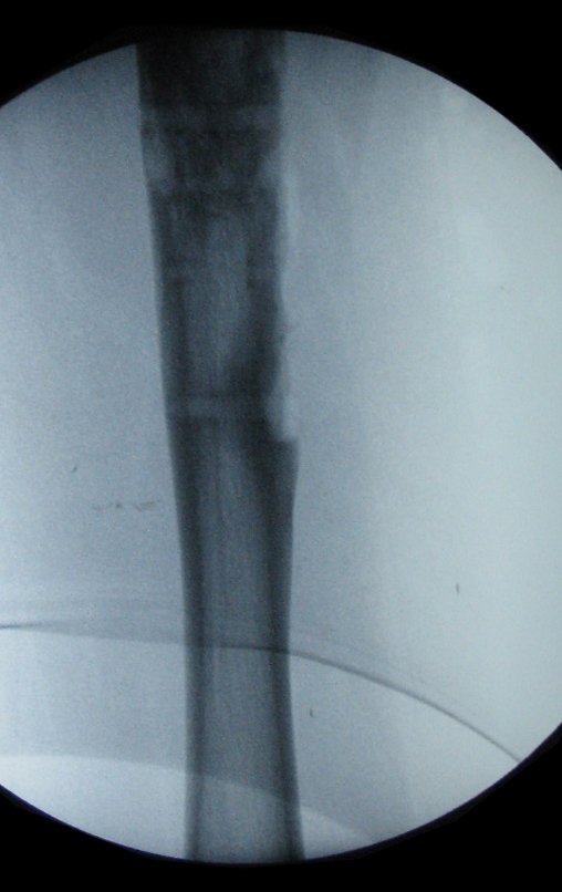
Nesta imagem em perfil do quadril podemos ver a marca dos parafusos no osso.
In this profile image of the hip we can see the mark of the screws in the bone.
Outra imagem mostrando a marca da retirada dos parafusos.
Another image showing the mark of the screws being removed.
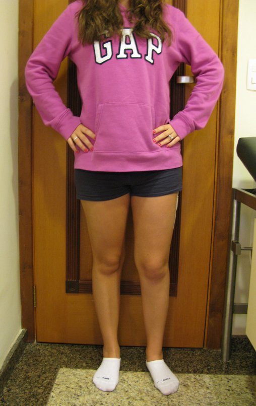
Paciente em posição ortostática.
Patient in orthostatic position.
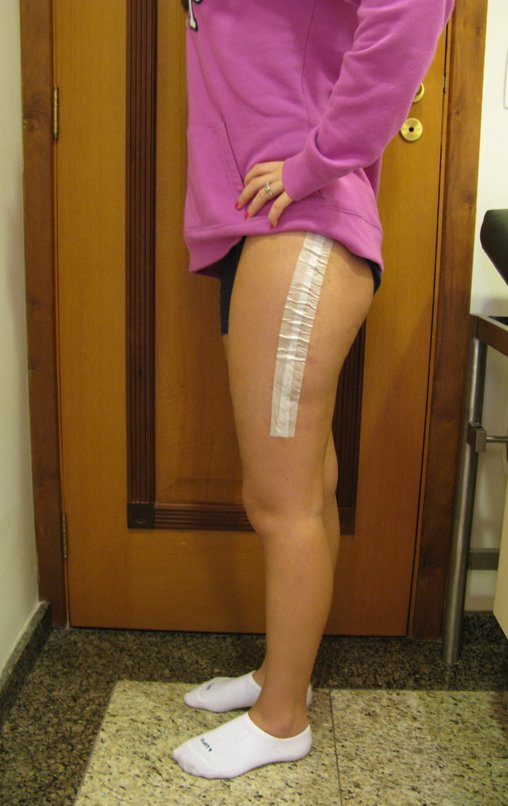
Paciente de perfil mostrando o curativo.
Patient in profile showing the dressing.
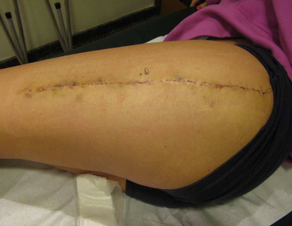
Cicatriz após a cirurgia.
Scar after surgery.
Patient returned to the office on 08/27/2014, after removing the dressing and stitches. According to the video, we can see the patient’s good recovery.

Author: Prof. Dr. Pedro Péricles Ribeiro Baptista

 Orthopedic Oncosurgery at the Dr. Arnaldo Vieira de Carvalho Cancer Institute

Hello! How can we assist you?