This digital library houses the book on Oncology and Orthopedic Oncosurgery.

It includes academic lectures, presentations from national and international congresses, published papers, case discussions, performed surgical procedures, and proprietary techniques developed.

The digital format was chosen because the web allows the inclusion of texts with numerous visual resources, such as images and videos, which would not be possible in a printed book.

The content is intended for students, healthcare professionals, and the general public interested in the field.

Sarcoma of the Popliteal Region

Sarcoma de la Región Poplítea

Soft Tissue Sarcoma of the Popliteal Region Adjuvant Radiotherapy

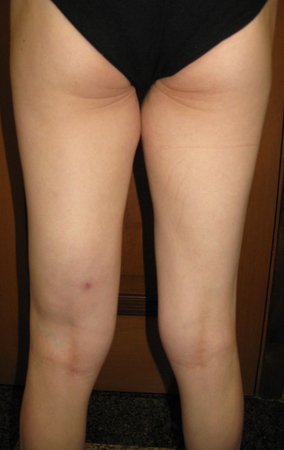
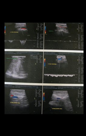
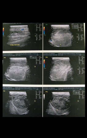
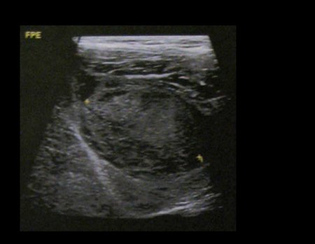
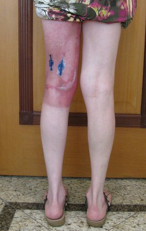
Sarcoma of the Popliteal Region. Female patient, 33 years old, reports a tumor on the posterior and distal aspect of the left thigh for a year, with greater growth in the last three months, during the end of pregnancy. After consultation with another professional, in which she underwent ultrasound examinations, bone scintigraphy, magnetic resonance imaging and biopsy, Figures 1-20, she was referred to us for evaluation and treatment.
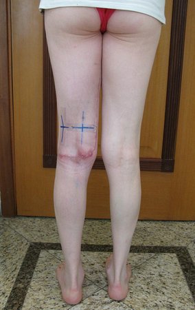
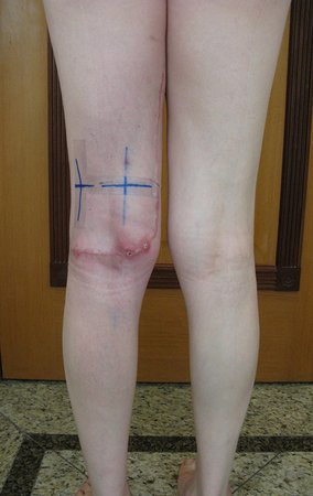
Figura 1: Observação clínica da face posterior da coxa esquerda, com abaulamento posterior no terço distal e cicatriz da biópsia realizada com agulha, em outro hospital.
Figure 1: Clinical observation of the posterior surface of the left thigh, with posterior bulging in the distal third and scar from the biopsy performed with a needle, in another hospital.
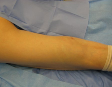
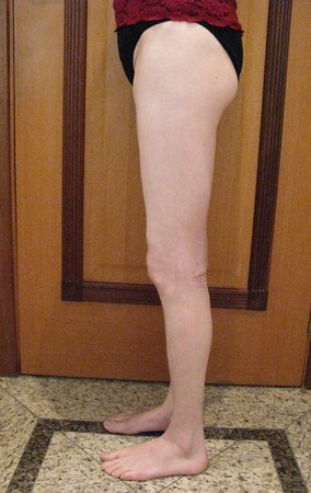
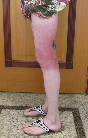
Figura 2: Perfil da coxa esquerda, com tumor abaulando o terço posterior e distal da coxa.
Figure 2: Profile of the left thigh, with tumor bulging the posterior and distal third of the thigh.
Figura 3: Ultrasonografia da coxa esquerda
Figure 3: Ultrasonography of the left thigh
Figura 4: Ultrasonografia da coxa esquerda
Figure 4: Ultrasonography of the left thigh
Figura 5: Ultrasonografia da coxa esquerda
Figure 5: Ultrasonography of the left thigh
Figura 6: Laudo da ultrasonografia da coxa esquerda.
Figure 6: Ultrasonography report of the left thigh.
Figura 7: Cintilografia óssea, fase angiográfica ou de fluxo.
Figure 7: Bone scintigraphy, angiographic or flow phase.
Figura 8: Cintilografia óssea, fase tardia.
Figure 8: Bone scintigraphy, late phase.
Figura 9: Laudo da cintilografia óssea.
Figure 9: Bone scintigraphy report.
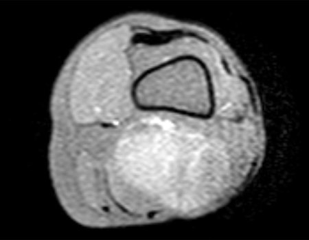
Figura 10: Rm cor T1, com lesão de tecidos moles no terço distal da coxa esquerda com baixo sinal.
Figure 10: T1 color MRI, with soft tissue lesion in the distal third of the left thigh with low signal intensity.
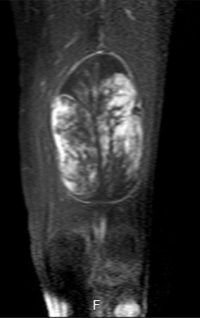
Figura 11: Rm sag T1 com supressão de gordura, lesão de tecidos moles na face posterior e distal da coxa esquerda.
Figure 11: MRI sag T1 with fat suppression, soft tissue injury on the posterior and distal aspect of the left thigh.
Figura 12: Rm ax T1 com saturação de gordura, lesão de tecidos moles posterior da coxa esquerda.
Figure 12: MRI ax T1 with fat saturation, posterior soft tissue injury of the left thigh.
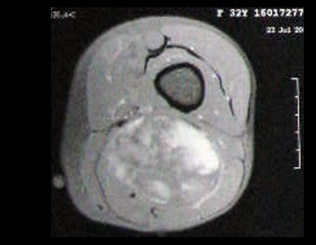
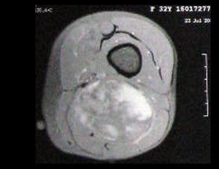
Figura 13: Rm ax T1 com saturação de gordura e contraste, lesão heterogênea, com áreas de baixo sinal, sinal intermediário e alto sinal, captação de contraste no interior da lesão, revelando neoplasia agresiva.
Figure 13: MRI ax T1 with fat saturation and contrast, heterogeneous lesion, with areas of low signal, intermediate signal and high signal, contrast uptake inside the lesion, revealing aggressive neoplasia.
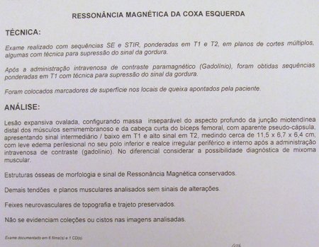
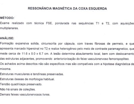
Figura 14: Laudo da ressonância magnética.
Figure 14: MRI report.
Figura 15: Radiografia de tórax PA, normal.
Figure 15: PA chest x-ray, normal.
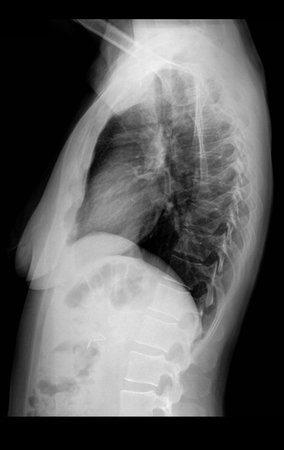
Figura 16: Radiografia de tórax perfil, normal.
Figure 16: Normal lateral chest x-ray.
Figura 17: Tomografia do tórax sem alteração. Sem sinais de lesão secundária.
Figure 17: Chest tomography without changes. No signs of secondary injury.
Figura 18: Radiografia do fêmur frente.
Figure 18: X-ray of the front femur.
Figura 19: Radiografia do fêmur, perfil revela lesão de tecidos moles com aumento de densidade na face posterior e distal da coxa.
Figure 19: X-ray of the femur, profile view reveals soft tissue injury with increased density on the posterior and distal aspect of the thigh.
Figura 20: Radiografia de bacia frente, normal.
Figure 20: X-ray of the front pelvis, normal.
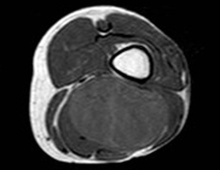
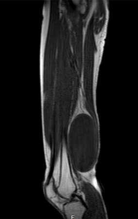
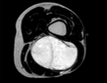
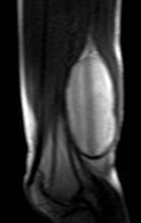
It is instructive to comment that some requested tests could be waived. Bone scintigraphy for this soft tissue injury is an exam that does not add value, as well as radiography of the pelvis, since the lesion is not in this location, the best imaging test to evaluate soft tissue injury is magnetic resonance imaging. new resonance, seeking to obtain better definition of the images, without granulation and in all routine weightings, aiming to evaluate in detail the relationship of the lesion with the vascular and nevus bundles. Figures 21 to 33.
Figura 21: Rm ax, lesão com baixo sinal em T1.
Figure 21: Rm ax, lesion with low signal on T1.
Figura 22: Rm cor, lesão com baixo sinal em T1.
Figure 22: Color MRI, lesion with low signal on T1.
Figura 23: Rm sag, lesão com baixo sinal em T1.
Figure 23: Rm sag, lesion with low signal on T1.
Figura 24: Rm ax, lesão com alto sinal em T2 spir.
Figure 24: MRI ax, lesion with high signal on T2 spir.
Figura 25: Rm cor, lesão com alto sinal em T2.
Figure 25: Color MRI, lesion with high signal on T2.
Figura 26: Rm sag, lesão com alto sinal em T2.
Figure 26: Rm sag, lesion with high signal on T2.
Figura 27: Rm apresenta lesão com sinal intermediário e alto sinal em ax T1 com supressão de gordura.
Figure 27: Rm presents a lesion with intermediate signal and high signal on ax T1 with fat suppression.
Figura 28: Rm ax FFE.
Figure 28: Rm ax FFE.
Figura 29: Rm ax T1 spir, com baixo sinal e captação de contraste.
Figure 29: Rm ax T1 spir, with low signal and contrast uptake.
Figura 30: Rm cor T1 spir, com baixo sinal, vascularização e captação de contraste.
Figure 30: MRI color T1 spir, with low signal, vascularization and contrast uptake.
Figura 31: Rm sag T1 spir, com baixo sinal e captação de contraste.
Figure 31: Rm sag T1 spir, with low signal and contrast uptake.
Figura 32: Laudo da segunda ressonância magnética.
Figure 32: Report of the second magnetic resonance imaging.
Figura 33: Laudo da biópsia.
Figure 33: Biopsy report.
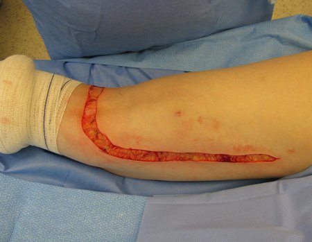
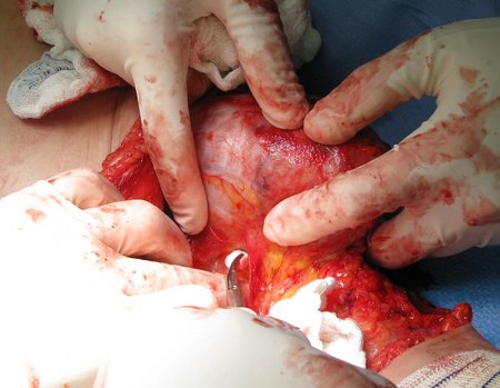
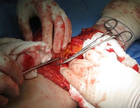
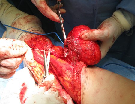
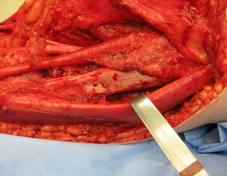
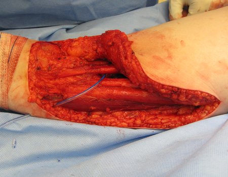
Tumor resection surgery was performed on 10/28/2014. We indicate an incision that begins at the posterior fold of the knee and goes proximally over the path of the internal saphenous muscle, to provide access to the entire path of the artery, vein and femoral nerve, figures 34 to 55.
Figura 34: Paciente em decúbito prono, cuidados de asepssia e antissepsia.
Figure 34: Patient in prone position, asepsis and antisepsis care.
Figura 35: Incisão póstero medial da coxa, curvando-se horizontalmente sobre a interlinha articular do joelho, sem uso de garroteamento do membro.
Figure 35: Posteromedial thigh incision, curving horizontally over the knee joint line, without using a tourniquet of the limb.
Figura 36: Hemostasia cuidadosa com eletrocautério e abertura do subcutãneo e fascia medial.
Figure 36: Careful hemostasis with electrocautery and opening of the subcutaneous tissue and medial fascia.
Figura 37: Prossegue-se dissecando todo o subcutâneo da incisão cirúrgica e cauterizando os vasos sangrantes, para uma cirurgia segura.
Figure 37: Continue dissecting the entire subcutaneous layer of the surgical incision and cauterizing the bleeding vessels, for safe surgery.
Figura 38: Afasta-se o músculo bíceps femoral, expondo o nervo ciático.
Figure 38: The biceps femoris muscle moves away, exposing the sciatic nerve.
Figura 39: Abertura do perineuro e liberação cuidadosa do nervo ciático, liberando-o do tumor.
Figure 39: Opening the perineurium and carefully releasing the sciatic nerve, freeing it from the tumor.
Figura 40: Dissecção do septo para liberação do polo distal da lesão.
Figure 40: Dissection of the septum to release the distal pole of the lesion.
Figura 41: Ligadura dos vasos nutrícios do tumor.
Figure 41: Ligation of the tumor's nutrient vessels.
Figura 42: Continua-se liberando a lesão no plano profundo, afastando-se os vasos femorais e expondo a extremidade distal do músculo envolvido para sua secção.
Figure 42: Continue releasing the lesion in the deep plane, moving the femoral vessels apart and exposing the distal end of the muscle involved for sectioning.
Figura 43: Exposição do segmento do músculo semimembranoso acometido pela lesão.
Figure 43: Exposure of the semimembranous muscle segment affected by the injury.
Figura 44: O corte do músculo afetado é feito preferencialmente com o eletrocautério, mantendo-se uma margem de tecido macroscopicamente sadio.
Figure 44: The affected muscle is cut preferably using electrocautery, maintaining a margin of macroscopically healthy tissue.
Figura 45: Separa-se o feixe muscular e prende-se as duas extremidades com pinças, para a secção e ligadura.
Figure 45: The muscle bundle is separated and the two ends are clamped with forceps for sectioning and ligation.
Figura 46: A ligadura deve ser realizada nas duas extremidades, para uma cirurgia limpa e oncológica .
Figure 46: The ligation must be performed on both ends, for a clean and oncological surgery.
Figura 48: Secção do segmento muscular envolvido, mantendo uma boa margem de tecido macroscopicamente sadio junto com a peça.
Figure 47: The muscle is sectioned and must be ligated into small bundles to ensure hemostasis.
Figura 48: Secção do segmento muscular envolvido, mantendo uma boa margem de tecido macroscopicamente sadio junto com a peça.
Figure 48: Section of the involved muscle segment, maintaining a good margin of macroscopically healthy tissue along with the piece.
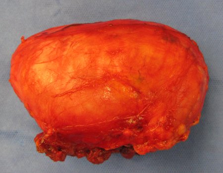
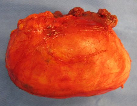
Figura 49: Peça ressecada, vista profunda.
Figure 49: Dried piece, deep view.
Figura 50: Peça ressecada, vista lateral.
Figure 50: Dried piece, side view.
Figura 51: Peça ressecada, vista medial.
Figure 51: Resected piece, medial view.
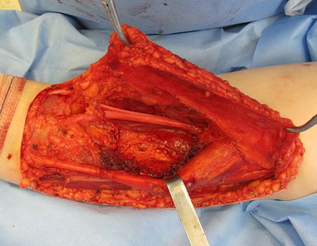
Figura 52: Leito operatório após a retirada do tumor, com a margem possível nesta região.
Figure 52: Operative bed after removal of the tumor, with the possible margin in this region.
Figura 53: Clips metálicos são colocados para delimitar o sítio anterior do tumor, fascilitando ao radioterapeuta.
Figure 53: Metal clips are placed to delimit the previous site of the tumor, facilitating the radiotherapist.
Figura 54: Mesmo com uma boa hemostasia, a colocação de dreno aspirativo é bem indicada.
Figure 54: Even with good hemostasis, the placement of an aspiration drain is well indicated.
Figura 55: Fechamento cuidadoso por planos.
Figure 55: Careful closure by planes.
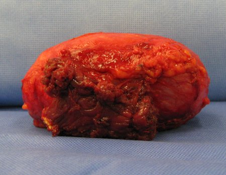
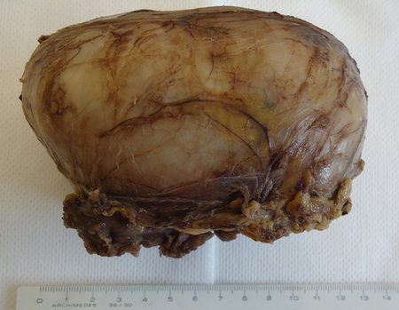
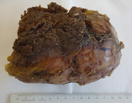
The resected piece was sent for pathological anatomical study, figures 56 to 61.
Figura 56: Peça ressecada em bloco, com a margem oncológica possível, dada a localização do tumor.
Figure 56: Piece resected en bloc, with the possible oncological margin, given the location of the tumor.
Figura 57: Peça ressecada em bloco, com a margem oncológica possível, dada a localização do tumor, vista medial
Figure 57: Piece resected en bloc, with the possible oncological margin, given the location of the tumor, medial view
Figura 58: Cortes da peça cirúrgica, marcada com nankin, para estudo da margem.
Figure 58: Sections of the surgical specimen, marked with nankin, to study the margin.
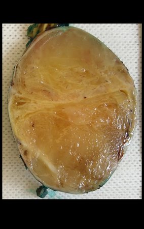
Figura 59: Peça cortada ao meio, para estudo da macroscopia, lado A.
Figure 59: Piece cut in half, for macroscopic study, side A.
Figura 60: Peça cortada ao meio, para estudo da macroscopia, lado B.
Figure 60: Piece cut in half, for macroscopic study, side B.
Figura 61: Laudo da peça cirúrgica.
Figure 61: Report of the surgical specimen.
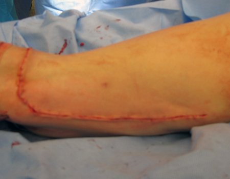
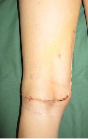
Figura 62: Pós operatório de duas semanas.
Figure 62: Two weeks post-operative.
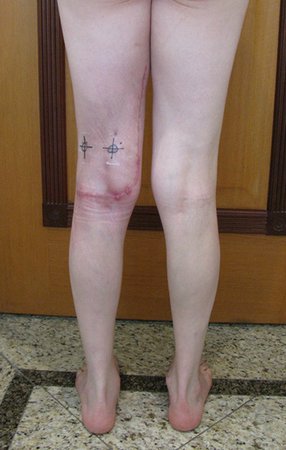
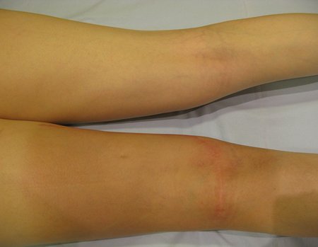
Figura 63: Pós operatório de um mês. Aspecto cosmético da cicatriz
Figure 63: One month post-operative. Cosmetic appearance of the scar
Figura 64: Pós operatório de um mês, perfil com carga. Boa função.
Figure 64: One month post-operative, profile with weight. Good function.
Figura 65: Pós operatório de um mês, flexão com carga.
Figure 65: One month post-operative, flexion with weight.
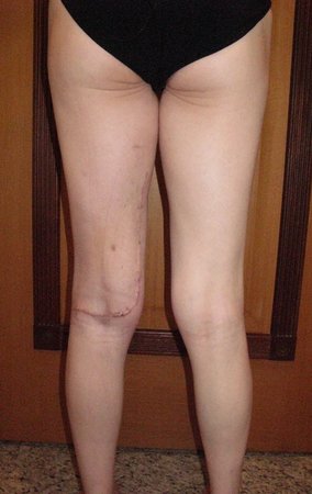
Figura 66: Pós operatório de sete semanas, aguardando cicatrização adequada e planejamento para inicio de radioterapia adjuvante.
Figure 66: Seven weeks post-operative, awaiting adequate healing and planning for the start of adjuvant radiotherapy.
Figura 67: Pós operatório de sete semanas, em programação para radioterapia adjuvante, dezembro de 2014.
Figure 67: Seven weeks post-operative, scheduled for adjuvant radiotherapy, December 2014.
After discussing the case again with the oncologists, adjuvant chemotherapy (after surgery) was not indicated, nor was neoadjuvant use recommended (before surgery). With the healing of the surgical wound progressing satisfactorily, we indicated the consolidation of local treatment with adjuvant radiotherapy.
Figura 68: Paciente após dois meses da cirurgia, em 30/12/2014, com boa cicatrização, apta ao tratamento re radioterapia adjuvante.
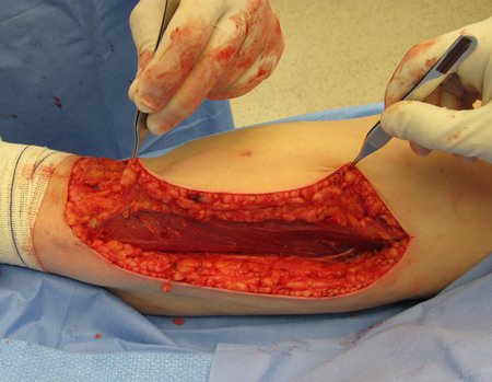
Figure 68: Patient two months after surgery, on 12/30/2014, with good healing, suitable for adjuvant radiotherapy treatment.
Figura 69: A radioterapia tem sua efiácia no tratamento local, mas também apresenta efeito colateral, como se tivesse ocorrido uma exposição solar exagerada, uma ¨queimadura¨.
Figure 69: Radiotherapy is effective in local treatment, but it also has side effects, as if excessive sun exposure had occurred, a ¨burn¨.
Figura 70: Aspecto da pele após aplicações de radioterapia, em 26/01/2015
Figure 70: Appearance of the skin after radiotherapy applications, on 01/26/2015
Figura 71: Aspecto da pele lateral da coxa, após aplicações de radioterapia, em 26/01/2015.
Figure 71: Appearance of the lateral skin of the thigh, after radiotherapy applications, on 01/26/2015.
Figura 72: Em detalhe, visão da face posterior da coxa, após término da radioterapia adjuvante.
Figure 72: In detail, view of the posterior aspect of the thigh, after completion of adjuvant radiotherapy.
Figura 73: Após o término das sessões de radioterapia, ocorre recuperação do tecido, Aspecto após a radioterapia, em 15/04/2015
Figure 73: After the end of radiotherapy sessions, tissue recovery occurs, Appearance after radiotherapy, on 04/15/2015
Figura 74: Evidente melhora progressiva do aspecto cosmético, em 11/05/2015.
Figure 74: Evident progressive improvement in the cosmetic appearance, on 05/11/2015.
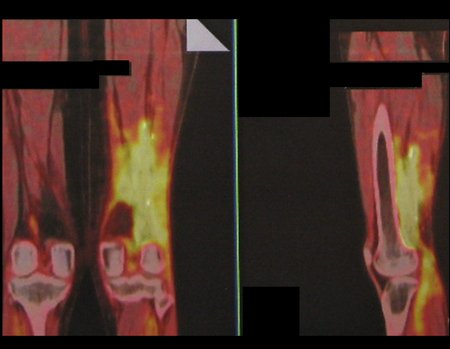
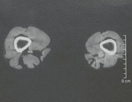
Figura 75: Pet-cet em novembro de 2015, após um ano da cirurgia. Este exame representa a fotografia inicial para o controle comparativo do acompanhamento pós tratamento.
Figure 75: Pet-cet in November 2015, one year after surgery. This examination represents the initial photograph for comparative control of post-treatment follow-up.
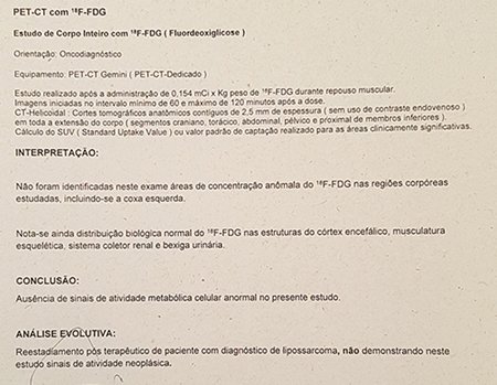
Figura 76: Laudo do Pet-Cet, com uma interpretação equivocada, sugerindo "atividade da doença de base". Na realidade trata-se da expressão inflamatória residual da radioterapia, SUV 2.0, baixo. Os "focos de calcificação" nada mais são do que os clips de demarcação do leito operatório, para orientação da radioterapia.
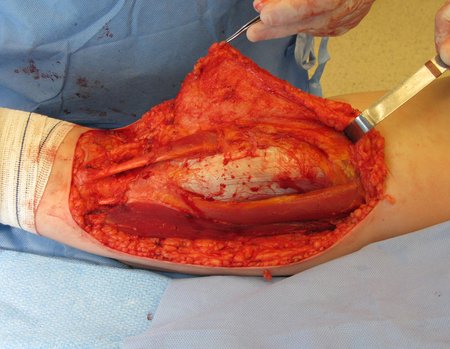
Figure 76: Pet-Cet report, with a mistaken interpretation, suggesting "activity of the underlying disease". In reality, it is the residual inflammatory expression of radiotherapy, SUV 2.0, low. The "calcification foci" are nothing more than the demarcation clips of the surgical bed, to guide radiotherapy.
Figura 77: Rm sagital após um ano, sem sinais de recorrência.
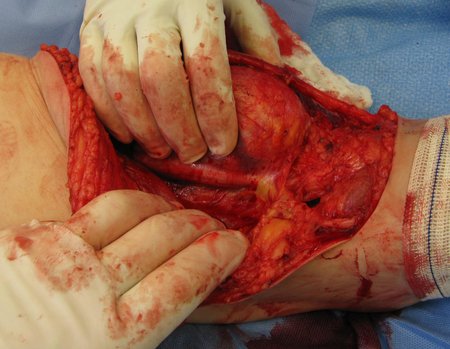
Figure 77: Sagittal MRI after one year, with no signs of recurrence.
Figura 78: Rm coronal após um ano. As setas assinalam a presença dos clips metálicos marcadores do leito cirúrgico para a radioterapia.
Figure 78: Coronal Rm after one year. The arrows indicate the presence of metallic clips marking the surgical bed for radiotherapy.
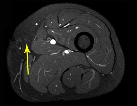
Figura 79: Rm axial após um ano da cirurgia, sem sinal de recidiva. A seta amarela assinala a cicatriz do acesso cirúrgico.
Figure 79: Axial MRI one year after surgery, with no sign of recurrence. The yellow arrow marks the surgical access scar.
Figura 80: Flexão simétrica, com carga total.
Figure 80: Symmetrical bending, with full load.
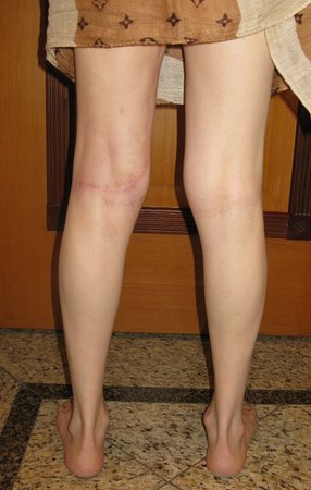
Figura 81: Pele posterior da coxa com hiperpigmentação sequelar da radioterapia. Evolução do aspecto cosmético em dezembro de 2015.
Figure 81: Posterior skin of the thigh with hyperpigmentation resulting from radiotherapy. Evolution of the cosmetic appearance in December 2015.
Figura 82: Avaliação em maio de 2016, após um ano e sete meses da cirurgia.
Figure 82: Assessment in May 2016, one year and seven months after surgery.
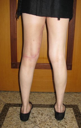
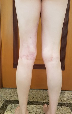
Figura 83: Recuperação bastante satisfatória do aspecto cosmético, em fevereiro de 2017.
Figure 83: Very satisfactory recovery of the cosmetic appearance, in February 2017.
Figura 84: Rm axial em fevereiro de 2017, após 2 e1/2 anos da cirurgia.
Figure 84: Axial MRI in February 2017, 2 and 1/2 years after surgery.
Figura 85: Laudo da ressonância de controle de fevereiro de 2016, sem sinais de recidiva.
Figure 85: Control MRI report from February 2016, with no signs of recurrence.
Analysis of the history, clinical picture and images of a homogeneous, compact lesion, with precise limits, producing mature bone, allowed the diagnosis of osteoma, with resection of this lesion for aesthetic reasons. The surgery was performed under general anesthesia and local infiltration to reduce bleeding (figures 10 to 20).

Author: Prof. Dr. Pedro Péricles Ribeiro Baptista

 Orthopedic Oncosurgery at the Dr. Arnaldo Vieira de Carvalho Cancer Institute

Hello! How can we assist you?