This digital library houses the book on Oncology and Orthopedic Oncosurgery.

It includes academic lectures, presentations from national and international congresses, published papers, case discussions, performed surgical procedures, and proprietary techniques developed.

The digital format was chosen because the web allows the inclusion of texts with numerous visual resources, such as images and videos, which would not be possible in a printed book.

The content is intended for students, healthcare professionals, and the general public interested in the field.

Sacral Chondrosarcoma

29a83a8a9641bb860a679d7e5ba52d26 3

Sacral chondrosarcoma - Partial sacrectomy - Videolaparoscopy

Sacral chondrosarcoma. Male patient, tradesman, 50 years old, with a tumor in the left sacral iliac region, since he was 5 years old, with progressive increase and worsening for 4 months, with bladder sphincter compression, which requires him to use an indwelling bladder catheter, Figures 1 – 17.
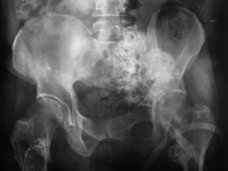
Figura 1: Radiografia de bacia frente revelando multiplos osteocondromas na bacia e nas metáfises proximais dos dois fêmures. Na Região sacro ilíaca a esquerda observa-se uma grande lesão acometende o hemisacro e a pelve, com características de degeneração sarcomatosa para condrossarcoma.
Figure 1: Front pelvic X-ray revealing multiple osteochondromas in the pelvis and proximal metaphyses of both femurs. In the sacral iliac region on the left, a large lesion is observed affecting the hemisacrum and pelvis, with characteristics of sarcomatous degeneration to chondrosarcoma.
Figura 2: Radiografia de bacia em obturatriz, ressaltando o condrossarcoma na região sacro-ilíaca esquerda.
Figure 2: X-ray of the pelvis in obturatrix, highlighting the chondrosarcoma in the left sacroiliac region.
Figura 3: Radiografia obliqua da bacia, que evidencia condrossarcoma envolvendo a articulação sacro ilíaca esquerda, com comprometimento da topografia das raízes nos forames sacrais.
Figure 3: Oblique radiography of the pelvis, which shows chondrosarcoma involving the left sacroiliac joint, with compromised topography of the roots in the sacral foramina.
Figura 4: Radiografia em perfil do sacro evidenciando condrossarcoma.
Figure 4: Lateral x-ray of the sacrum showing chondrosarcoma.
Figura 5: Radiografia da hemipelve esquerda e região sacral, afetada pelo condrossarcoma.
Figure 5: Radiograph of the left hemipelvis and sacral region, affected by chondrosarcoma.
Figura 6: Tomografia axial evidenciando o limite proximal da lesão infiltrando a musculatura para vertebral esquerda com calcificação próxima a face externa da lâmina.
Figure 6: Axial tomography showing the proximal limit of the lesion infiltrating the left vertebral musculature with calcification close to the external surface of the lamina.
Figura 7: Tomografia com corte ao nível de S1, mostra o aumento da lesão, presa à asa do ilíaco esquerdo, com inúmeras calcificações.
Figure 7: Tomography with section at the level of S1, shows the increase in the lesion, attached to the wing of the left iliac, with numerous calcifications.
Figura 8: Tomografia com corte mais distal, mostra a lesão afetando a face externa da lâmina e protrusão do tumor em direção à pele.
Figure 8: Tomography with a more distal section, shows the lesion affecting the external surface of the slide and protrusion of the tumor towards the skin.
Figura 9: Tomografia com lesão crescendo por sobre a asa do ilíaco esquerdo.
Figure 9: Tomography with lesion growing over the left iliac wing.
Figura 10: Tomografia com lesão acomentendo a parede externa do hemisacro e pelve esquerda.
Figure 10: Tomography with lesion affecting the external wall of the hemisacrum and left pelvis.
Figura 11: Tomografia com lesão em corte mais distal, com superficie anfractuosa.
Figure 11: Tomography with lesion in a more distal section, with an anfractuous surface.
Figura 12: Lesão infiltrando o cóxix e comprimindo a pele, com focos de calcificação em tecidos cartilaginoso irregular.
Figure 12: Lesion infiltrating the coxix and compressing the skin, with foci of calcification in irregular cartilaginous tissues.
Figura 13: Condrosarcoma comprimindo o nervo ciático.
Figure 13: Chondrosarcoma compressing the sciatic nerve.
Figura 14: Lesão com aumento da capa cartilaginosa, logo abaixo da pele, sugestivo de malignização.
Figure 14: Lesion with increase in the cartilaginous layer, just below the skin, suggestive of malignancy.
Figura 15: A lesão envolve a porção distal do cóxix.
Figure 15: The lesion involves the distal portion of the thigh.
Figura 16: Tomografia com corte mais distal mostrando a extremidade do cóxix, e a grande massa tumoral aderida sobre ele.
Figure 16: Tomography with a more distal section showing the end of the cox, and the large tumor mass attached to it.
Figura 17: A porção distal do cóxix e região sem evidência de lesão tumoral.
Figure 17: The distal portion of the coxix and region without evidence of tumor lesion.
After studying the x-rays and tomography, an MRI examination was requested, Figures 18 – 31.
Figura 18: Rm sag T1, lesão heterogênia acomentendo região anterior e posterior do sacro, com focos de calcificação, superficie irregular e espessa capa cartilagionsa.
Figure 18: Rm sag T1, heterogeneous lesion affecting the anterior and posterior region of the sacrum, with foci of calcification, irregular surface and thick cartilaginous layer.
Figura 19: Rm sag T1, lesão na região anterior e posterior do sacro e acometendo parte de S3.
Figure 19: Rm sag T1, lesion in the anterior and posterior region of the sacrum and affecting part of S3.
Figura 20: Rm sag T2, lesão heterogênia acomentendo região anterior e posterior do sacro, com focos de calcificação, superficie irregular e espessa capa cartilagionsa.
Figure 20: MRI sag T2, heterogeneous lesion affecting the anterior and posterior region of the sacrum, with foci of calcification, irregular surface and thick cartilaginous layer.
Figura 21: Rm sag T2, lesão heterogênia acomentendo região anterior e posterior do sacro.
Figure 21: Rm sag T2, heterogeneous lesion affecting the anterior and posterior region of the sacrum.
Figura 22: Rm sag T2 stir, mostrando a lesão heterogênia acomentendo região mais posterior do sacro.
Figure 22: MRI sag T2 stir, showing the heterogeneous lesion affecting the most posterior region of the sacrum.
Figura 23: Rm sag T2 stir, mostrando a lesão na região de S2, S3.
Figure 23: Rm sag T2 stir, showing the lesion in the S2, S3 region.
Figura 24: Rm axial T2, lesão na região da musculatura para vertebral.
Figure 24: Axial T2 MRI, lesion in the region of the vertebral muscles.
Figura 25: Rm axial T2 , mostrando a lesão na região posterior do ilíaco esquerdo com alto periférico e baixo sinal no centro da periferia.
Figure 25: Axial T2 MRI, showing the lesion in the posterior region of the left iliac with high peripheral signal and low signal in the center of the periphery.
Figura 26: Rm axial T2 , mostrando o acometimento do tumor na região posterior da coluna.
Figure 26: Axial T2 MRI, showing the tumor involvement in the posterior region of the spine.
Figura 27: Rm axial T2 ,mostrando o grande volume do tumor na região posterior da coluna.
Figure 27: Axial T2 MRI, showing the large volume of the tumor in the posterior region of the spine.
Figura 28: Rm axial T2, lesão heterogênea na região sacro-ilíaca esquerda.
Figure 28: Axial T2 MRI, heterogeneous lesion in the left sacroiliac region.
Figura 29: Rm axial T2 ,mostrando a agressividade do tumor na região posterior da coluna.
Figure 29: Axial T2 MRI, showing the aggressiveness of the tumor in the posterior region of the spine.
Figura 30: Rm cor T2 , mostrando o grande volume do tumor na região posterior do ilíaco.
Figure 30: T2 color MRI, showing the large volume of the tumor in the posterior iliac region.
Figura 31: Rm cor T2, mostrando a extensão da lesão.
Figure 31: T2 color MRI, showing the extent of the lesion.
The patient was referred to another doctor who requested a new tomography, with 3D reconstruction to better study the case, Figures 32 – 45.
Figura 32: TC em 3D da bacia, frente.
Figure 32: 3D CT of the pelvis, front.
Figura 33: TC em 3D da bacia em rotação, evidenciando a lesão na região postero-lateral esquerda.
Figure 33: 3D CT of the pelvis in rotation, showing the lesion in the left posterolateral region.
Figura 34: TC em 3D da bacia em perfil do lado esquerdo.
Figure 34: 3D CT of the pelvis in profile on the left side.
Figura 35: TC em 3D da bacia em perfil do lado direito.
Figure 35: 3D CT of the basin in profile on the right side.
Figura 36: TC em 3D da bacia vista posterior.
Figure 36: 3D CT of the pelvis posterior view.
Figura 37: TC em densidade para tecidos moles, mostrando o comprometimento das lâminas e do processo espinhoso de S3 e da região Sacro-Ilíaca.
Figure 37: Soft tissue density CT, showing involvement of the laminae and spinous process of S3 and the Sacro-Iliac region.
Figura 38: Lesão localizada no processo espinhoso de S2 e região posterior do Ilíaco esquerdo.
Figure 38: Lesion located in the spinous process of S2 and posterior region of the left iliac.
Figura 39: TC para densidade óssea, acometendo a região do hemi-sacro e Ilíaco esquerdo.
Figure 39: CT for bone density, affecting the hemi-sacrum and left iliac region.
Figura 40: TC em densidade para partes moles, acometendo a extremidade do cóccix.
Figure 40: Density CT for soft tissues, affecting the end of the coccyx.
Figura 41: TC coronal, mostrando a lesão afetada na região Sacro-Ilíaca esquerda na altura de L5 - S1.
Figure 41: Coronal CT, showing the affected lesion in the left Sacro-Iliac region at the level of L5 - S1.
Figura 42: TC cor, corte mais posterior mostrando o comprometimento da musculatura glútea do lado esquerdo.
Figure 42: Color CT, more posterior section showing involvement of the gluteal muscles on the left side.
Figura 43: TC cor em 3D, pós-operatório de 2 meses evidenciando a ressecção da lesão.
Figure 43: 3D color CT, 2 months postoperatively, showing resection of the lesion.
Figura 44: TC coronal da região lombo-sacra, pós operatório de 2 meses.
Figure 44: Coronal CT of the lumbosacral region, 2 months postoperatively.
Figura 45: TC sagital da região lombo-sacra, pós operatório de 2 meses.
Figure 45: Sagittal CT of the lumbosacral region, 2 months postoperatively.
The patient underwent surgery with the Gynecology, General Surgery and Orthopedic Oncology team on 05/30/2014, Figures 46 – 162
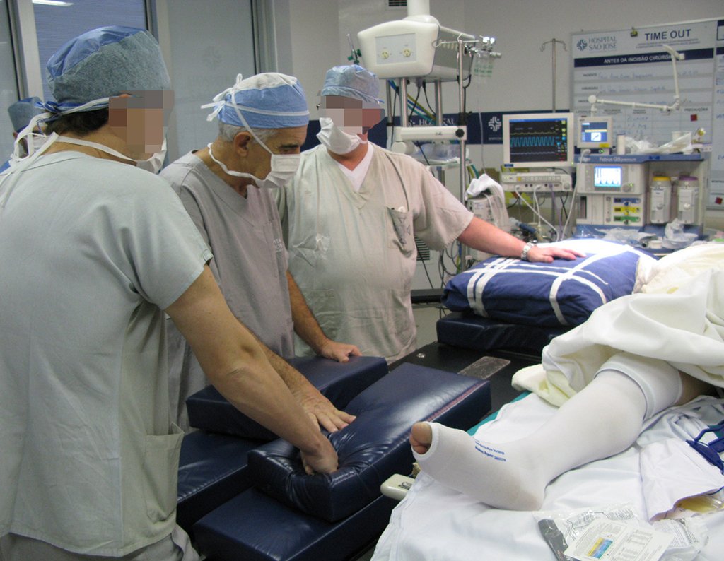
Figura 46: Preparação para anestesia.
Figure 46: Preparation for anesthesia.
Figura 47: Montagem de campos (não estéreis) para apoio no posicionamento do paciente.
Figure 47: Assembling drapes (non-sterile) to support patient positioning.
Figura 48: São utilizados lençóis e /ou campos cirúrgicos não estéreis para a montagem do apoio.
Figure 48: Non-sterile sheets and/or surgical drapes are used to assemble the support.
Figura 49: Os campos são dobrados sobre si mesmos fazendo um apoio de aproximadamente 15 a 20 centímetros de altura.
Figure 49: The fields are folded on themselves, creating a support approximately 15 to 20 centimeters high.
Figura 50: O conjunto é envolto em um outro campo e lacrado com fita crepe para facilitar a manipulação.
Figure 50: The set is wrapped in another field and sealed with masking tape to facilitate handling.
Figura 51: Finalizando a montagem do apoio com fita grepe.
Figure 51: Finishing the assembly of the support with masking tape.
Figura 52: Montagem da mesa cirúrgica junto com o apoio a ser utilizado. Retira-se o colchão que ficaria sob o sacro e adiciona-se o apoio recém produzido na região lombar acima do local a ser operado. Com isso deixaremos um grande espaço vazio abaixo do sacro que permitirá manipulação no intra operatório durante o decúbito dorsal
Figure 52: Assembling the surgical table together with the support to be used. The mattress that would be under the sacrum is removed and the newly produced support is added to the lumbar region above the area to be operated. This will leave a large empty space below the sacrum that will allow intraoperative manipulation during the supine position.
Figura 53: Paciente já anestesiado, aguardando para ser posicionado na mesa cirúrgica.
Figure 53: Patient already anesthetized, waiting to be positioned on the surgical table.
Figura 54: Paciente sendo removido da maca para a mesa cirúrgica. Atente para o degrau que ficará abaixo da coluna lombar, permitindo a manipulação posterior.
Figure 54: Patient being removed from the stretcher to the surgical table. Pay attention to the step that will be below the lumbar spine, allowing later manipulation.
Figura 55: paciente já sondado e com meias elásticas pronto para a passagem para a mesa cirúrgica.
Figure 55: patient already probed and wearing elastic stockings ready to move to the surgical table.
Figure 56: Patient being positioned on the surgical table. The lower limbs will be supported by gynecological leggings and the distal portion of the table will be removed.
Figure 57: Adjusting the position of the right leg.
Figura 58: Paciente em posição ginecológica já sem o apoio distal da mesa. Seu peso está distribuído entre as perneiras e o apoio feito com os campos cirúrgicos na região lombar acima do sacro.
Figure 58: Patient in gynecological position without the distal support of the table. Your weight is distributed between the leggings and the support provided by the surgical drapes in the lumbar region above the sacrum.
Figura 59: Os membros superiores serão colocados sobre o tórax, com os braços cruzados. Isto permitirá melhor acesso a equipe de vídeo-laparoscopia.
Figure 59: The upper limbs will be placed on the chest, with the arms crossed. This will allow better access to the video-laparoscopic team.
Figure 60: Performing asepsis. The patient is suspended by the lower limbs while the lumbar region above the area to be operated remains on the previously placed drape support. Note that the sacral region will be prepared for surgical manipulation during the supine position.
Figura 61: Colocação de campo cirúrgico na região dorsal do paciente.
Figure 61: Placement of a surgical drape on the patient’s dorsal region.
Figura 62: Após assepsia na região perineal e sacral, são colocados campos estéreis no espaço entre a mesa e o paciente, espaço este essencial para a realização da técnica descrita e produzido pelo apoio de campos previamente descrito.
Figure 62: After asepsis in the perineal and sacral region, sterile drapes are placed in the space between the table and the patient, a space essential for carrying out the described technique and produced by the drape support previously described.
Figure 63: Adjustment of the surgical field.
Figure 64: Patient in gynecological position, with the sacral region already aseptic. The weight is being supported by the gynecological leggings and lumbar support.
Figura 65: A equipe da ginecologia começa o procedimento cirúrgico com um portal feito na região abdominal.
Figure 65: The gynecology team begins the surgical procedure with a portal made in the abdominal region.
Figura 66: Visualização da região abdominal por via Laparoscópica.
Figure 66: Laparoscopic visualization of the abdominal region.
Figura 67: A primeira abordagem é feita pela equipe ginecológica que tem papel essencial na dissecção dos vasos ilíacos e pélvicos, além de proporcionar a passagem das serras flexíveis como se verá a seguir
Figure 67: The first approach is carried out by the gynecological team, which plays an essential role in the dissection of the iliac and pelvic vessels, in addition to providing the passage of flexible saws, as will be seen below
Figura 68: Visão laparoscópica onde o ginecologista acessa o retroperitôneo.
Figure 68: Laparoscopic view where the gynecologist accesses the retroperitoneum.
Figura 69: A video cirurgia permite visualizar melhor o espaço retroperitoneal. Está região é de difícil acesso a céu aberto pela via abdominal.
Figure 69: Video surgery allows you to better visualize the retroperitoneal space. This region is difficult to access in the open via the abdominal route.
Figura 70: Aqui o Ginecologista realiza um importante passo do procedimento. Ele disceca os vasos anteriores ao tumor ligando e cauterizando com facilidade.
Figure 70: Here the Gynecologist performs an important step in the procedure. It discates the vessels anterior to the tumor by ligating and cauterizing them with ease.
Figura 71: Os Vasos Iliacos serão visualizados e os ramos necessários são ligados.
Figure 71: The Iliacus Vessels will be visualized and the necessary branches are ligated.
Figura 72: Com as pinças laparoscópicas o ginecologista faz a homeostase em pequenos espaços, onde a mão do cirurgião teria dificuldade de chegar.
Figure 72: With laparoscopic forceps, the gynecologist performs homeostasis in small spaces, where the surgeon's hand would have difficulty reaching.
Figura 73: Passo a passo os tecidos anteriores ao sacro são dissecados com delicada anastomose.
Figure 73: Step by step, the tissues anterior to the sacrum are dissected with delicate anastomosis.
Figura 74: descrição da cirurgia vídeo Laparoscópica.
Figure 74: description of Laparoscopic video surgery.
Figura 75: descrição da cirurgia vídeo Laparoscópica.
Figure 75: description of Laparoscopic video surgery.
Figura 76: descrição da cirurgia vídeo Laparoscópica.
Figure 76: description of Laparoscopic video surgery.
Figura 77: descrição da cirurgia vídeo Laparoscópica.
Figure 77: description of Laparoscopic video surgery.
Figura 78: A mobilização dos grandes vasos após a ligadura dos seus ramos permite visualizar os tecidos mais profundos.
Figure 78: The mobilization of the large vessels after ligating their branches allows the deeper tissues to be visualized.
Figura 79: O sigmoide é tracionado, e podemos visualizar o melhor caminho até a parede anterior do sacro.
Figure 79: The sigmoid is pulled, and we can visualize the best path to the anterior wall of the sacrum.
Figura 80: A artéria glútea superior esquerda, ramo da Artéria Iliaca interna é identificada.
Figure 80: The left superior gluteal artery, a branch of the internal Iliaca Artery, is identified.
Figura 81: São realizadas ligaduras com fios na artería glútea superior esquerda .
Figure 81: Wire ligatures are placed on the left superior gluteal artery.
Figura 82: Secção da arteria glútea e visualização do espaço profundo a ela.
Figure 82: Section of the gluteal artery and visualization of the space deep to it.
Figura 83: Visualização do espaço onde sera início da ressecção sacral.
Figure 83: Visualization of the space where the sacral resection will begin.
Figura 84: Ampliação do espaço onde será introduzido o primeiro cateter de ressecção.
Figure 84: Enlargement of the space where the first resection catheter will be introduced.
Figura 85: Afastamento dos vasos nessa região para evitar lesão vascular na introdução dos cateteres.
Figure 85: Spacement of vessels in this region to avoid vascular injury when introducing catheters.
Figura 86: Espaço pronto para receber o primeiro cateter de ressecção.
Figure 86: Space ready to receive the first resection catheter.
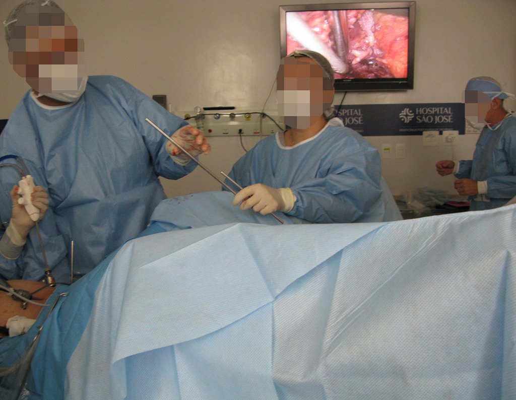
Figura 87: O primeiro cateter de ressecção é um tubo cilindrico de aproximadamente 45 cm que transfixará o paciente entrando pelo portal videolapaoscópico, e sairá pela região sacral previamente preparada com assepsia cirrúrgica, e exposta a manipulação, apesar do decúbito dorsal, pelo apoio feito com campos estéreis descrito no início da cirurgia. O objetivo dele é passar de maneira segura a serra metálica flexível (serra de gilhe) para a posterior osteotomia por via dorsal.
Figure 87: The first resection catheter is a cylindrical tube of approximately 45 cm that will transfix the patient, entering through the videolapaoscopic portal, and exiting through the sacral region previously prepared with surgical asepsis, and exposed to manipulation, despite the supine position, due to the support provided with Sterile fields described at the beginning of surgery. Its objective is to safely pass the flexible metal saw (gilhe saw) for the subsequent osteotomy via the dorsal route.
Figura 88: O primeiro cateter de ressecção é então introduzido pelo abdomem, repare no longo comprimento desse instrumental, isso será relevante.
Figure 88: The first resection catheter is then introduced through the abdomen, note the long length of this instrument, this will be relevant.
Figura 89: Ao introduzi-lo, o ginecologista irá acompanhar seu movimento intra abdominal pelo vídeo posicionado em outro portal.
Figure 89: When introducing it, the gynecologist will monitor its intra-abdominal movement using the video positioned on another portal.
Figura 90: Repare que o cateter toca o sítio cirúrgico anterior do sacro previamente preparado para recebe-lo, e já planejado na melhor posição para ressecção desejada. Caso o Ortopedista queira, ele pode confirmar a posição utilizando o intensificador de imagens.
Figure 90: Note that the catheter touches the anterior surgical site of the sacrum previously prepared to receive it, and already planned in the best position for the desired resection. If the Orthopedist wishes, he can confirm the position using the image intensifier.
Figura 91: Neste momento o ortopedista assume a cirurgia e introduz o cateter transfixando o sacro, ou a região adjacente que seja adequada a ressecção. O cateter entra de anterior para posterior e vai fazer saliência na pele da região lombosacra.
Figure 91: At this point, the orthopedist takes over the surgery and introduces the catheter transfixing the sacrum, or the adjacent region that is suitable for resection. The catheter enters from anterior to posterior and protrudes into the skin of the lumbosacral region.
Figura 92: Caso o cateter não seja suficientemente longo para transfixar o paciente de pele a pele, um outro cateter semelhante é utilizado para empurra-lo sob visão video laparoscópica.
Figure 92: If the catheter is not long enough to transfix the patient from skin to skin, another similar catheter is used to push it under laparoscopic video vision.
Figura 93: O mandril do cateter é retirado e agora ele possui um lumem.
Figure 93: The catheter mandrel is removed and it now has a lumen.
Figura 94: Primeiro cateter de ressecção posicionado corretamente visto pela visão videolaparoscopica da porção anterior do retroperitoneo. Ao lado do cateter encontra-se o sigmóide
Figure 94: First correctly positioned resection catheter seen by videolaparoscopic view of the anterior portion of the retroperitoneum. Next to the catheter is the sigmoid
Figura 95: Nesse momento a região lombosacra é acessada mesmo com o paciente em decÚbito dorsal, pois o mesmo encontra-se suspenso pelo apoio de campos previamente realizado. O cateter de ressecção está saliente na pele, e uma incisão de 2 cm é realizada para expor sua extermidade.
Figure 95: At this moment, the lumbosacral region is accessed even with the patient in the supine position, as the patient is suspended by the previously performed field support. The resection catheter is protruding from the skin, and a 2 cm incision is made to expose its tip.
Figura 96: descrição da cirurgia vídeo Laparoscópica.
Figure 96: description of Laparoscopic video surgery.
Figura 97: descrição da cirurgia vídeo Laparoscópica.
Figure 97: description of Laparoscopic video surgery.
Figura 98: descrição da cirurgia vídeo Laparoscópica.
Figure 98: description of Laparoscopic video surgery.
Figura 99: Preparando a passagem da serra de Gigle junto com a canula.
Figure 99: Preparing the passage of the Gigle saw together with the cannula.
Figura 100: Introdução da canula junto com serra de Gigle na região abdominal.
Figure 100: Introduction of the cannula together with a Gigle saw in the abdominal region.
Figura 101: Passagem total da serra de Gigle.
Figure 101: Full passage of the Gigle mountain range.
Figura 102: Passagem total da serra de Gigle na região abdominal e saindo na região posterior da coluna próximo ao sacro.
Figure 102: Full passage of the Gigle saw in the abdominal region and exiting in the posterior region of the spine close to the sacrum.
Figura 103: Saída da serra de Gigle na região posterior da coluna próximo ao sacro.
Figure 103: Gigle's saw exit in the posterior region of the spine close to the sacrum.
Figura 104: Visualização da canula com o a serra de Gigle.
Figure 104: Visualization of the cannula with the Gigle saw.
Figura 105: descrição da cirurgia vídeo Laparoscópica.
Figure 105: description of Laparoscopic video surgery.
Figura 106: descrição da cirurgia vídeo Laparoscópica.
Figure 106: description of Laparoscopic video surgery.
Figura 107: descrição da cirurgia vídeo Laparoscópica.
Figure 107: description of Laparoscopic video surgery.
Figura 108: descrição da cirurgia vídeo Laparoscópica.
Figure 108: description of Laparoscopic video surgery.
Figura 109: descrição da cirurgia vídeo Laparoscópica.
Figure 109: description of Laparoscopic video surgery.
Figura 110: descrição da cirurgia vídeo Laparoscópica.
Figure 110: description of Laparoscopic video surgery.
Figura 111: descrição da cirurgia vídeo Laparoscópica.
Figure 111: description of Laparoscopic video surgery.
Figura 112: descrição da cirurgia vídeo Laparoscópica.
Figure 112: description of Laparoscopic video surgery.
Figura 113: descrição da cirurgia vídeo Laparoscópica.
Figure 113: description of Laparoscopic video surgery.
Figura 114: descrição da cirurgia vídeo Laparoscópica.
Figure 114: description of Laparoscopic video surgery.
Figura 115: descrição da cirurgia vídeo Laparoscópica.
Figure 115: description of Laparoscopic video surgery.
Figura 116: descrição da cirurgia vídeo Laparoscópica.
Figure 116: description of Laparoscopic video surgery.
Figura 117: descrição da cirurgia vídeo Laparoscópica.
Figure 117: description of Laparoscopic video surgery.
Figura 118: descrição da cirurgia vídeo Laparoscópica.
Figure 118: description of Laparoscopic video surgery.
Figura 119: Colostomia ou Ileostomia provisória.
Figure 119: Temporary colostomy or ileostomy.
Figura 120: Alça intestinal sendo removida.
Figure 120: Intestinal loop being removed.
Figura 121: Alça intestinal sendo removida para o meio externo para colocação da bolsa de colostomia.
Figure 121: Intestinal loop being removed to the external environment for placement of the colostomy bag.
Figura 122: descrição da cirurgia vídeo Laparoscópica.
Figure 122: description of Laparoscopic video surgery.
Figura 123: Finalizando a sutura da colostomia.
Figure 123: Finishing the colostomy suture.
Figura 124: Sutura final da alça intestinal.
Figure 124: Final suture of the intestinal loop.
Figura 125: descrição da cirurgia vídeo Laparoscópica.
Figure 125: description of Laparoscopic video surgery.
Figura 126: descrição da cirurgia vídeo Laparoscópica.
Figure 126: description of Laparoscopic video surgery.
Figura 127: descrição da cirurgia vídeo Laparoscópica.
Figure 127: description of Laparoscopic video surgery.
Video Laparoscopy.
Figura 128: descrição da cirurgia vídeo Laparoscópica.
Figure 128: description of Laparoscopic video surgery.
Figura 129: Paciente foi posicionado em decúbito ventral para realização da retirada do tumor na região do sacro pela equipe da Ortopedia.
Figure 129: The patient was positioned in the prone position to remove the tumor in the sacral region by the Orthopedics team.
Figura 130: Incisão na região posterior da coluna próximo ao sacro.
Figure 130: Incision in the posterior region of the spine close to the sacrum.
Figura 131: Localização da passagem da serra de Gigle .
Figure 131: Location of the Gigle mountain pass.
Figura 132: Incisão na região posterior da coluna vertebral.
Figure 132: Incision in the posterior region of the spine.
Figura 133: Aumento da incisão cirúrgica para melhor visualização da lesão.
Figure 133: Enlargement of the surgical incision for better visualization of the lesion.
Figura 134: Outra visão mais aproximada.
Figure 134: Another closer view.
Figura 135: Ampliação da incisão cirúrgica.
Figure 135: Enlargement of the surgical incision.
Figura 136
Figure 136
Figura 137: Ressecando a lesão.
Figure 137: Resecting the lesion.
Figura 138: Ampliando a incisão.
Figure 138: Enlarging the incision.
Figura 139: desbridando da lesão.
Figure 139: debriding the lesion.
Figura 140: Visualização da lesão na região do sacro.
Figure 140: Visualization of the lesion in the sacral region.
Figura 141: Reposicionado a Serra de Gigle.
Figure 141: Repositioned the Gigle Mountain.
Figura 142: Visualização da lesão.
Figure 142: Visualization of the lesion.
Figura 143: Visualização da lesão.
Figure 143: Visualization of the lesion.
Figura 144
Figure 144
Figura 145
Figure 145
Figura 146
Figure 146
Figura 147: Demonstração do aparelho para ressecção de pedículo.
Figure 147: Demonstration of the device for pedicle resection.
Figura 148
Figure 148
Figura 149
Figure 149
Figura 150
Figure 150
Figura 151: Posicionando a Serra de Gigle para ressecar a lesão.
Figure 151: Positioning the Gigle Saw to resect the lesion.
Figura 152: Ressecção da lesão.
Figure 152: Resection of the lesion.
Figura 153: Peça ressecada.
Figure 153: Dried piece.
Figura 154: Visualização da peça ressecada e mensurada com uma régua.
Figure 154: Visualization of the dried piece and measured with a ruler.
Figura 155: Visualização da peça ressecada e mensurada com uma seringa de 20ml.
Figure 155: Visualization of the dried piece and measured with a 20ml syringe.
Figura 156: Peça ressecada em outro ângulo,sendo mensurada com uma régua 12cm.
Figure 156: Piece resected at another angle, being measured with a 12cm ruler.
Figura 157: Peça ressecada.
Figure 157: Dried piece.
Figura 158: Visualização da peça ressecada em outro ângulo, e mensurada com uma seringa de 20ml.
Figure 158: View of the resected piece from another angle, and measured with a 20ml syringe.
Figura 159: Colocação de hemostático de partes moles.
Figure 159: Placement of soft tissue hemostat.
Figura 160: Visualização da sutura
Figure 160: Visualization of the suture
Figura 161: Aspecto da sutura.
Figure 161: Appearance of the suture.
Figura 162: Aspecto final do curativo.
Figure 162: Final appearance of the dressing.

Video 4 : 4 days post-operative.

Post-operative ……. Figures 163 – 188
Figura 163: Rx de bacia frente, pós operatório.
Figure 163: X-ray of the front pelvis, post-operatively.
Figura 164: TC cor em 3D, frente.
Figure 164: 3D color CT, front.
Figura 165: TC cor em 3D, vista posterior.
Figure 165: 3D color CT, posterior view.
Figura 166: TC cor em 3D, perfil.
Figure 166: 3D color CT, profile.
Figura 167: TC em 3D vista posterior, evidenciando a ressecção do hemi-sacro do lado esquerdo.
Figure 167: 3D CT posterior view, showing the resection of the hemi-sacrum on the left side.
Figura 168: TC em 3D vista de frente com rotação.....
Figure 168: 3D CT viewed from the front with rotation.....
Figura 169: TC evidenciando o local da ressecção da lesão em S1.
Figure 169: CT showing the site of resection of the lesion in S1.
Figura 170: TC evidenciando o local da ressecção da lesão.
Figure 170: CT showing the site of lesion resection.
Figura 171: TC.
Figure 171: CT.
Figura 172: TC evidenciando ....
Figure 172: CT showing....
Figura 173: TC evidenciando a .....
Figure 173: CT showing the .....
Figura 174: TC evidenciando a ressecção da hemi-pelve.
Figure 174: CT showing resection of the hemi-pelvis.
Figura 175: TC.
Figure 175: CT.
Figura 176: TC com vista mais aproximada da região onde houve a ressecção.
Figure 176: CT scan with a closer view of the region where the resection took place.
Figura 177: TC ?
Figure 177: TC ?
Figura 178: TC
Figure 178: TC
Figura 179
Figure 179
Figura 180
Figure 180
Figura 181
Figure 181
Figura 182
Figure 182
Figura 183
Figure 183
Figura 184
Figure 184
Figura 185
Figure 185
Figura 186
Figure 186
Figura 187: TC 3D vista posterior, evidenciando a ressecção do hemi-sacro, e uma lesão na coluna na região de L4 e L5..
Figure 187: 3D CT posterior view, showing the resection of the hemi-sacrum, and a lesion in the spine in the L4 and L5 region.
Figura 188: TC 3D vista lateral, evidenciando uma lesão na coluna na região de L4 e L5.
Figure 188: 3D CT lateral view, showing a lesion in the spine in the L4 and L5 region.
Return to the Office 1 month after surgery.
Patient returned to the office on 12/22/2014, 6 months after surgery. Figure 189 – 195
Figura 189: Aspecto da cicatrização da ferida pós operatória.
Figure 189: Aspect of post-operative wound healing.
Figura 190: Paciente em pós operatório em boas condições de sáude, que venho dirigindo de Taubaté á São Paulo para o retorno no consultório.
Figure 190: Post-operative patient in good health, who I have been driving from Taubaté to São Paulo to return to the office.
Figura 191: Paciente em pós operatório de ....evidencaindo a boa postura ortostática.
Figure 191: Post-operative patient for .... showing good orthostatic posture.
Figura 192: Paciente em pós operatório de ....realizando exame físico.
Figure 192: Post-operative patient...undergoing physical examination.
Figura 193: Paciente em pós operatório de ....realizando exame físico de agachamento.
Figure 193: Patient post-operatively... performing a physical examination in a squat.
Figura 195: Paciente mostrando a bolsa de colostomia.
Figure 195: Patient showing the colostomy bag.

Video : Patient walking 6 months postoperatively.

Video 5

Author: Prof. Dr. Pedro Péricles Ribeiro Baptista

 Orthopedic Oncosurgery at the Dr. Arnaldo Vieira de Carvalho Cancer Institute

Hello! How can we assist you?