This digital library houses the book on Oncology and Orthopedic Oncosurgery.

It includes academic lectures, presentations from national and international congresses, published papers, case discussions, performed surgical procedures, and proprietary techniques developed.

The digital format was chosen because the web allows the inclusion of texts with numerous visual resources, such as images and videos, which would not be possible in a printed book.

The content is intended for students, healthcare professionals, and the general public interested in the field.

Radial Giant Cell Tumor

288d7e4b1fa80acd699c8287ec363e12 3

Radial Giant Cell Tumor - Obtaining an autologous fibula graft - Wrist reconstruction technique

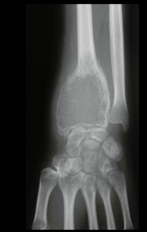
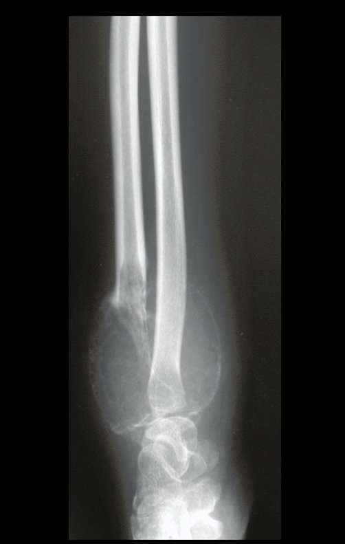
Radial Giant Cell Tumor. Giant cell tumor of the radius: Female patient, 26 years old, with pain in her left wrist for two months. She initially sought care in January 2000, when x-rays and tomography were taken, showing the following images (Figures 1 to 6).
Figura 1: Lesão de rarefação óssea epifisária do rádio, com abaulamento da cortical radial e anfractuosidades. A cortical do lado ulnar encontra-se preservada.
FiguraaFigure 1: Radial epiphyseal bone rarefaction lesion, with bulging of the radial cortex and anfractuosities. The cortex on the ulnar side is preserved. 1: Lesão de rarefação óssea epifisária do rádio, com abaulamento da cortical radial e anfractuosidades. A cortical do lado ulnar encontra-se preservada.
Figura 2: Radiografia evidenciando a insuflação da cortical, em lesão lítica epifisária do rádio.
Figure 2: Radiograph showing cortical inflation in a lytic epiphyseal lesion of the radius.
Figura 4: TC, densidade para tecido ósseo, com a cortical insuflada, sem tumor extracortical.
Figure 3: Tomography, with soft tissue density, shows the lytic epiphyseal lesion of the radius, with the cortex inflated, without extracortical tumor. Figure 4: CT, density for bone tissue, with the cortex inflated, without extracortical tumor.
Figura 6: TC, densidade para tecidos moles, evidencia a lesão delimitada externamente pela cortical, sobrepondo-se à parede da cortical proximal, tanto volar como dorsal, porém sem tumor extracortical e sem invasão de tecidos moles.
Figure 5: CT, soft tissue density, shows the lesion surrounded by thin cortical bone, overlapping the proximal bone wall, but without soft tissue invasion. Figure 6: CT, soft tissue density, shows the lesion externally delimited by the cortex, overlapping the proximal cortical wall, both volar and dorsal, but without extracortical tumor and without soft tissue invasion.

Em maio de 2005, com a suspeita de tumor de células gigantes, foi encaminhada para um segundo hospital, cinco meses do primeiro atendimento. Novo encaminhamento foi feito, agora para nosso atendimento, em outubro daquele ano (Figuras 7 à 10).

Figura 7: Radiografia frente, após cinco meses.
Figure 7: Front X-ray, after five months.
Figura 8: Radiografia perfil, após cinco meses, quando a paciente é avaliada em outro hospital.
Figure 8: Profile x-ray, after five months, when the patient is evaluated in another hospital.
Figura 9: Após dez meses. Piora da lesão, com fratura da cortical e afundamento da superfície articular.
Figure 9: After ten months. Worsening of the injury, with cortical fracture and sinking of the articular surface.
Figura 10: RX, após dez meses, progressão do tumor, com erosão circunferencial da cortical, deformando a articulação do punho.
Figure 10: X-ray, after ten months, tumor progression, with circumferential cortical erosion, deforming the wrist joint.
Figura 11: Grande erosão da cortical radial e também do lado ulnar, que se encontrava preservada dez meses antes.
Figure 11: Major erosion of the radial cortex and also of the ulnar side, which was preserved ten months earlier.
Figura 12: Aspecto radiográfico em 01/11/2000, antes da biópsia. Grande agressividade local do tumor.
Figure 12: Radiographic appearance on 11/01/2000, before the biopsy. Great local aggressiveness of the tumor.
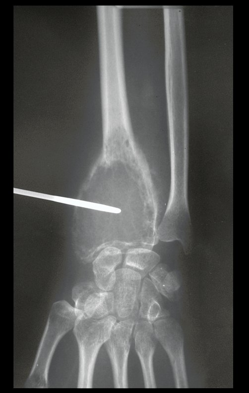
Figura 13: Controle radiográfico da biópsia, com agulha de Jamshid.
Figure 13: Radiographic control of the biopsy, with a Jamshid needle.
Figura 14: Radiografia com régua, planejamento da ressecção.
Figure 14: Radiograph with ruler, resection planning.
Figura 15: Radiografia, com régua, da fíbula que será utilizada na reconstrução do punho.
Figure 15: Radiograph, with a ruler, of the fibula that will be used in wrist reconstruction.
Figura 16: Aspecto volar pré-operatório do punho. A paciente precisa fazer rotação no ombro, pois tem limitação da supinação.
Figure 16: Preoperative volar aspect of the wrist. The patient needs to rotate the shoulder, as she has limited supination.
Figura 17: Tumor no punho, visão dorsal.
Figure 17: Tumor on the wrist, dorsal view.
Figura 18: Punho em posição neutra.
Figure 18: Wrist in neutral position.
The histological diagnosis of the biopsy confirmed the clinical suspicion of giant cell tumor (Figures 19 to 24).
Figura 19: Histologia revelando neoplasia benigna, com inúmeras células gigantes em meio a estroma de células, cujos núcleos se assemelham aos núcleos das células gigantes. TGC
Figure 19: Histology revealing a benign neoplasm, with numerous giant cells amidst the stroma of cells, whose nuclei resemble the nuclei of giant cells. TGC
Figura 20: Em maior aumento, observamos que os núcleos das células gigantes são semelhantes aos núcleos das células do estroma.
Figure 20: At higher magnification, we observe that the giant cell nuclei are similar to the stromal cell nuclei.
Figura 21: As células gigantes parecem ser um aglomerado de células do estroma, que se aglutinam, característico do TGC.
Figure 21: Giant cells appear to be a cluster of stromal cells that clump together, characteristic of TGC.
Figura 22: Células do estroma do TGC.
Figure 22: TGC stromal cells.
Figura 23: Aglomeração das células do estroma em a; que se aglutinam por fusão de seus citoplasmas em b e formam uma célula gigante em c.
Figure 23: Agglomeration of stromal cells in a; which coalesce by fusion of their cytoplasms in b and form a giant cell in c.
Figura 24: Campo contendo estroma, células aglutinadas e o sincício, formando as células gigantes.
Figure 24: Field containing stroma, agglutinated cells and the syncytium, forming giant cells.
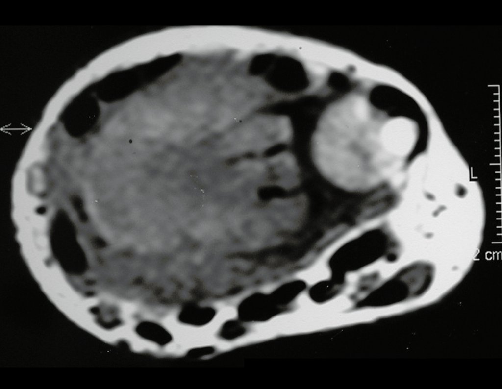
The magnetic resonance study shows a lesion of bone rarefaction, with areas of necrosis, erosion, inflation and cortical infraction fracture, with deformity and sinking of the articular surface (figures 25 to 36).
Figura 25: RM cor t1, lesão de rarefação óssea, com áreas de necrose, erosão, insuflação e fratura infracção da cortical, com deformidade e afundamento da superfície articular.
Figure 25: MRI color t1, bone rarefaction lesion, with areas of necrosis, erosion, inflation and cortical infraction fracture, with deformity and sinking of the articular surface.
Figura 26: RM cor t1, com supressão de gordura.
Figure 26: T1 color MRI, with fat suppression.
Figura 27: RM ax t1, lesão com sinal intermediário, empurrando ligamentos e tendões, que fazem impressão no tumor. Não há infiltração em tecidos moles. Este tumor geralmente empurra os tecidos moles, sem infiltrá-lo.
Figure 27: MRI ax t1, lesion with intermediate signal, pushing ligaments and tendons, which make an impression on the tumor. There is no infiltration into soft tissues. This tumor usually pushes against soft tissue without infiltrating it.
Figura 28: RM cor t2, com supressão de gordura, alto sinal.
Figure 28: T2 color MRI, with fat suppression, high signal.
Figura 29: RM cor FFE, sem evidência de focos de ossificação.
Figure 29: FFE color MRI, without evidence of ossification foci.
Figura 26: RM cor t1, com supressão de gordura.
Figure 30: MRI color t1, with fat suppression, lesion with high signal.
Figura 32: RM ax t1, com saturação de gordura, destaca que os tendões estão livres.
Figure 31: MRI ax t1, with fat suppression, high signal. Figure 32: MRI ax t1, with fat saturation, highlights that the tendons are free.
Figura 33: RM sag DP, lesão com alto sinal.
Figure 33: MRI sag DP, lesion with high signal.
Figura 34: RM cor t1 com captação de contraste apenas na periferia da lesão.
Figure 34: T1 color MRI with contrast uptake only at the periphery of the lesion.
Figura 35: RM ax t1, lesão com captação de contraste na periferia e em septos do tumor.
Figure 35: MRI ax t1, lesion with contrast uptake in the periphery and in tumor septa.
Figura 36: RM ax t1, contraste apenas na periferia e em septos do tumor.
Figure 36: MRI ax t1, contrast only in the periphery and in tumor septa.
Figura 37: Após a biópsia, aspecto clínico pré-operatório do punho esquerdo.
Figure 37: After biopsy, preoperative clinical appearance of the left wrist.

Com este estudo, confirmamos a agressividade local do tumor, e a grande destruição da extremidade distal do rádio esquerdo, com fratura por infracção, deformidade articular e encurtamento do rádio. Nesta situação o tratamento intralesional deve ser contraindicado, devido à alta possibilidade de recidiva local, além da perda da função. Oncologicamente está indicada a ressecção segmentar do rádio, e reconstrução do punho. A artrodese pode ser uma alternativa, mas preferimos preservar a função e a anatomia do punho, sempre que possível. Neste caso, optamos por reconstruir com enxerto autólogo não vascularizado, obtido do segmento proximal da fíbula ipsilateral, que mimetiza melhor o rádio.
A cirurgia é realizada em decúbito dorsal horizontal, preferentemente sem uso de garroteamento do membro. Os detalhes da ressecção são mostrados nas figuras 38 a 57.

Figura 38: Paciente em DDH, com o MSE apoiado em mesa, após assepsia e antissepsia.
Figure 38: Patient in DDH, with the MSE supported on a table, after asepsis and antisepsis.
Figura 39: Incisão dorsal, arciforme, iniciando 1,0 cm distal a apófise estiloide ulnar, dirigindo-se à apófise estiloide do rádio, curvando-se e prolongando-se proximamente, acompanhando a projeção cutânea do rádio.
Figure 39: Dorsal, arcuate incision, starting 1.0 cm distal to the ulnar styloid process, heading towards the styloid process of the radius, curving and extending closely, following the cutaneous projection of the radius.
Figura 40: Abertura da pele, tecido subcutâneo e fáscia do antebraço. Os túneis extensores são abertos longitudinalmente, liberando os tendões adjacentes ao tumor, unicamente por suas bainhas.
Figure 40: Opening of the skin, subcutaneous tissue and fascia of the forearm. The extensor tunnels are opened longitudinally, releasing the tendons adjacent to the tumor, solely through their sheaths.
Figura 41: Tenotomia do braquiestilorradial, próximo à sua inserção na apófise estiloide do rádio. afastamento dos tendões extensores, expondo-se o segmento radial acometido.
Figure 41: Brachystyloradialis tenotomy, close to its insertion into the radial styloid process. removal of the extensor tendons, exposing the affected radial segment.
Figura 42: Ligadura de vasos nutrícios dorsais do rádio.
Figure 42: Ligation of dorsal nutrient vessels of the radius.
Figura 43: Hemostasia e exposição do segmento distal do rádio a ser ressecado.
Figure 42: Ligation of dorsal nutrient vessels of the radius.
Figura 44: Osteotomia do rádio, 1,5 cm proximal ao tumor, protegendo-se os tecidos moles com afastador de Bennet.
Figure 44: Osteotomy of the radius, 1.5 cm proximal to the tumor, protecting the soft tissues with a Bennet retractor.
B90c46963248e6d7aab1e0f429743ca0 3
Figure 45: After the osteotomy, the posterior periosteum is released, partially detaching the interosseous membrane of the radius.
Figura 46: O segmento distal do rádio é liberado cuidadosamente, e levantado com uma pinça para acesso e liberação volar.
Figure 46: The distal segment of the radius is carefully released and lifted with forceps for volar access and release.
Figura 47: Realiza-se a capsulotomia rádio cárpica, procurando-se preservar o máximo possível do complexo capsular, o que facilitará a reconstrução entre o carpo e o enxerto fibular.
Figure 47: Radiocarpal capsulotomy is performed, seeking to preserve as much of the capsular complex as possible, which will facilitate reconstruction between the carpus and the fibular graft.
Figura 48: Identifica-se as estruturas volares e libera-se a radioulnar distal.
Figure 48: The volar structures are identified and the distal radioulnar structure is released.
Figura 49: O pronador quadrado, aderido à superfície volar do rádio, constitui a margem oncológica volar e deve ser ressecado em bloco com o segmento ósseo.
Figure 49: The pronator quadratus, attached to the volar surface of the radius, constitutes the volar oncological margin and must be resected en bloc with the bone segment.
Figura 50: Revisão da hemostasia do leito operatório após a ressecção.
Figure 50: Review of hemostasis in the operating bed after resection.
Figura 51: Face volar da peça ressecada, com o músculo pronador quadrado como margem.
Figure 51: Volar face of the resected specimen, with the pronator quadratus muscle as a margin.
Figura 52: Face articular, sem lesão da superfície, os sulcos dos túneis tendinosos salientam os limites oncológicos da ressecção.
Figure 52: Joint face, without surface injury, the grooves of the tendon tunnels highlight the oncological limits of the resection.
Figura 53: Face da osteotomia evidência macroscopicamente a ressecção com margem oncológica circunferencialmente.
Figure 53: Osteotomy face macroscopically showing resection with circumferential oncological margin.
Figura 54: Face radial integra, com boa margem oncológica e impressão dos sulcos tendinosos.
Figure 54: Full radial face, with good oncological margin and impression of the tendon grooves.
Figura 56: Face dorsal, com os sulcos dos tenões extensores.
Figure 55: Ulnar surface, complete. Figure 56: Dorsal side, with the grooves of the extensor tenons.
Figura 57: Aspecto macroscópico do corte da peça cirúrgica, sem tumor extracortical e, portanto, sem invasão de tecidos moles, conforme afirmamos anteriormente.
Figure 57: Macroscopic appearance of the section of the surgical specimen, without extracortical tumor and, therefore, without invasion of soft tissues, as previously stated.
After resection of the distal segment of the radius and careful review of hemostasis, we placed a compress moistened with saline on the surgical bed and temporarily bandaged it with a crepe bandage. We move on to the second stage: Obtaining the autologous, non-vascularized graft, obtained from the proximal segment of the ipsilateral fibula. It is preferable to use the graft obtained from the same side, as the lateral surface of the fibula and the insertion tuberosity of the lateral collateral ligament better mimic the dorsal surface of the radius on the same side. In this way, the apex of the fibula will replace the radial styloid. It is important to obtain this graft with a joint capsule, from the proximal tibio-fibular joint, around the entire circumference of the fibular epiphysis. This way we will have soft tissues for the circumferential reconstruction of the new fibulocarpal joint. Next, we detail the obtaining of the fibula graft (Figures 58 to 75).
Figura 58: Joelho esquerdo em discreta flexão, para a retirada do enxerto.
Figure 58: Left knee in slight flexion, for graft removal.
Figura 59: Inicia-se posteriormente ao tendão do bíceps, 5 cms acima da interlinha articular, em direção à cabeça da fíbula e prolongando-se sobre ela por cerca de 12 cms.
Figure 59: It starts posterior to the biceps tendon, 5 cm above the joint line, towards the head of the fibula and extending over it for around 12 cm.
Figura 60: Abertura da pele e subcutâneo, hemostasia e exposição do perimísio dos músculos extensor e flexor longo dos dedos. A= septo que divide os compartimentos anterior e lateral da perna, e B= visualização do tecido gorduroso que envolve o nervo ciático poplíteo externo, sob a aponevrose.
Figure 60: Opening of the skin and subcutaneous tissue, hemostasis and exposure of the perimysium of the extensor and flexor digitorum longus muscles. A= septum that divides the anterior and lateral compartments of the leg, and B= visualization of the fatty tissue that surrounds the external popliteal sciatic nerve, under the aponeurosis.
Figura 61: O septo deve ser aberto cuidadosamente, para expor as seguintes estruturas: 1= tendão longo do bíceps; 2= músculo extensor longo dos dedos; 3= músculo flexor longo dos dedos e 4= nervo ciático.
Figure 61: The septum must be carefully opened to expose the following structures: 1= long biceps tendon; 2= ​​extensor digitorum longus muscle; 3= flexor digitorum longus muscle and 4= sciatic nerve.
Figura 62: Abertura do septo e afastamento do extensor e flexor dos dedos, expondo a diáfise da fíbula, e início do isolamento do nervo ciático.
Figure 62: Opening of the septum and removal of the extensor and flexor fingers, exposing the fibula shaft, and beginning of isolation of the sciatic nerve.
Figura 63: A diáfise da fíbula e o nervo ciático são dissecados. O nervo deve ser manipulado cuidadosamente, sem estirá-lo.
Figure 63: The fibula shaft and sciatic nerve are dissected. The nerve must be handled carefully without stretching it.
Figura 64: A extremidade proximal da fíbula é ressecada, desinserindo-se os músculos extensor longo dos dedos, extensor longo do hálux, flexor longo dos dedos, sóleo e membrana interóssea. O tendão do bíceps femoral e o ligamento colateral lateral são seccionados, deixando-se 1,0 cm para facilitar a reinserção na capsuloplastia do punho.
Figure 64: The proximal end of the fibula is resected, removing the extensor digitorum longus, extensor hallucis longus, flexor digitorum longus, soleus and interosseous membrane muscles. The biceps femoris tendon and the lateral collateral ligament are sectioned, leaving 1.0 cm to facilitate reinsertion in wrist capsuloplasty.
Figura 65: Dimensiona-se o tamanho do segmento a ser ressecado para enxerto.
Figure 65: Dimension the size of the segment to be resected for grafting.
Figura 66: Realizamos a osteotomia neste momento, visando ter mobilidade do segmento proximal, facilitando a capsulotomia circunferencial da extremidade proximal da fíbula. Esta osteotomia deve ser reta, perpendicular à diáfise.
Figure 66: We perform the osteotomy at this point, aiming to have mobility of the proximal segment, facilitating the circumferential capsulotomy of the proximal end of the fibula. This osteotomy must be straight, perpendicular to the diaphysis.
Figura 67: Liberação da articulação tibio-fibular proximal, seccionando-se os ligamentos anterior, posterior, e parte do ligamento arqueado, que servirão para ancoragem na reconstrução do punho.
Figure 67: Release of the proximal tibiofibular joint, sectioning the anterior and posterior ligaments and part of the arcuate ligament, which will serve as anchorage in wrist reconstruction.
Figura 68: Liberação circunferencial da cápsula, preservando-se cerca de um centímetro, para facilitar a capsuloplastia entre a fíbula e o carpo.
Figure 68: Circumferential release of the capsule, preserving approximately one centimeter, to facilitate capsuloplasty between the fibula and carpus.
Figura 69: Leito operatório após a ressecção e hemostasia. 1= vasos interósseos; 2= artéria e veia tíbial anterior e 3= nervo ciático.
Figure 69: Operative bed after resection and hemostasis. 1= interosseous vessels; 2= ​​anterior tibial artery and vein and 3= sciatic nerve.
Figura 70: O enxerto, obtido da extremidade proximal da fíbula, deve possuir coto da cápsula articular em toda a sua circunferência, para possibilitar uma reconstrução adequada do punho.
Figure 70: The graft, obtained from the proximal end of the fibula, must have a stump of the joint capsule along its entire circumference, to enable adequate reconstruction of the wrist.
Figura 71: Segmento proximal da fíbula, com cápsula articular, que será utilizado na reconstrução do punho.
Figure 71: Proximal segment of the fibula, with joint capsule, which will be used in wrist reconstruction.
Figura 72: Retirada de enxerto ósseo esponjoso através de janela na faceta articular da tíbia, (articulação tíbio-fibular proximal).
Figure 72: Removal of cancellous bone graft through a window in the articular facet of the tibia (proximal tibio-fibular joint).
Figura 73: Reinserção do ligamento colateral lateral, nesta janela ósteo-cartilaginosa com ponto de ethibond 2.
Figure 73: Reinsertion of the lateral collateral ligament, in this osteo-cartilaginous window with ethibond stitch 2.
Figura 74: Fechamento da área de obtenção do enxerto fibular.
Figure 74: Closing the area to obtain the fibular graft.
Figura 75: Enxerto obtido para substituir o segmento ressecado. Observe o tecido capsular preservado no enxerto, em toda a sua circunferência. Uma placa DCP com sete orifícios será utilizada para a fixação do enxerto ao rádio remanescente.
Figure 75: Graft obtained to replace the resected segment. Note the capsular tissue preserved in the graft, throughout its circumference. A DCP plate with seven holes will be used to fix the graft to the remaining radius.
Figura 76: A reconstrução do punho inicia-se pela capsuloplastia da face volar, suturando-se o remanescente da cápsula articular do punho com a do enxerto fibular.
Figure 76: Wrist reconstruction begins with capsuloplasty of the volar face, suturing the remainder of the wrist joint capsule with that of the fibular graft.
Figura 77: Primeiro ponto unindo os extremos laterais e volares do enxerto e do punho.
Figure 77: First point joining the lateral and volar ends of the graft and the wrist.
Figura 78: Segundo ponto, continuando a capsuloplastia lateral.
Figure 78: Second point, continuing the lateral capsuloplasty.
Figura 79: Completa-se a capsuloplastia circunferencialmente, criando-se uma neo articulação fíbulo-carpal.
Figure 79: The capsuloplasty is completed circumferentially, creating a new fibulocarpal joint.
Figura 80: Após a sutura do enxerto, toma-se a medida do comprimento necessário, para que o comprimento do rádio seja restabelecido.
Figure 80: After suturing the graft, the necessary length is measured so that the length of the radius is reestablished.
Figura 81: Realiza-se a osteotomia do enxerto, adequando o comprimento necessário para preencher a falha.
Figure 81: The graft osteotomy is performed, adapting the necessary length to fill the gap.
Figura 82: Neste momento, estuda-se a prono-supinação e realiza-se uma marcação da rotação adequada, em que será fixado o enxerto ao rádio.
Figure 82: At this point, pronosupination is studied and the appropriate rotation is marked, in which the graft will be fixed to the radius.
Figura 83: Marcação com bisturi elétrico, do grau de rotação para a fixação.
Figure 83: Marking with an electric scalpel the degree of rotation for fixation.
Figura 84: Modelagem da placa e posicionamento, deixando três orifícios para a fixação dos parafusos distais no enxerto.
Figure 84: Modeling of the plate and positioning, leaving three holes for fixing the distal screws in the graft.
Figura 85: Apresentação para a passagem dos parafusos, protegendo-se os tecidos moles com afastador de Bennet.
Figure 85: Presentation for passing the screws, protecting the soft tissues with a Bennet retractor.
Figura 86: Seis parafusos, três distais e três proximais, são fixados, deixando-se o sétimo orifício sem fixação. Após a fixação, coloca-se o enxerto ósseo autólogo obtido da tíbia proximal.
Figure 86: Six screws, three distal and three proximal, are fixed, leaving the seventh hole unfixed. After fixation, the autologous bone graft obtained from the proximal tibia is placed.
Figura 87: A seguir, realiza-se o fechamento, criando-se polias tendinosas para a reconstrução dos túneis, visando propiciar a melhor função do punho.
Figure 87: Next, closure is performed, creating tendon pulleys to reconstruct the tunnels, aiming to provide better wrist function.
Figura 88: Aspecto volar da ferida operatória.
Figure 88: Volar aspect of the surgical wound.
Figura 89: Aspecto dorsal da ferida operatória.
Figure 89: Dorsal aspect of the surgical wound.
Figura 90: Radiografia, frente, da fixação e do enxerto no pós operatório imediato.
Figure 90: Radiograph, front, of the fixation and graft in the immediate post-operative period.
Figura 91: Radiografia, perfil, da fixação e do enxerto no pós operatório imediato.
Figure 91: Radiograph, profile, of the fixation and graft in the immediate post-operative period.
Figura 92: Imobilização pós-operatória com gesso braquio-palmar, por três semanas.
Figure 92: Postoperative immobilization with brachio-palmar plaster for three weeks.
Figura 94: Radiografia perfil, da peça cirúrgica.
Figure 93: Front x-ray of the surgical specimen. Figure 94: Profile x-ray of the surgical specimen.
Figura 95: Radiografia frente, após três semanas, com imobilização. Esta imobilização é retirada e inicia-se movimentação ativa do antebraço, flexo-extensão e prono-supinação.
Figure 95: Front X-ray, after three weeks, with immobilization. This immobilization is removed and active movement of the forearm, flexion-extension and pronation-supination begins.
Figura 96: Radiografia perfil, após três semanas com imobilização. Esta imobilização é retirada e inicia-se movimentação ativa do antebraço, flexo-extensão e prono-supinação.
Figure 96: Profile x-ray, after three weeks of immobilization. This immobilization is removed and active movement of the forearm, flexion-extension and pronosupination begins.
Figura 97: Radiografia frente, após três semanas sem gesso, enxerto com maior rarefação, indicando que o processo de reabsorção e neoformação óssea encontra-se em andamento.
Figure 97: Front X-ray, after three weeks without plaster, graft with greater rarefaction, indicating that the process of bone resorption and new formation is underway.
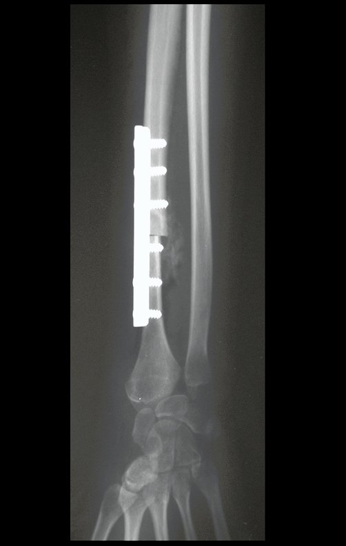
Figura 98: Radiografia perfil, após três semanas sem gesso, enxerto com maior rarefação, indicando que o processo de reabsorção e neoformação óssea encontra-se em andamento.
Figure 98: Profile x-ray, after three weeks without plaster, graft with greater rarefaction, indicating that the process of bone resorption and new formation is underway.
Figura 100: Supinação de 00, no pré-operatório.
Figure 99: 900 pronation preoperatively. Figure 100: Supination of 00, preoperatively.
Figura 101: Para se conseguir apoiar a face dorsal do punho, é necessária a rotação do ombro. Aspecto pré-operatório.
Figure 101: To be able to support the dorsal surface of the wrist, shoulder rotation is necessary. Preoperative appearance.
Figura 103: Início da fisioterapia após três semanas de imobilização. Extensão dos dedos.
Figure 102: Beginning of physiotherapy after three weeks of immobilization. Flexion of the fingers. Figure 103: Beginning of physiotherapy after three weeks of immobilization. Finger extension.
Figura 104: Aspecto da cicatrização e alinhamento do punho esquerdo. Após três semanas da cirurgia.
Figure 104: Appearance of healing and alignment of the left wrist. Three weeks after surgery.
Figura 105: Flexão dos dedos, após sete semanas.
Figure 105: Flexion of the fingers, after seven weeks.
Figura 106: Extensão dos dedos, após sete semanas.
Figure 106: Finger extension after seven weeks.
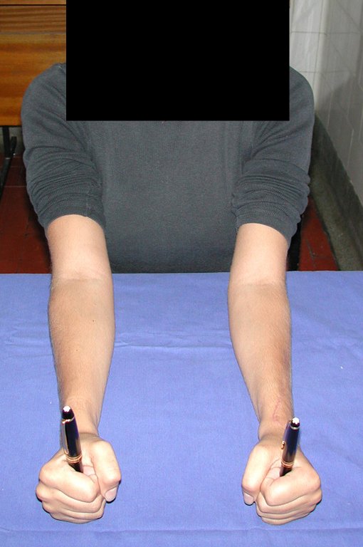
Figura 107: Pronação após sete semanas.
Figure 107: Pronation after seven weeks.
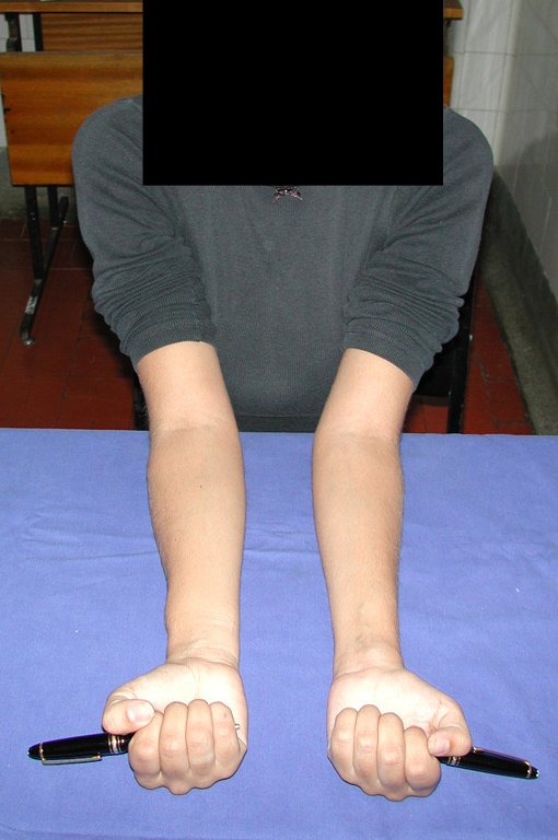
Figura 108: Supinação após sete semanas.
Figure 108: Supination after seven weeks.
Figura 109: Após três meses, boa maturação da cicatriz operatória, melhor flexão dos dedos.
Figure 109: After three months, good maturation of the surgical scar, better flexion of the fingers.
Figura 110: Extensão após três meses, maturação da cicatriz.
Figure 110: Extension after three months, scar maturation.
Figura 111: Pronação após três meses, aspecto simétrico dos punhos.
Figure 111: Pronation after three months, symmetrical appearance of the wrists.
Figura 112: Supinação após três meses, trofismo simétrico dos punhos.
Figure 112: Supination after three months, symmetrical trophism of the wrists.
Figura 113: Boa função com o punho esquerdo, que é o dominante desta paciente.
Figure 113: Good function with the left wrist, which is this patient's dominant wrist.
Figura 114: Trofismo e simetria dos punhos.
Figure 114: Trophism and symmetry of the wrists.
Figura 115: Mensuração da força do punho direito.
Figure 115: Measurement of right wrist strength.
Figura 116: Força simétrica dos punhos, após três meses.
Figure 116: Symmetrical wrist strength after three months.
Figura 117: Consolidação após seis meses, calo homogêneo.
Figure 117: Consolidation after six months, homogeneous callus.
Figura 118: Radiografia em perfil, após seis meses.
Figure 118: Profile x-ray, after six months.
Figura 119: Aspecto após seis meses.
Figure 119: Appearance after six months.
Figura 120: Paciente seis meses depois da cirurgia.
Figure 120: Patient six months after surgery.
Figura 121: Função de flexão dorsal, após seis meses.
Figure 121: Dorsal flexion function, after six months.
Figura 122: Função de flexão volar, após seis meses.
Figure 122: Volar flexion function, after six months.
Figura 123: Força de preensão, após seis meses.
Figure 123: Grip strength after six months.
Figura 124: Rotação neutra dos punhos simétrica.
Figure 124: Symmetrical neutral rotation of the wrists.
Figura 125: Supinação simétrica dos punhos.
Figure 125: Symmetrical supination of the wrists.
Figura 126: Pronação simétrica dos punhos.
Figure 126: Symmetrical pronation of the wrists.
Video 1: Active and symmetrical supination and pronation of the wrists, six months after surgery. Good function.
Video 2: Active and symmetrical flexion and extension of the fingers, after six months. Good function.
Bdc6c33585d0cf5d2a8cb83141aFigura 127: Após nove meses.cd037f 3
Figure 127: After nine months.
Figura 128: Pronação após nove meses.
Figure 128: Pronation after nine months.
Figura 129: Supinação após nove meses.
Figure 129: Supination after nine months.
Figura 130: Pronação após dez meses.
Figure 130: Pronation after ten months.
Figura 131: Supinação após dez meses.
Figure 131: Supination after ten months.
Figura 132: Pós-operatório de dois anos, pronação.
Figure 132: Two years post-operative, pronation.
Figura 133: Pós-operatório de dois anos, supinação.
Figure 133: Two years post-operative, supination.
Figura 134: Pós-operatório de dois anos, desvio radial.
Figure 134: Two years postoperative, radial deviation.
Figura 135: Pós-operatório de dois anos, desvio ulnar.
Figure 135: Two years postoperative, ulnar deviation.
Figura 136: Pós-operatório de dois anos, boa flexão dorsal.
Figure 136: Two years postoperative, good dorsal flexion.
Figura 137: Melhora significativa da flexão volar, após dois anos da cirurgia de transposição da fíbula para o rádio.
Figure 137: Significant improvement in volar flexion, two years after surgery to transpose the fibula to the radius.
Figura 138: Paciente sem limitação da pronação, em 29/09/2015 pós-operatório de 15 anos.
Figure 138: Patient without limitation of pronation, on 09/29/2015, 15 years after surgery.
Figura 139: Supinação simétrica e normal, após 15 anos da cirurgia.
Figure 139: Symmetrical and normal supination, 15 years after surgery.
Figura 140: Flexão dorsal em 29/09/2015, após quinze anos de pós operatório
Figure 140: Dorsal flexion on 09/29/2015, fifteen years after surgery
Figura 141: Flexão volar em 29/09/2015, após quinze anos.
Figure 141: Volar flexion on 09/29/2015, after fifteen years.
Figura 142: Boa função e simetria em atitude espontânea com os punhos.
Figure 142: Good function and symmetry in a spontaneous attitude with the fists.
Figura 143: Assinando a autorização do uso de imagens, boa estética e função após quinze anos.
Figure 143: Signing authorization to use images, good aesthetics and function after fifteen years.
Figura 144: Cicatriz da perna esquerda, área doadora do enxerto de fíbula. A paciente aplicou laser recentemente visando deixar a cicatriz mais estreita.
Figure 144: Scar on the left leg, fibula graft donor area. The patient recently had a laser applied to make the scar narrower.
Figura 145: Boa estabilidade e função normal do joelho, no lado doador do enxerto de fíbula.
Figure 145: Good stability and normal function of the knee, on the donor side of the fibula graft.
Video 3: Symmetrical pronation and supination, fifteen years after surgery.
Video 4: Flexion and extension on September 29, 2015, fifteen years after surgery.
Video 5: Indifferent and spontaneous attitude of the fists. Good symmetry and function on 15/29/2015. Fifteen years post-operative.
Video 6: Good muscle strength and stability of the left wrist, lifting significant weight after 19 years of surgery, on August 22, 2019.

Author: Prof. Dr. Pedro Péricles Ribeiro Baptista

 Orthopedic Oncosurgery at the Dr. Arnaldo Vieira de Carvalho Cancer Institute

Hello! How can we assist you?