This digital library houses the book on Oncology and Orthopedic Oncosurgery.

It includes academic lectures, presentations from national and international congresses, published papers, case discussions, performed surgical procedures, and proprietary techniques developed.

The digital format was chosen because the web allows the inclusion of texts with numerous visual resources, such as images and videos, which would not be possible in a printed book.

The content is intended for students, healthcare professionals, and the general public interested in the field.

Osteosarcoma with pulmonary metastasis

73c730319cf839f143bf40954448ce39 2

Humeral osteosarcoma with lung metastasis Polyethylene Endoprosthesis - Metastectomy

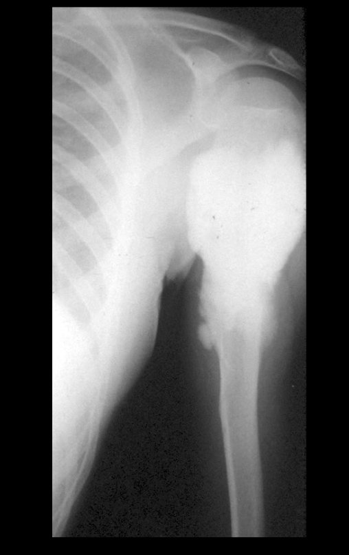
Osteosarcoma with pulmonary metastasis. Patient born on 06/07/1976, treated in 1987, at the age of eleven, with a history of two previous surgical interventions, curettage of a lesion in the left proximal humerus, a year ago.

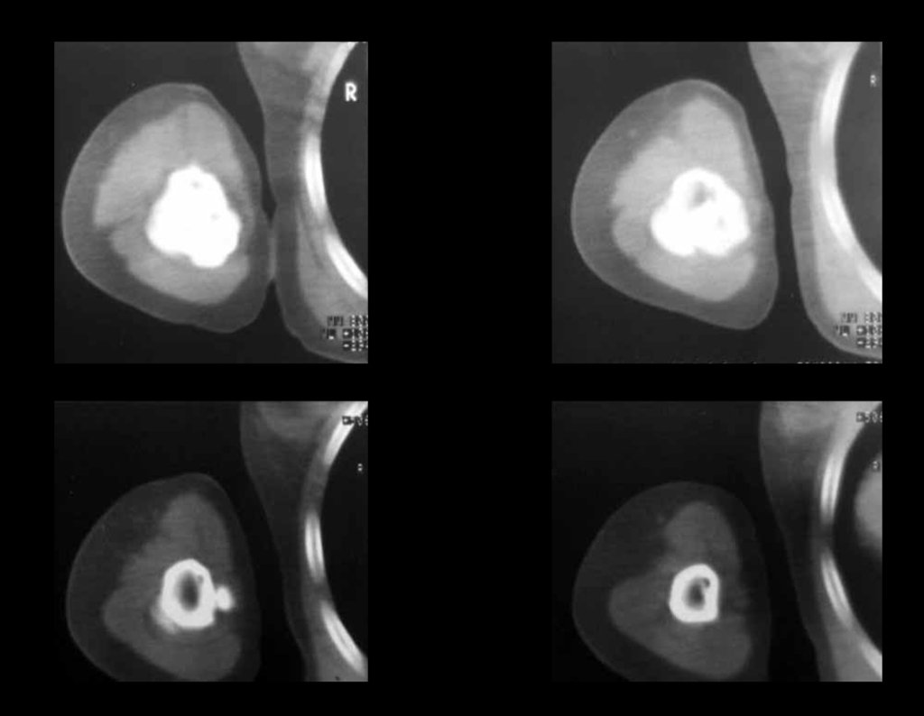
The pathological anatomical report diagnosed paraosteal osteosarcoma, which due to surgical manipulation had invaded the spinal canal and already presented two metastatic nodules to the lung, figures 1 to 5.

Figura 1: Radiografia do úmero esquerdo, frente, com lesão condensante, metadiafisária.
Figure 1: Radiograph of the left humerus, front, with condensing lesion, metadiaphyseal.
Figura 2: Radiografia do úmero esquerdo, perfil, com lesão invadindo o canal medular.
Figure 2: Radiograph of the left humerus, profile, with the lesion invading the spinal canal.
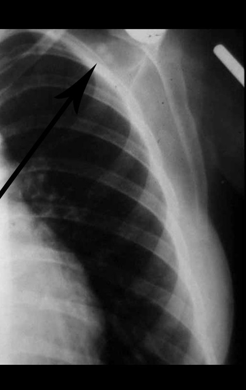
Figura 3: Radiografia do tórax, frente, destaca dois nódulos metastáticos no pulmão direito. À esquerda a seta inferior aponta a lesão no úmero esquerdo. A seta superior chama atenção para o ombro, sem evidencia de neoplasia.
Figure 3: Chest X-ray, front, highlights two metastatic nodules in the right lung. On the left, the lower arrow points to the injury in the left humerus. The upper arrow draws attention to the shoulder, with no evidence of neoplasia.
Figura 4: Cortes tomográficos mostrando o comprometimento tanto da superfície como da medular óssea.
Figure 4: Tomographic sections showing involvement of both the surface and the bone marrow.
Figura 5: A cirurgia inadequada de curetagem propiciou a invasão deste tumor que inicialmente era apenas de superfície.
Figure 5: Inadequate curettage surgery led to the invasion of this tumor, which initially was only superficial.
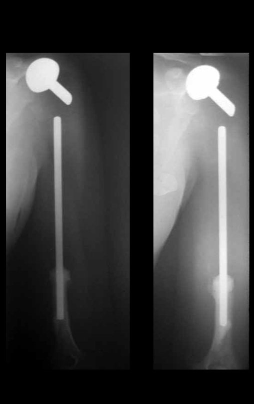
Figura 6: Pós operatório imediato de ressecção de 2/3 proximais do úmero e reconstrução com endoprótese não convencional de polietileno.
Figure 6: Immediate post-operative resection of the proximal 2/3 of the humerus and reconstruction with non-conventional polyethylene endoprosthesis.
Figura 7: A seta salienta a região proximal do úmero, aparentemente sem lesão neoplásica.
Figure 7: The arrow highlights the proximal region of the humerus, apparently without neoplastic lesion.
Figura 8: Corte da peça evidenciando a neoplasia de superfície e a infiltração medular ocorrida após as curetagens, inadvertidas.
Figure 8: Section of the piece showing the surface neoplasia and the medullary infiltration that occurred after the inadvertent curettages.
Figura 9: Radiografia da peça cirúrgica, ressecada em bloco.
Figure 9: Radiograph of the surgical specimen, resected en bloc.
Figura 10: Radiografia do tórax com os dois nódulos pulmonares, após a prótese do úmero e antes da ressecção das metástases.
Figure 10: Chest X-ray with the two pulmonary nodules, after humerus prosthesis and before resection of metastases.
Figura 11: Radiografia do tórax após a toracotomia para ressecção dos nódulos metastáticos. A seta inferior aponta o dreno de tórax e a seta superior salienta o ombro esquerdo sem lesão.
Figure 11: Chest X-ray after thoracotomy for resection of metastatic nodules. The lower arrow points to the chest tube and the upper arrow highlights the uninjured left shoulder.
Figura 12: Pós operatório de três meses.
Figure 12: Three months post-operative.
Figura 13: Após três meses da ressecção do tumor do úmero e das metástases pulmonares.
Figure 13: Three months after resection of the humerus tumor and lung metastases.
Figura 14: Função do MSE, após três meses.
Figure 14: MSE function, after three months.
Figura 15: Radiografia após o terceiro mês da cirurgia. Aparecimento de dois nódulos próximos à cirurgia do ombro.
Figure 15: Radiograph after the third month of surgery. Appearance of two nodules close to shoulder surgery.
Figura 16: Radiografia do tórax, após o terceiro mês das cirurgias do tumor e da toracotomia, revelando o aparecimento de dois nódulos no ombro, evidenciando a recorrência local da neoplasia.
Figure 16: Chest X-ray, after the third month of tumor surgery and thoracotomy, revealing the appearance of two nodules in the shoulder, demonstrating the local recurrence of the neoplasm.
Figura 17: Em outubro de 1988 decidimos ressecar os dois nódulos do ombro, que haviam quadruplicado o seu tamanho.
Figure 17: In October 1988 we decided to resect the two shoulder nodules, which had quadrupled in size.
Figura 18: Nódulos ressecados em bloco.
Figure 18: Nodules resected en bloc.
Figura 19: Pós operatório após três anos.
Figure 19: Post-operative period after three years.
Figura 20: Controle radiográfico após três anos de acompanhamento, em 19/04/1990.
Figure 20: Radiographic control after three years of follow-up, on 04/19/1990.
Figura 21: Paciente em 20/04/1995, após sete anos.
Figure 21: Patient on 04/20/1995, after seven years.
22: Paciente em 15/07/1997, com nove anos e sete meses de acompanhamento.
22: Patient on 07/15/1997, with nine years and seven months of follow-up.
Figura 23: Em 09/02/1999, após doze anos.
Figure 23: On 02/09/1999, after twelve years.
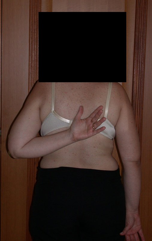
Figura 24: Boa rotação interna, em 09/02/1999, após doze anos.
Figure 24: Good internal rotation, on 02/09/1999, after twelve years.
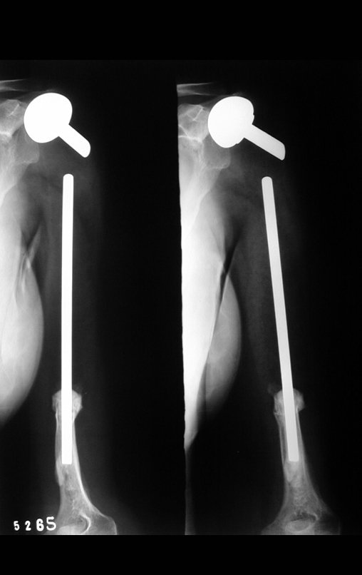
Figura 25: Radiografia, frente, em 09/02/1999, após doze anos.
Figure 25: Radiograph, front, on 02/09/1999, after twelve years.
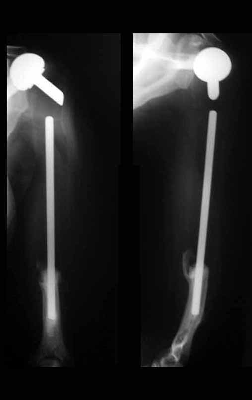
Figura 26: Radiografia, perfil, após doze anos.
Figure 26: X-ray, profile, after twelve years.
Figura 27: Radiografia em 2001, após 14 anos, sem intercorrência.
Figure 27: Radiograph in 2001, after 14 years, without complications.
Figura 28: Perfil do tórax normal, após 14 anos.
Figure 28: Normal chest profile after 14 years.
Figura 29: Prótese bem, após 14 anos.
Figure 29: Prosthetic well, after 14 years.
Figura 30: Paciente em 2001, após 14 anos. bem.
Figure 30: Patient in 2001, after 14 years. good.
Figura 31: Função após 14 anos.
Figure 31: Function after 14 years.
Figura 32: Em 2001, sem intercorrência.
Figure 32: In 2001, without complications.
Figura 33: Boa rotação interna, após 14 anos da cirurgia.
Figure 33: Good internal rotation, 14 years after surgery.
Figura 34: Controle em 20/08/2002, após 15 anos.
Figure 34: Control on 08/20/2002, after 15 years.
Figura 35: Paciente grávida, sem intercorrência, após 15 anos.
Figure 35: Pregnant patient, without complications, after 15 years.
Figura 36: Bem, após 15 anos das cirurgias.
Figure 36: Well, after 15 years of surgery.
Figura 37: Função em 08/07/2003, após 16 anos.
Figure 37: Function on 07/08/2003, after 16 years.
Figura 38: Segurando a filha com o braço operado.
Figure 38: Holding the daughter with the operated arm.
Figura 39: Boa função, sem intercorrência, em 08/07/2003, após 16 anos.
Figure 39: Good function, without complications, on 07/08/2003, after 16 years.
Figura 40: Função em 28/10/2004.
Figure 40: Function on 10/28/2004.
Figura 41: Rotação interna em 28/10/2004
Figure 41: Internal rotation on 10/28/2004
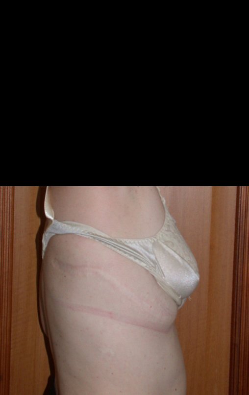
Figura 42: Cicatriz da toracotomia após 17 anos.
Figure 42: Thoracotomy scar after 17 years.
Figura 43: Segurando a filha com o MSE, boa função, após 17 anos.
Figure 43: Holding daughter with MSE, good function, after 17 years.
Figura 44: Em 23/10/2006, após 19 anos.
Figure 44: On 10/23/2006, after 19 years.
Figura 45: Rotação interna normal, após 19 anos.
Figure 45: Normal internal rotation, after 19 years.
Figura 46: Radiografia da reconstrução do úmero com prótese de polietileno, após 19 anos.
Figure 46: Radiograph of humerus reconstruction with polyethylene prosthesis, after 19 years.
Figura 47: Boa força do MSE e excelente função.
Figure 47: Good MSE strength and excellent function.
Video 1: Function after 19 years, on 10/23/2006.
Figura 48: Cintilografia de 06/08/2014, após 27 anos de acompanhamento.
Figure 48: Scintigraphy on 08/06/2014, after 27 years of follow-up.
Figura 49: Rx de tórax normal, em 06/08/2014.
Figure 49: Normal chest X-ray, on 08/06/2014.
Figura 50: Radiografia de tórax, AP, sem alterações, em 06/08/2014, após 27 anos de acompanhamento.
Figure 50: Chest X-ray, AP, without changes, on 08/06/2014, after 27 years of follow-up.
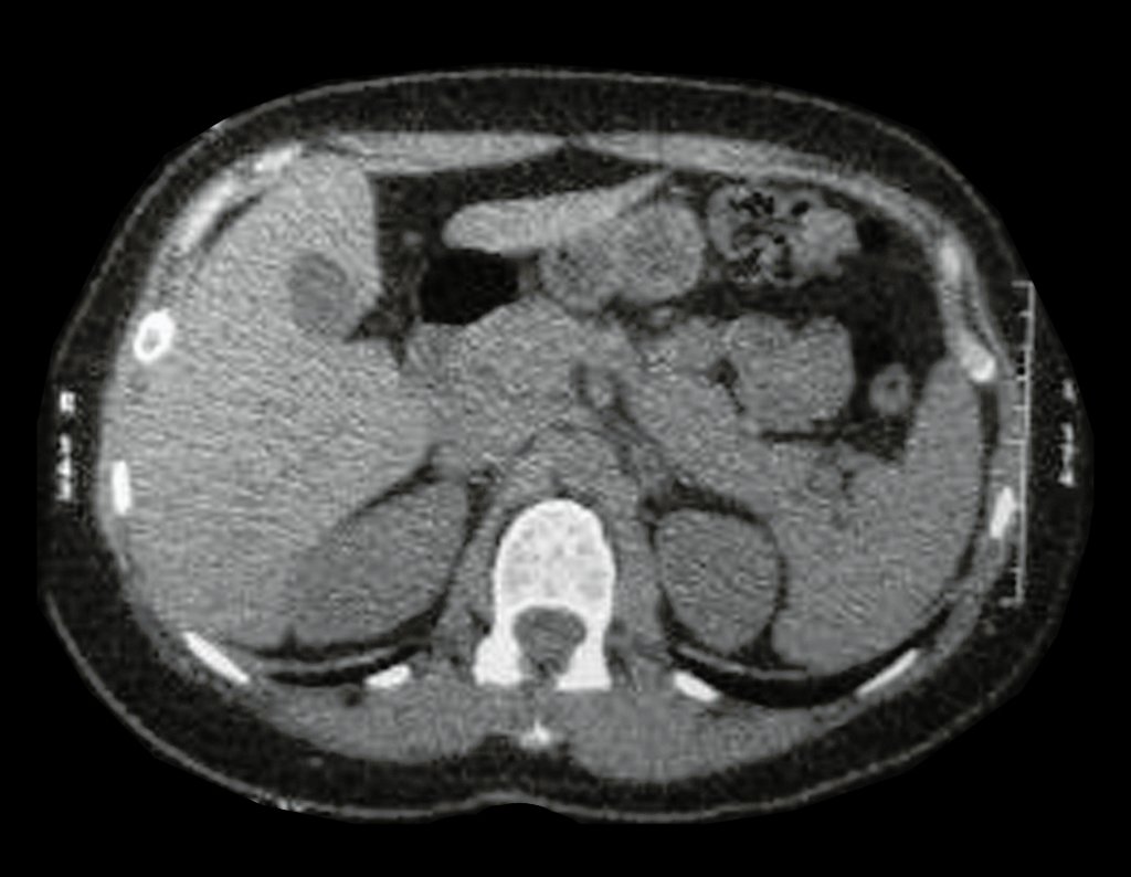
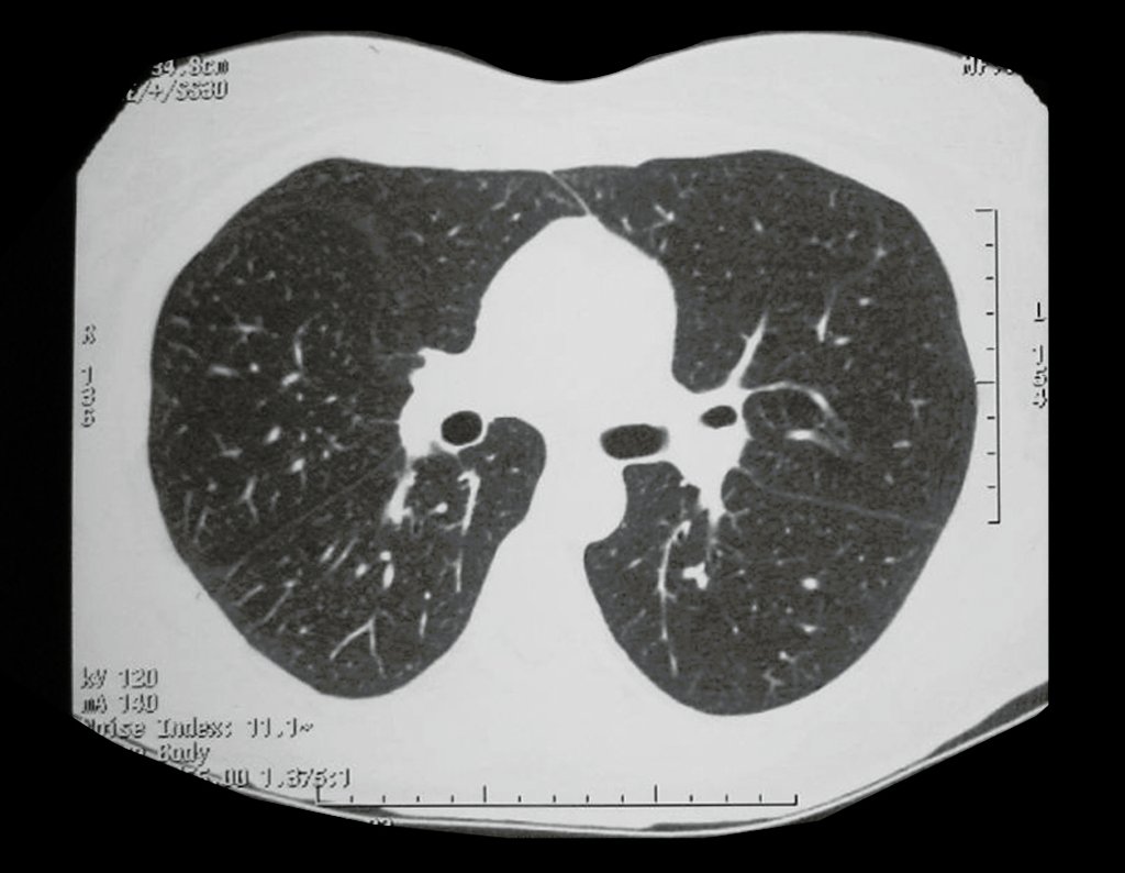
Figura 51: Tomografia de tórax, em 2014, sem anormalidade.
Figure 51: Chest tomography, in 2014, without abnormality.
Figura 52: Tomografia de 2014 sem anormalidades.
Figure 52: Tomography from 2014 without abnormalities.
Figura 53: Campos pulmonares livres, 06/08/2014.
Figure 53: Free lung fields, 08/06/2014.
Figura 54: TC de tórax normal, 06/08/2014.
Figure 54: Normal chest CT, 08/06/2014.
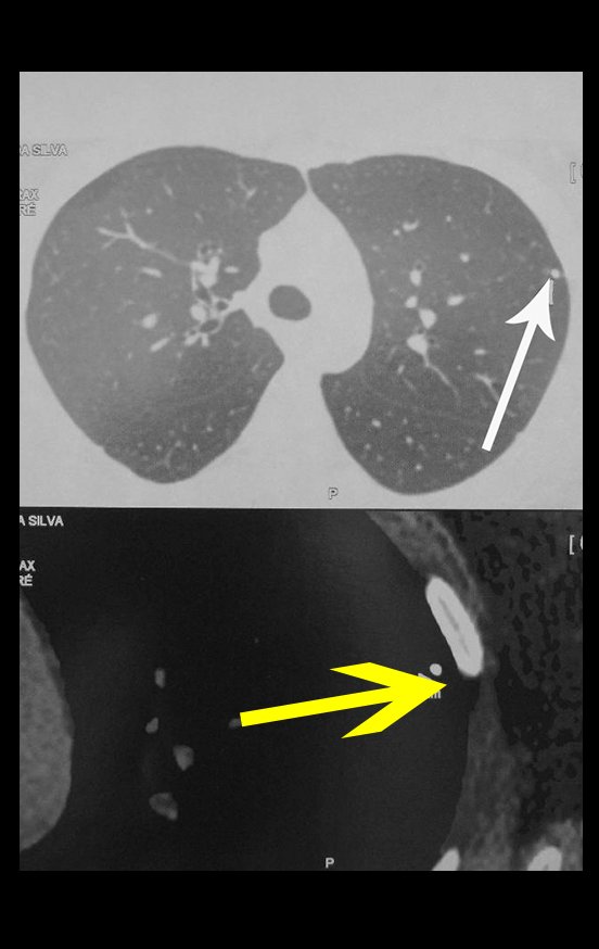
It is important to emphasize that the patient undergoing oncological monitoring suffers at each control assessment. Therefore, it is prudent to prevent them, regarding the small calcified pulmonary nodule of the primary tuberculosis complex, which is normal and present in the vast majority of the population. This way we will be saving unnecessary suffering for both the patient and their families.
Figura 55: No estudo desta tomografia de tórax evidencia-se o pequeno nódulo calcificado, "cicatriz" do complexo primário da tuberculose.
Figure 55: In the study of this chest tomography, the small calcified nodule, "scar" of the primary tuberculosis complex, is evident.
Figura 56: Laudo da Tomografia pulmonar.
Figure 56: Lung CT report.
Figura 57: Cintilografia óssea, frente, sem anormalidades.
Figure 57: Bone scintigraphy, front, without abnormalities.
Figura 58: Cintilografia óssea, vista posterior.
Figure 58: Bone scintigraphy, posterior view.
Figura 59: Laudo da Cintilografia, após 27 anos, sem anormalidade.
Figure 59: Scintigraphy report, after 27 years, without abnormality.
Figura 60: Radiografia de controle da prótese, após 27 anos, bem.
Figure 60: Control X-ray of the prosthesis, after 27 years, well.
Figura 61: Paciente em 06/08/2014, após 27 anos.
Figure 61: Patient on 08/06/2014, after 27 years.
Figura 62: Função após 27 anos.
Figure 62: Function after 27 years.
Figura 63: Boa rotação interna após 27 anos da prótese.
Figure 63: Good internal rotation after 27 years of prosthesis.
Video 2: Function on 08/06/2014, with 27 years of follow-up of osteosarcoma resection of the left humerus and reconstruction with non-conventional polyethylene endoprosthesis.
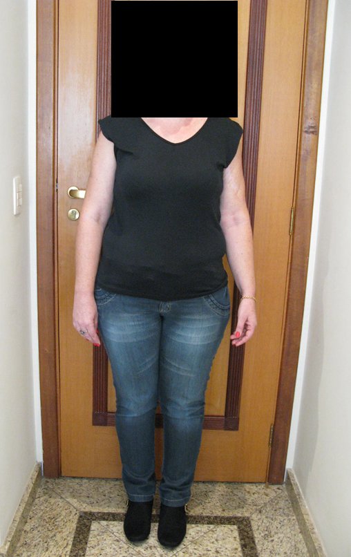
Figura 64: Bom esquema corporal. Membros simétricos.
Figure 64: Good body layout. Symmetrical limbs.
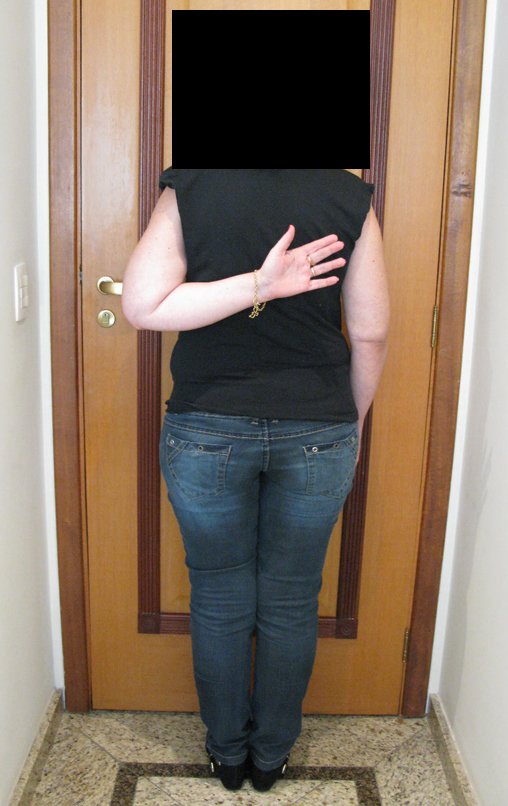
Figura 65: Boa função do membro operado.
Figure 65: Good function of the operated limb.
Figura 66: Prótese de úmero bem, inalterada após 31 anos da cirurgia.
Figure 66: Good humeral prosthesis, unchanged 31 years after surgery.
Figura 67: Controle pulmonar sem anormalidade. Em destaque o nódulo periférico calcificado, cicatriz da primo infecção da tuberculose.
Figure 67: Lung control without abnormality. Highlighting the calcified peripheral nodule, a scar from the first tuberculosis infection.
In 2018, the patient was doing well after 31 years of treatment.

Author: Prof. Dr. Pedro Péricles Ribeiro Baptista

 Orthopedic Oncosurgery at the Dr. Arnaldo Vieira de Carvalho Cancer Institute

Hello! How can we assist you?