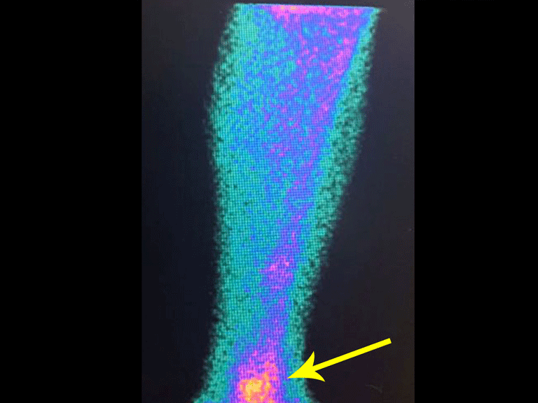
Osteoid osteoma and foreign body.
The coronal and sagittal sections, with density for bone and soft tissues continued to show this radio-opaque image which, in these sections revealed to be linear, figures 23 to 26. Metallic artifact???
Upon outpatient return six weeks after removal of the foreign body, the patient reported the disappearance of the acute and disabling pain but reported the persistence of nighttime pain caused by the osteoid osteoma.
Surgery was scheduled to excise the tumor, following the initial planning with the aid of gammagraphy, to be performed shortly.
Authors of the case
Author: Prof. Dr. Pedro Péricles Ribeiro Baptista
Orthopedic Oncosurgery at the Dr. Arnaldo Vieira de Carvalho Cancer Institute
Office : Rua General Jardim, 846 – Cj 41 – Cep: 01223-010 Higienópolis São Paulo – SP
Phone: +55 11 3231-4638 Cell:+55 11 99863-5577 Email: drpprb@gmail.com


