This digital library houses the book on Oncology and Orthopedic Oncosurgery.

It includes academic lectures, presentations from national and international congresses, published papers, case discussions, performed surgical procedures, and proprietary techniques developed.

The digital format was chosen because the web allows the inclusion of texts with numerous visual resources, such as images and videos, which would not be possible in a printed book.

The content is intended for students, healthcare professionals, and the general public interested in the field.

Myxoid liposarcoma

043a1c71577f6249b071679bce73d008 2

Myxoid Liposarcoma

Myxoid liposarcoma. Male patient, 28 years old, reports pain and progressive bulging in the posterior region of the right thigh, distally, 08 months ago, denies local trauma. Initially he sought an orthopedic service, a biopsy was performed and myxoid liposarcoma was diagnosed.

Referred to our service, we found an extensive tumor on the posterior surface of the thigh, starting in the middle third to the joint line, without pain or neurological deficit.

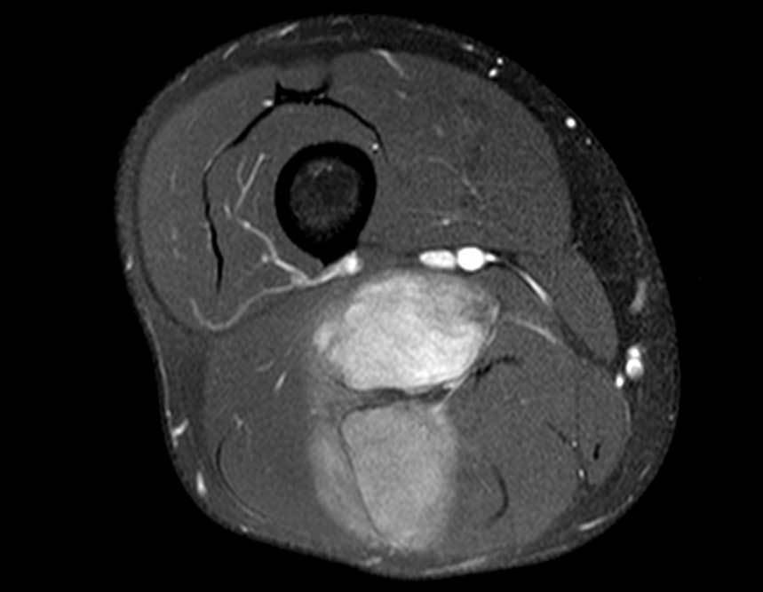
Magnetic resonance imaging, figures 1 to 14, demonstrate the heterogeneity of the lesion and its aggressiveness, showing the large extent of the lesion and its proximity to the vascular-nervous bundle.

Figura 02: Seta vermelha mostra a divisão do nervo ciativo e a seta em azul o feixe femoral vásculonervoso.
Figure 01: Arrow points to the sciatic nerve, in the proximal region of the lesion.
Figure 02: Red arrow shows the division of the sciative nerve and the blue arrow shows the femoral vascular-nervous bundle.
Figura 04: Extensão da lesão para região posterior do joelho.
Figure 03: Red arrows show the nerves in close contact with the tumor septum (yellow arrow).
Figure 04: Extension of the injury to the posterior region of the knee.
Figura 05: Seta em amarelo aponta para o marcador, destacando a proximidade do tumor com o nervo fibular.
Figure 05: Yellow arrow points to the marker, highlighting the proximity of the tumor to the peroneal nerve.
Figura 7: Lesão septada em intimo contato com feixe vasculonervoso.
Figure 6: Post-contrast, showing the proximal region of the lesion, adjacent to the sciatic nerve.
Figure 7: Septate lesion in close contact with the vascular-nervous bundle.
Figura 9: Grande heterogeneidade da lesão.
Figure 8: Intimacy of the nerve with the lesion after contrast.
Figure 9: Great heterogeneity of the lesion.
Figura 10: Grande heterogeneidade da lesão.
Figure 10: Heterogeneity of the lesion extending to the distal third of the femur.
Figura 11: Corte sagital, demonstrando toda a extensão da lesão.
Figure 11: Sagittal section, demonstrating the entire extent of the lesion.
Figura 12: Lesão septada e agressiva, caracterizada após contraste.
Figure 12: Septate and aggressive lesion, characterized after contrast.
Figura 13: Proximidade da lesão com o nervo ciático, numa visão sagital.
Figure 13: Proximity of the lesion to the sciatic nerve, in a sagittal view.
Figura 14: Corte coronal mostrando a agressividade da neoplasia.
Figure 14: Coronal section showing the aggressiveness of the neoplasm.
Clinically, he presented bulging in the posterior region of the right thigh, with pain on palpation and exertion, currently starting with paresthesia in the left leg, without neurological deficit, figures 15 to 18.
Figura 15: Vista posterior com o trajeto da biópsia e abaulamento na coxa direita.
Figure 15: Posterior view with the biopsy path and bulging in the right thigh.
Figura 16: Fotografia em perfil do membro acometido.
Figure 16: Profile photograph of the affected limb.
Figura 17: demonstração clínica da função do paciente, ao realizar agachamento.
Figure 17: clinical demonstration of the patient's function when performing squats.
Figura 18: seta em amarelo aponta o local da biópsia realizada em outro serviço.
Figure 18: yellow arrow points to the location of the biopsy performed in another service.

After carrying out anamnesis, physical examination and complementary tests on the patient, we put forward some diagnostic hypotheses. As the patient underwent a biopsy at another service, we discussed the biopsy slide with the local pathology team who analyzed the material and after discussing, we arrived at the diagnosis of chondromyxoid liposarcoma.

We talk to the patient about their pathology and treatment (resection + complementation with local radiotherapy at the discretion of the radiotherapist), we schedule the surgical resection with them accordingly. The following images explain the surgical step by step.

Figura 19: seta em vermelho com enfoque para o trajeto da biópsia.
Figure 19: red arrow focusing on the biopsy path.
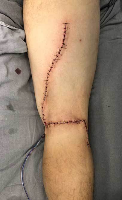
Figura 20: incisão cirúrgica envolvendo a região posterior da coxa, em direção à região poplítea medial, com ressecção do trajeto da biópisa, seguindo transversalmente pela prega poplítea até face póstero lateral do joelho.
Figure 20: surgical incision involving the posterior region of the thigh, towards the medial popliteal region, with resection of the biopsy path, running transversely through the popliteal fold to the posterolateral aspect of the knee.
Figura 21: dissecção da pele e tecido subcutâneo, com controle total da hemostasia, sem uso de nenhum tipo de compressão ou garrote cirúrgico.
Figure 21: dissection of the skin and subcutaneous tissue, with total control of hemostasis, without the use of any type of compression or surgical tourniquet.
Figura 22: dissecção até planos musculares, visualizando o tumor, indicado pela seta preta. A seta em amarelo mostra a intimidade do tumor com o nervo fibular profundo.
Figure 22: dissection to muscular planes, visualizing the tumor, indicated by the black arrow. The yellow arrow shows the proximity of the tumor to the deep peroneal nerve.
Figura 23: Imagem evidencia dissecção da bifurcação do nervo ciático (demonstrado pela pinça), liberando o tumor do seu trajeto.
Figure 23: Image shows dissection of the sciatic nerve bifurcation (demonstrated by forceps), releasing the tumor from its path.
Figura 24: liberação do tumor do feixe nervoso, sem danificar as estruturas nobres, preservando à anatomia do nervo.
Figure 24: release of the tumor from the nerve bundle, without damaging the noble structures, preserving the nerve anatomy.
Figura 25: após retirada do tumor, reparem na hemostasia cirúrgica, a integridade dos tecidos e a anatomia dos nervos fibulares, representado pelas setas amarelas e do nervo tibial na seta azul.
Figure 25: after removing the tumor, note the surgical hemostasis, the integrity of the tissues and the anatomy of the peroneal nerves, represented by the yellow arrows and the tibial nerve in the blue arrow.
Figura 26: sutura cirúrgica no final do procedimento com colocação de dreno porto-vac 3.2 mm.
Figure 26: surgical suture at the end of the procedure with placement of a 3.2 mm porto-vac drain.
Figuras 27
Figures 27
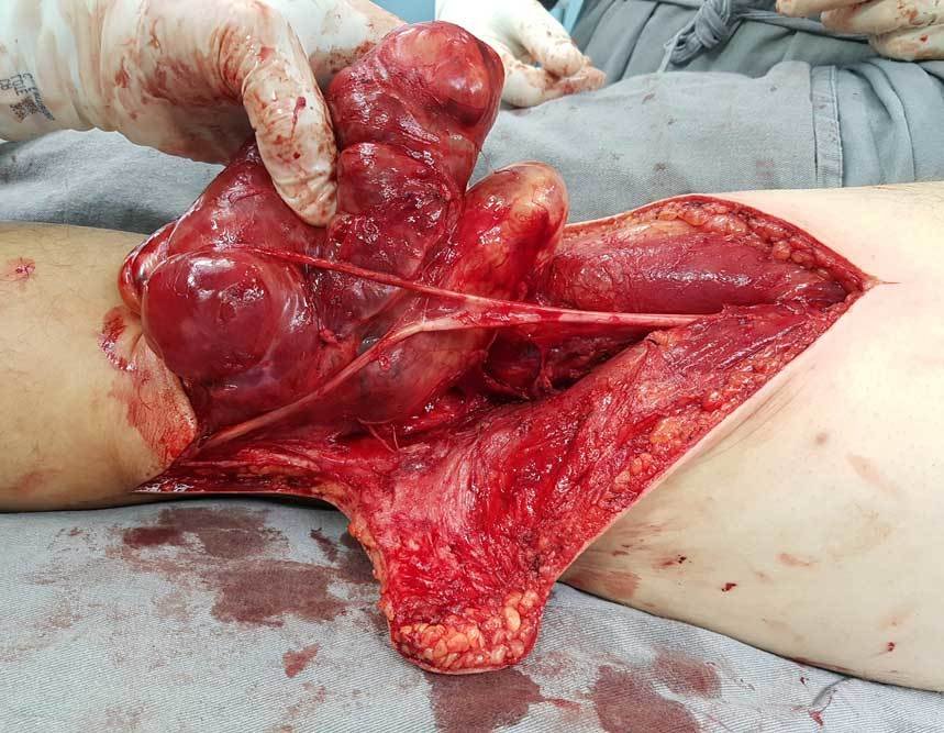
e 28: demonstram a peça cirúrgica na face anterior e posterior. Notem que o tumor não invade as partes moles e sim se deforma respeitando o trajeto do nervo em sua bifurcação, representado pela seta preta.
and 28: demonstrate the surgical specimen on the anterior and posterior surface. Note that the tumor does not invade the soft tissues but rather deforms respecting the path of the nerve at its bifurcation, represented by the black arrow.
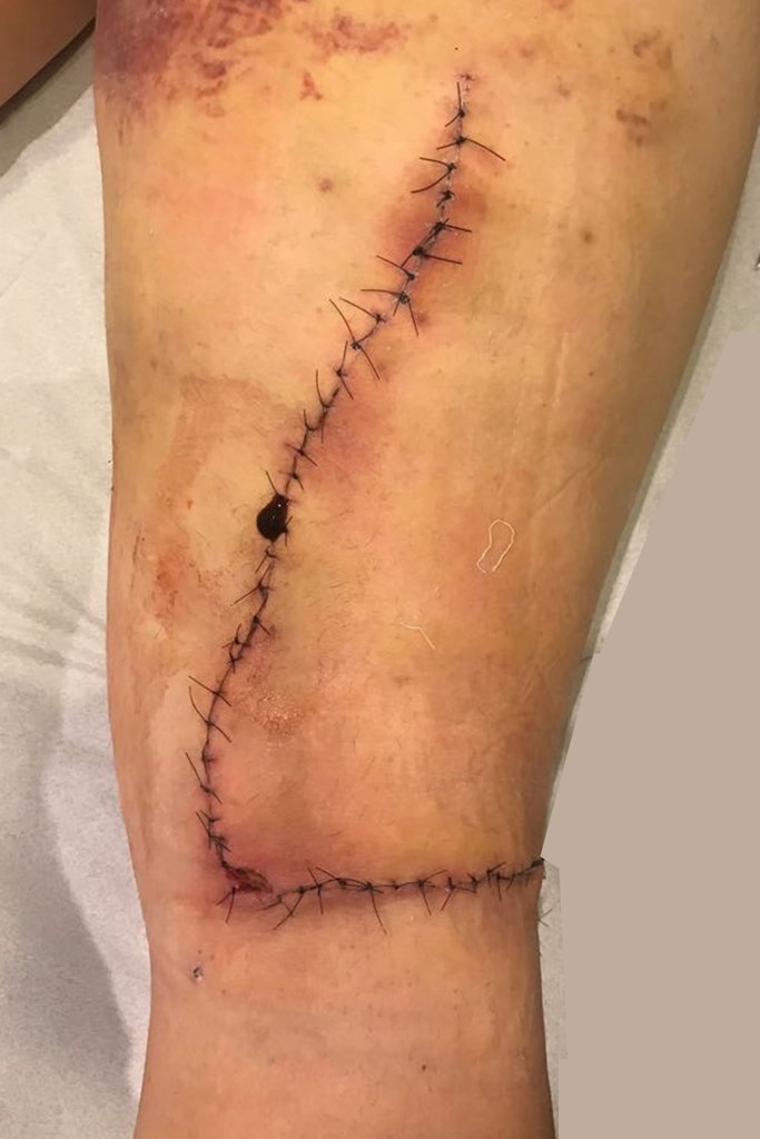
Figura 29: paciente no 7º PO, mostrando o processo inflamatório normal e esperado da cicatrização
Figure 29: patient on the 7th POD, showing the normal and expected inflammatory process of healing
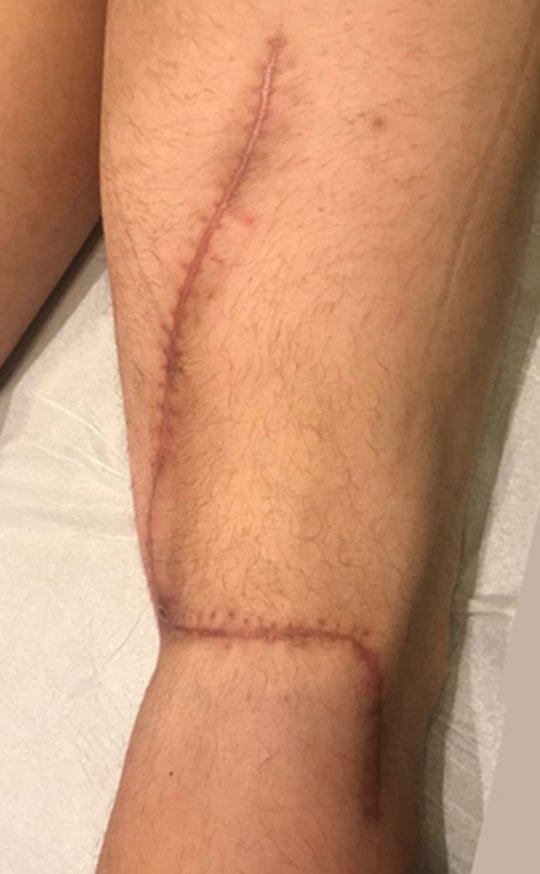
Figura 30: Evolução da ferida após retirada dos pontos, com 14 dias de pós operatório.
Figure 30: Evolution of the wound after removing the stitches, 14 days after surgery.
Figuras 31
Figures 31
e 32: 4 semanas de pós operatório, mostrando a ótima cicatrização da ferida e em região medial a ferida ainda demanda de pouco tempo para cicatrizar totalmente. Reparem que a cicatriz não possui nenhum sinal flogistico local.
and 32: 4 weeks post-operatively, showing excellent wound healing and in the medial region the wound still requires little time to fully heal. Note that the scar does not have any local phlogistic signs.
Video 1: Patient 4 weeks after surgery. Referred for adjuvant radiotherapy.

Patient in the post-operative segment, returns for consultation, without complaints at the moment, with good function of the operated limb and carrying out all his work activities as a Personal Trainer.

 
Figuras 33
Figures 33
34: Imagem clinica da região posterior da coxa, com 4 meses de pós-operatório e 30 dias do término da radioterapia adjuvante, mostrando ótima cicatrização da ferida .
34: Clinical image of the posterior region of the thigh, 4 months postoperatively and 30 days after the end of adjuvant radiotherapy, showing excellent wound healing.
Figura 35: vista posterior do paciente com carga total nos membros inferiores.
Figure 35: posterior view of the patient with full weight on the lower limbs.
Figura 36: vista em perfil do paciente com carga toral nos membros inferiores
Figure 36: profile view of the patient with torsional weight on the lower limbs
Figura 37: paciente realizando agachamento sem apoio, com carga total nos membros inferiores.
Figure 37: patient performing squats without support, with full load on the lower limbs.
Figura 38: Paciente em carga monopodal no membro operado, sem apoio, realizando carga toral no membro inferior direito.
Figure 38: Patient carrying a single leg load on the operated limb, without support, performing a torsional load on the right lower limb.
Video 2: Patient after adjuvant radiotherapy, four months after surgery. Good healing and excellent function.

Author: Prof. Dr. Pedro Péricles Ribeiro Baptista

 Orthopedic Oncosurgery at the Dr. Arnaldo Vieira de Carvalho Cancer Institute

Hello! How can we assist you?