This digital library houses the book on Oncology and Orthopedic Oncosurgery.

It includes academic lectures, presentations from national and international congresses, published papers, case discussions, performed surgical procedures, and proprietary techniques developed.

The digital format was chosen because the web allows the inclusion of texts with numerous visual resources, such as images and videos, which would not be possible in a printed book.

The content is intended for students, healthcare professionals, and the general public interested in the field.

Kidney Cancer Metastasis

4a914e5c38172ae9b61780ffbd0b2f90 3

Bone Metastasis of Hypernephroma - (Kidney Cancer)

Kidney Cancer Metastasis. Hypernephroma bone metastasis, also known as metastatic kidney cancer to the bone, is a serious complication of kidney cancer. Hypernephroma, or renal cell carcinoma, is a common form of cancer that originates in the kidneys. When bone metastasis occurs, cancer cells spread from the kidneys to the bones, potentially affecting several areas of the skeleton.

This spread of cancer to the bones can cause a series of symptoms, such as persistent bone pain, pathological fractures and impaired mobility. Furthermore, bone metastasis from hypernephroma can lead to serious complications such as spinal cord compression that require immediate medical intervention.

Treatment of bone metastasis from hypernephroma generally involves a multidisciplinary approach, including surgery, radiotherapy, targeted therapy, and/or immunotherapy. The aim of treatment is to control the spread of cancer, alleviate symptoms and improve the patient’s quality of life.

However, bone metastasis from hypernephroma is often a challenging condition to treat and can have a significant impact on patient survival and prognosis. Therefore, a careful and coordinated approach between a specialized medical team is essential to provide the best possible care and ensure the patient’s well-being.

Figura 1: Rm axial T1.
Figure 1: T1 axial MRI.
Figura 2: Rm axial T1 Spir.
Figure 2: Axial Rm T1 Spir.
Figura 3: Rm axial T1 spir.
Figure 3: Axial Rm T1 spir.
Figura 4: Rm axial T2 spir.
Figure 4: Axial Rm T2 spir.
Figura 5: Rm cor T1
Figure 5: Rm color T1
Figura 6: Rm cor T2 spir.
Figure 6: Rm color T2 spir.
Figura 7: Rm sag dp spir.
Figure 7: Rm sag dp spir.
Figura 8: Laudo da Rm do quadril direito.
Figure 8: MRI report of the right hip.
Figura 9: Rm sag. T1
Figure 9: Rm sag. T1
Figura 10: Rm sag. T1.
Figure 10: Rm sag. T1.
Figura 11: Rm sag. T1.
Figure 11: Rm sag. T1.
Figura 12: Laudo da Rm da coluna cervical, toraxica e lombossacra.
Figure 12: MRI report of the cervical, thoracic and lumbosacral spine.
Figura 13
Figure 13
Figura 14
Figure 14
Figura 15: Laudo da Cintilografia.
Figure 15: Scintigraphy report.
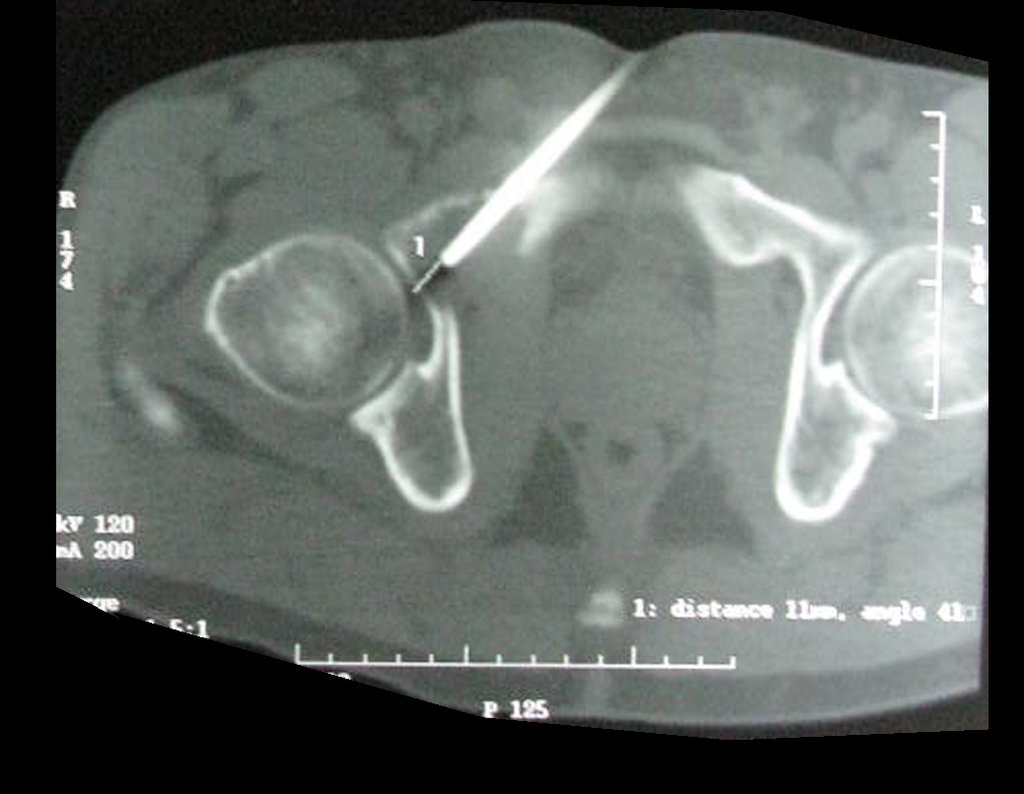
Figura 16: TC mostrando o local da biópsia.
Figure 16: CT showing the biopsy site.
Figura 17: TC mostrando o local da biópsia.
Figure 17: CT showing the biopsy site.
Figura 18
Figure 18
Figura 19: Laudo da TC.
Figure 19: CT report.
Figura 29: Pet-cet .
Figure 29: Pet-cet.
Figura 30: Pet-cet .
Figure 30: Pet-cet.
Figura 20: Pet-cet evidenciando a lesão no Isquio do lado direito.
Figure 20: Pet-cet showing the injury to the ischium on the right side.
Figura 21: Pet-cet evidenciando a lesão no Isquio do lado direito, marcação em círculo.
Figure 21: Pet-cet showing the injury to the ischium on the right side, circle marking.
Figura 22: Pet-cet evidenciando o local acometido pelo tumor.
Figure 22: Pet-cet showing the location affected by the tumor.
Figura 24: Pet-cet, evidenciando a lesão,vista lateral.
Figure 24: Pet-cet, showing the injury, side view.
Figura 25: Pet-cet .
Figure 25: Pet-cet.
Figura 26: Pet-cet .
Figure 26: Pet-cet.
Figura 27: Pet-cet .
Figure 27: Pet-cet.
Figura 28: Pet-cet .
Figure 28: Pet-cet.
Figura 29: Pet-cet .
Figure 29: Pet-cet.
Figura 30: Pet-cet .
Figure 30: Pet-cet.
Figura 31: Tc 2015.
Figure 31: Tc 2015.
Figura 32: Tc 2015.
Figure 32: Tc 2015.
Figura 33: Tc 2015.
Figure 33: Tc 2015.
Figura 34: Tc 2015.
Figure 34: Tc 2015.
Figura 35: Tc 2015.
Figure 35: Tc 2015.
Figura 36: Laudo da Tc.
Figure 36: Tc report.
Figura 37: RM cor T1.
Figure 37: T1 color MRI.
Figura 38: RM axial T1.
Figure 38: Axial T1 MRI.
Figura 39: RM sag. dp spir.
Figure 39: MR sag. dp spir.
Figura 40: RM cor T2.
Figure 40: T2 color MRI.
Figura 41: RM axial T2.
Figure 41: Axial T2 MRI.
Figura 42: Laudo da Rm da bacia.
Figure 42: Basin Rm report.
Figura 43: Laudo do Anatomopatológico
Figure 43: Anatomopathological Report
Legenda: Figura 44: Laudo do Imunofenotipagem.
Caption: Figure 44: Immunophenotyping Report.
Figura 45: Incisão cirúrgica na região inquinal do lado direito.
Figure 45: Surgical incision in the inquinal region on the right side.
Figura 46: Incisão cirúrgica na região inquinal, sendo expandida.
Figure 46: Surgical incision in the inquinal region, being expanded.
Figura 47: Incisão cirúrgica na região inquinal.
Figure 47: Surgical incision in the inquinal region.
Figura 48: Incisão cirúrgica na região inquinal.
Figure 48: Surgical incision in the inquinal region.
Figura 49
Figure 49
Figura 50
Figure 50
Figura 51
Figure 51
Figura 52
Figure 52
Figura 53: Tumor removido.
Figure 53: Tumor removed.
Figura 54: Evidenciamos o local de onde foi removido o tumor.
Figure 54: We highlight the location where the tumor was removed.
Figura 55
Figure 55
Figura 56: Colocação de cera de osso no local da lesão removida.
Figure 56: Placement of bone wax at the site of the removed lesion.
Figura 57: Colocação de cera de osso.
Figure 57: Bone wax placement.
Figura 58: Colocação de hemostático de partes moles.
Figure 58: Placement of soft tissue hemostat.
Figura 59
Figure 59
Figura 60: Fechamento da incisão cirúrgica.
Figure 60: Closure of the surgical incision.
Figura 61: Fechamento da incisão cirúrgica.
Figure 61: Closure of the surgical incision.
Figura 62: Curativo da incisão cirúrgica.
Figure 62: Surgical incision dressing.
Figura 63: Radigrafia pós-operatório imediato do quadril direito frente.
Figure 63: Immediate postoperative radiograph of the front right hip.
Figura 64: Radigrafia pós-operatório imediato do quadril direito frente.
Figure 64: Immediate postoperative radiograph of the front right hip.
Figura 65: Radigrafia pós-operatório imediato do quadril direito oblíqua.
Figure 65: Immediate postoperative oblique radiograph of the right hip.
Figura 66: Anatomo Patológico pós cirúrgia.
Figure 66: Pathological anatomy after surgery.
Video 1 : Patient on the eighth day after surgery to resect the lesion and reconstruct it with cement.
Figura 67: Paciente no pós - operatório de 8 dias.
Figure 67: Patient 8 days post-operatively.
Figura 68: Paciente em posição monopodal.
Figure 68: Patient in single-leg position.
Figura 69: Ferida pós-operatório de 8 dias.
Figure 69: 8-day postoperative wound.
Figura 70: Paciente em posição bipodal.
Figure 70: Patient in bipedal position.
Figura 71
Figure 71
Figura 72
Figure 72
Figura 73
Figure 73
Figura 74: Laudo Imuno-Histoquímico.
Figure 74: Immunohistochemical Report.
Patient 18 days post-operatively.
Figura 75: Paciente no pós operatório de 18 dias.
Figure 75: Patient 18 days after surgery.
Figura 76: Paciente no pós operatório de 18 dias.
Figure 76: Patient 18 days after surgery.
Video 2 : Patient 18 days after surgery.

Author: Prof. Dr. Pedro Péricles Ribeiro Baptista

 Orthopedic Oncosurgery at the Dr. Arnaldo Vieira de Carvalho Cancer Institute

Hello! How can we assist you?