This digital library houses the book on Oncology and Orthopedic Oncosurgery.

It includes academic lectures, presentations from national and international congresses, published papers, case discussions, performed surgical procedures, and proprietary techniques developed.

The digital format was chosen because the web allows the inclusion of texts with numerous visual resources, such as images and videos, which would not be possible in a printed book.

The content is intended for students, healthcare professionals, and the general public interested in the field.

Intraneural Fibular Nerve Cyst

Cisto Intraneural do nervo fibular. Ganglion justaarticular.

Intraneural cyst of the peroneal nerve. Juxtaarticular ganglion.

Intraneural cyst. A male patient, 64 years old, complained of inability to perform dorsal flexion of the left ankle for eleven months, without comorbidities or history of trauma.

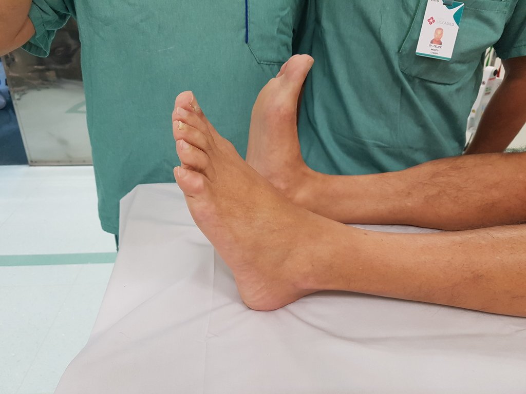
On clinical examination, the left foot showed a plantar drop, without active dorsiflexion or any radiographic changes in the knee or ankle, figures 1 to 3.

figura 01: Atitude em queda plantar do tornozelo esquerdo.
Figure 01: Plantar drop attitude of the left ankle.
Figura 02: Radiografia do joelho e tornozelo esquerdos, frente.
Figure 02: Radiograph of the left knee and ankle, front.
Figura 03: Radiografia do joelho e tornozelo esquerdos, perfil.
Figure 03: Radiograph of the left knee and ankle, profile.

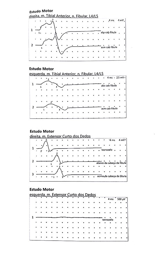
The knee resonance detected the presence of a lobulated and elongated ganglion cyst inside the peroneal nerve and the electroneuromyography reports the absence of action potentials of the peroneal nerve on the left, suggesting axoniotmesis, figures 4 to 7.

Figura 04: Rm de 31 de julho de 2018 revelando alteração no nervo fibular.
Figure 04: MRI from July 31, 2018 revealing changes in the peroneal nerve.
Figura Figura 05: Laudo da ressonância.
Figure Figure 05: Resonance report.
Figura 06: Eletroneuromiografia revelando alteração do nervo fibular do MIE.
Figure 06: Electroneuromyography revealing changes in the MIE peroneal nerve.
Figura 07: Laudo da eletroneuromiografia sugerindo axoniotmese ? !
Figure 07: Electroneuromyography report suggesting axonotmesis? !

Author: Prof. Dr. Pedro Péricles Ribeiro Baptista

 Orthopedic Oncosurgery at the Dr. Arnaldo Vieira de Carvalho Cancer Institute

Hello! How can we assist you?