This digital library houses the book on Oncology and Orthopedic Oncosurgery.

It includes academic lectures, presentations from national and international congresses, published papers, case discussions, performed surgical procedures, and proprietary techniques developed.

The digital format was chosen because the web allows the inclusion of texts with numerous visual resources, such as images and videos, which would not be possible in a printed book.

The content is intended for students, healthcare professionals, and the general public interested in the field.

Giant cell tumor of the tibia

030d13e49bb7d1add5ac5ea2e4a43231 3

Giant Cell Tumor of the Tibia Intraepiphyseal Prosthesis

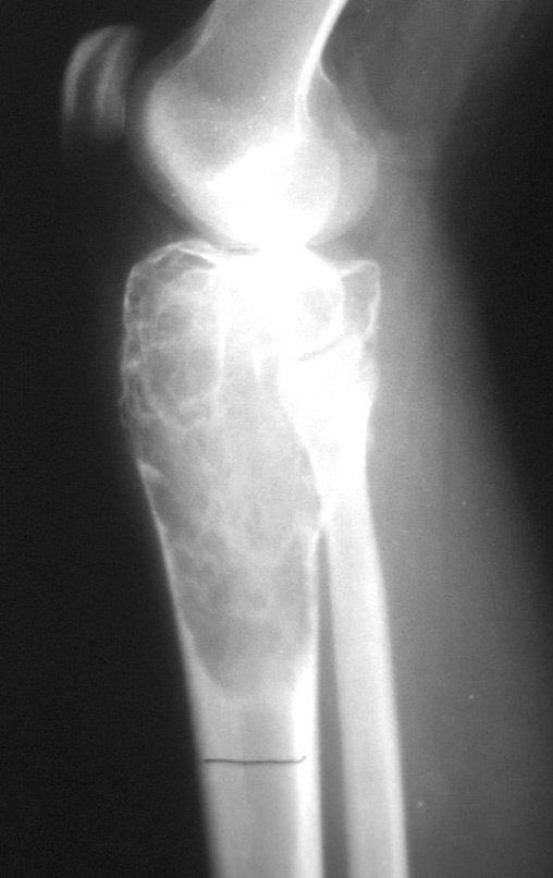
Giant cell tumor of the tibia. A patient born in April 1973, she presented with a bone rarefaction lesion in the right tibial epiphysis, when she was only fourteen years and six months old. In January 1988, she underwent surgery in Salvador – Ba and underwent curettage of the tumor. The anatomical pathological examination diagnosed it as a giant cell tumor, which is rare in the patient’s young age group. In February 1989, the patient underwent reoperation in the city of Vitória da Conquista, due to tumor recurrence. In June of the same year, she came to us due to a new recurrence, figures 1 to 3.
Figura 1: Paciente em julho de 1989, com duas intervenções cirúrgicas, por diferentes vias de acesso !!! e com recorrência da lesão. A seta aponta infiltração sub-cutânea do tumor.
Figure 1: Patient in July 1989, with two surgical interventions, through different access routes!!! and with recurrence of the lesion. The arrow points to subcutaneous infiltration of the tumor.
Figura 2: Radiografia, frente, com grande lesão de rarefação óssea ocupando toda a epífise proximal da tibia direita, com afundamento do planalto medial.
Figure 2: Radiograph, front, with a large bone rarefaction lesion occupying the entire proximal epiphysis of the right tibia, with sinking of the medial plateau.
Figura 3: Radiografia, perfil, com lesão na epífise da tibia e diáfise proximal.
Figure 3: Radiograph, profile, with injury to the tibial epiphysis and proximal diaphysis.
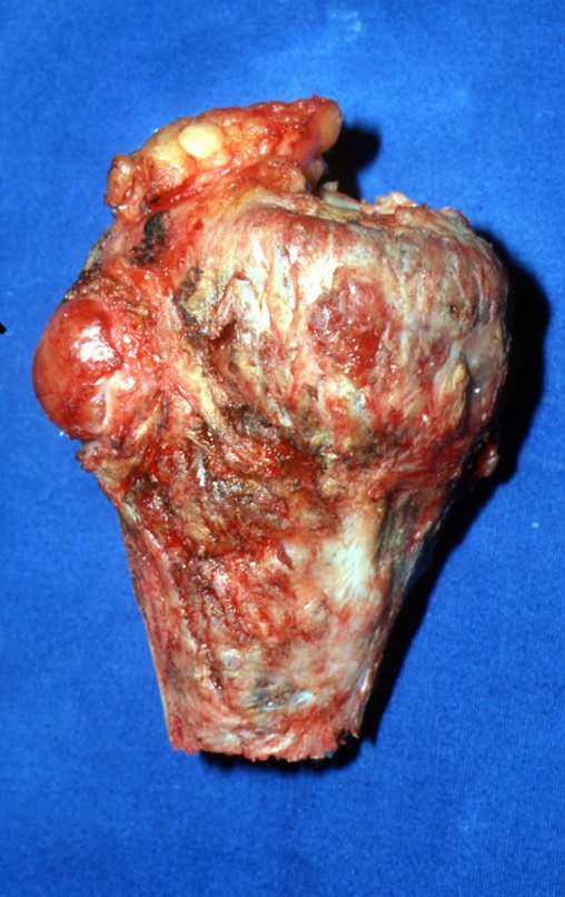
Despite the patient being very young, we opted for en bloc resection, due to two recurrences and tumor implantation in soft tissues. We designed a special prosthesis for this case, creating a model with an intra-epiphyseal component in the femur and replacement of the proximal third of the tibia, both components made of polyethylene. The resection was carried out en bloc, on 06/03/1989, with removal of the entire affected area, figures 4 to 15.
Figura 4: Acesso para patelar medial, deixando as incisões anteriores. Dissecção contornando o nódulo do sub cutâneo.
Figure 4: Access to the medial patellar, leaving the previous incisions. Dissection bypassing the subcutaneous nodule.
Figura 5: dissecção sub perióstica, contornando o nódulo tumoral.
Figure 5: subperiosteal dissection, bypassing the tumor nodule.
Figura 6: Rebatimento da pata de ganso como um tendão conjunto.
Figure 6: Reflection of the pes ansus as a joint tendon.
Figura 7: Mensuração do segmento a ser ressecado.
Figure 7: Measurement of the segment to be resected.
Figura 8: Marcação do nível da osteotomia e liberação do tibial anterior e tecidos moles inseridos no segmento proximal.
Figure 8: Marking the level of the osteotomy and release of the tibialis anterior and soft tissues inserted into the proximal segment.
Figura 9: Realização da osteotomia da tíbia e liberação do músculo solear e cápsula articular lateral, anterior e medialmente.
Figure 9: Performing the tibial osteotomy and releasing the soleus muscle and joint capsule laterally, anteriorly and medially.
Figura 10: Desinserção do semimembranoso e cápsula posterior.
Figure 10: Detachment of the semimembranosus and posterior capsule.
Figura 11: Peça ressecada em bloco, juntamente com o nódulo, vista de frente.
Figure 11: Piece resected in a block, together with the nodule, seen from the front.
Figura 12: Peça ressecada em bloco, juntamente com o nódulo, vista de perfil.
Figure 12: Piece resected as a block, together with the nodule, profile view.
The reconstruction will be carried out with the prosthesis designed especially for this patient, made of polyethylene and stainless steel and with an intraepiphyseal component in the femur, figures 13 to 15.
Figura 13: Componente intraepifisário femoral, vista de frente.
Figure 13: Femoral intraepiphyseal component, front view.
Figura 14: Componente intraepifisário femoral, vista de perfil.
Figure 14: Femoral intraepiphyseal component, profile view.
Figura 15: Componente tibial, com bloqueio em T, sem componente rotacional.
Figure 15: Tibial component, with T-lock, without rotational component.
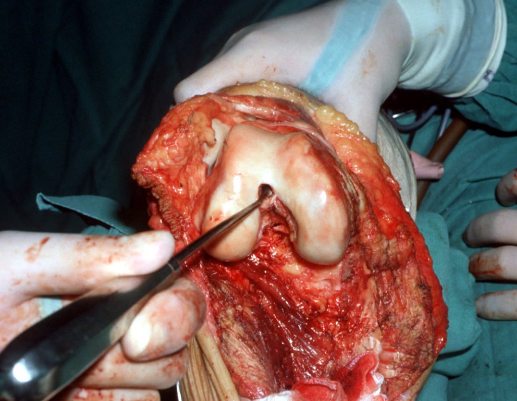
Figura 16: Exposição de todo o côndilo femoral.
Figure 16: Exposure of the entire femoral condyle.
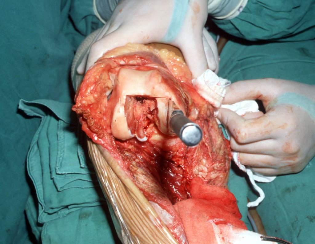
The patellofemoral cartilage must be preserved as much as possible. Using the curette, we locate the femoral canal, figure 16. The femoral component will be fitted between the femoral condyles. To do this, it is necessary to perform a rectangular osteotomy, allowing adequate positioning of the component, figures 17 and 18.
Figura 17: Haste da tíbia posicionada no canal femoral.
Figure 17: Tibial stem positioned in the femoral canal.
Figura 18: Realização dos cortes femorais com formão.
Figure 18: Making femoral cuts with a chisel.
This was the first intraepiphyseal prosthesis we performed. At that time, in 1989, we did not have cutting guides and the surgeon made the cuts by hand, using his skill to follow clinical parameters.
Figura 19: Marcação do retângulo, prévio às osteotomias.
Figure 19: Marking of the rectangle, prior to osteotomies.
Figura 20: Sulco intercondilar, esculpido com martelo e formão, para receber a prótese.
Figure 20: Intercondylar groove, carved with a hammer and chisel, to receive the prosthesis.
Figura 21: Prótese posicionada para realização do teste de prova, antes da cimentação.
Figure 21: Prosthesis positioned to carry out the proof test, before cementation.
Figura 22: Cimentação do componente intra epifisário femoral.
Figure 22: Cementation of the femoral intraepiphyseal component.
Figura 23: Preparação do canal tibial.
Figure 23: Preparation of the tibial canal.
Figura 24: Encaixe do componente tibial, testando a adequação dos componentes.
Figure 24: Fitting the tibial component, testing the adequacy of the components.
Figura 25: Revisão da hemostasia, antes da cimentação final.
Figure 25: Review of hemostasis, before final cementation.
Figura 26: Cimentação do componente tibial e reinserção do tibial anterior na prótese.
Figure 26: Cementation of the tibial component and reinsertion of the anterior tibialis into the prosthesis.
Figura 27: Radiografia, frente, no terceiro mês após a cirurgia.
Figure 27: Radiograph, front, in the third month after surgery.
Figura 28: Radiografia, perfil, após três meses da cirurgia.
Figure 28: X-ray, profile, three months after surgery.
Figura 29: Carga monopodal na perna operada, após três meses.
Figure 29: Single leg load on the operated leg, after three months.
Figura 30: Radiografia, frente, após um ano e dois meses da cirurgia.
Figure 30: Radiograph, front, one year and two months after surgery.
Figura 31: Radiografia, perfil, após um ano e dois meses.
Figure 31: X-ray, profile, after one year and two months.
Figura 32: Carga monopodal na perna operada, após um ano e dois meses.
Figure 32: Single leg load on the operated leg, after one year and two months.
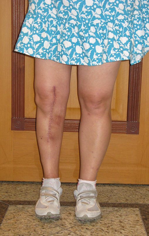
Patient residing in another state, dancing lambada, which was the rhythm of the time, figures 33 and 34. In January 2007, he sent this photo for our evaluation, figure 35. In February 2008, he suffered a fall resulting in a fracture of the femur, just above the femoral component, which was treated closedly, figure 36.
Figura 33: Boa função do membro.
Figure 33: Good limb function.
Figura 34: Boa flexão com carga, simétrica, após um ano e dois meses.
Figure 34: Good flexion with load, symmetrical, after one year and two months.
Figura 35: Foto de janeiro de 2007, após dezoito anos da cirurgia.
Figure 35: Photo from January 2007, eighteen years after surgery.
Figura 36: Em fevereiro de 2008, sofreu queda, apresentando fratura femoral, justo acima do componente femoral da prótese, que foi tratada incruentamentte.
Figure 36: In February 2008, he suffered a fall, presenting a femoral fracture, just above the femoral component of the prosthesis, which was treated closed.
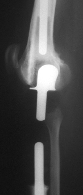
The fracture had malunion, with shortening and rotational deviation, resulting in protrusion of the femoral component, leading to functional limitation of the knee, requiring revision, figures 37 and 38. In 2009, the endoprosthesis was revised, figures 39 and 40 .
Figura 37: Fratura consolidada com desvio, com encurtamento e desvio rotacional.
Figure 37: Consolidated fracture with displacement, with shortening and rotational deviation.
Figura 38: Protrusão do componente femoral, necessitando de revisão, em Abril de 2009, após vinte anos da cirurgia.
Figure 38: Protrusion of the femoral component, requiring revision, in April 2009, twenty years after surgery.
Figura 39: Revisão da endoprótese em 2009. Paciente com carga total.
Figure 39: Revision of the endoprosthesis in 2009. Patient fully loaded.
Figura 40: Pós operatório de três semanas, apresentando cinquenta gráus de flexão do joelho.
Figure 40: Three weeks post-operative, showing fifty degrees of knee flexion.

Author: Prof. Dr. Pedro Péricles Ribeiro Baptista

 Orthopedic Oncosurgery at the Dr. Arnaldo Vieira de Carvalho Cancer Institute

Hello! How can we assist you?