This digital library houses the book on Oncology and Orthopedic Oncosurgery.

It includes academic lectures, presentations from national and international congresses, published papers, case discussions, performed surgical procedures, and proprietary techniques developed.

The digital format was chosen because the web allows the inclusion of texts with numerous visual resources, such as images and videos, which would not be possible in a printed book.

The content is intended for students, healthcare professionals, and the general public interested in the field.

Femur Fracture

043ea4eb48d314b91e95fb5052d3d8f0 3

Femur Fracture

Femur fracture. Female patient, 46 years old, victim of a car accident, suffered a closed fracture of the left femur (Figures 1 and 2).
Figura 1: Radiografia do fêmur esquerdo, frente, evidenciando fratura diafisária oblíqua curta.
Figure 1: Radiograph of the left femur, front, showing a short oblique diaphyseal fracture.
Figura 2: Radiografia frente, com detalhe da fratura em maior aumento e visualização parcial dos côndilos femorais.
Figure 2: Front radiograph, with detail of the fracture at higher magnification and partial visualization of the femoral condyles.
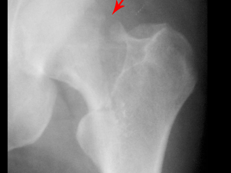
Figura 3: Radiografia focalizada do quadril esquerdo, destacando o colo femoral.
Figure 3: Focused radiograph of the left hip, highlighting the femoral neck.
These radiographs deserve some observations, shown in figures 4,5 and 6.
Figura 4: O paciente esta vestido, provavelmente com calça jeans, pois podemos observar o zipper da roupa. É preciso retirar a calça para observarmos possíveis lesões na pele.
Figure 4: The patient is dressed, probably in jeans, as we can see the zipper on his clothes. It is necessary to remove the pants to observe possible skin injuries.
Figura 5: Falta de visualização completa da articulação. Esta é uma prerrogativa necessária para analisar uma fratura. Devemos poder avaliar uma articulação acima e uma abaixo da lesão.
Figure 5: Lack of complete visualization of the joint. This is a necessary prerogative to analyze a fracture. We must be able to evaluate one joint above and one below the injury.
Figura 6: Não de pode avaliar adequadamente a articulação do quadril. Mal se pode observar a fratura do colo femoral.
Figure 6: The hip joint cannot be adequately assessed. The fracture of the femoral neck can barely be seen.
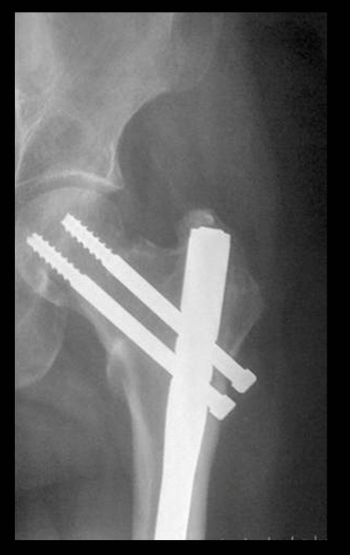
The fracture was fixed with a locked cephalofemoral nail (figures 7 and 8).
Figura 7: Fixação com haste céfalo-femoral. Em detalhe a diastase do colo.
Figure 7: Fixation with cephalofemoral nail. In detail, colonic diastasis.
Figura 8: Diastase na diáfise femoral, a seta amarela destaca o perfil e a seta vermelha o grande afastamento na imagem de frente. O parafuso de bloqueio impede a rotação, mas também a compressão dos fragmentos, dificultando a consolidação.
Figure 8: Diastasis in the femoral shaft, the yellow arrow highlights the profile and the red arrow the large gap in the front image. The locking screw prevents rotation, but also compression of the fragments, making consolidation difficult.
It evolved with pseudoarthrosis, deformity and shortening (Figure 9, 10 and 11)
Figura 9: Radiografia do quadril frente.
Figure 9: Radiograph of the front hip.
Figura 10: Radiografia do quadril perfil.
Figure 10: Radiograph of the hip in profile.
Figura 11: Pseudo-artrose da diáfise femoral e quebra do parafuso de bloqueio, o que era previsível.
Figure 11: Pseudo-arthrosis of the femoral shaft and breakage of the locking screw, which was predictable.
Figura 12: Paciente com deformidade em valgo e encurtamento.
Figure 12: Patient with valgus deformity and shortening.

Author: Prof. Dr. Pedro Péricles Ribeiro Baptista

 Orthopedic Oncosurgery at the Dr. Arnaldo Vieira de Carvalho Cancer Institute

Hello! How can we assist you?