This digital library houses the book on Oncology and Orthopedic Oncosurgery.

It includes academic lectures, presentations from national and international congresses, published papers, case discussions, performed surgical procedures, and proprietary techniques developed.

The digital format was chosen because the web allows the inclusion of texts with numerous visual resources, such as images and videos, which would not be possible in a printed book.

The content is intended for students, healthcare professionals, and the general public interested in the field.

Custom-made prostheses

Prótese Fabricada Sob Medida

Custom Made Femur Prosthesis

Femur Prosthesis. Prosthetics manufactured especially for the patient, as shown in figures 1 to 12, require authorization from the National Health Surveillance Agency – ANVISA , as per the document exemplified in figure 3,

Figura 1: Eescanometria em perfil destacando o antecurvatum. Projeção do trajeto falso que a haste reta da prótese ocasiona. Figura 2: Modelo disponível em outubro de 2015, à esquerda. Conecção redonda dos segmentos, setas de cor rocha, parafusos pequenos de fixação, setas em vermelho. À direita, novos módulos solicitados. Bloqueios retangulares nas conecções, setas amarelas A e B; parafusos de fixação, mais fortes, setas em branco. Conexão, retangular no encaixe do corpo da nova prótese, seta azul .
Figure 1: Scanometry in profile highlighting the antecurvatum. Projection of the false path caused by the straight stem of the prosthesis. Figure 2: Model available in October 2015, on the left. Round connection of segments, rock-colored arrows, small fixing screws, red arrows. On the right, new requested modules. Rectangular blocks in connections, yellow arrows A and B; fixing screws, stronger, blank arrows. Connection, rectangular in the fitting of the new prosthesis body, blue arrow.
Figura 3: Solicitação à ANVISA para autorização de confecção de modêlo especial de prótese. Figura 4: Prótese confeccionada sob encomenda, com os encaixes retangulares entre os módulos, bloqueando a rotação, setas amarelas A e B. As setas azuis assinalam o encaixe retangular do corpo da prótese. As setas brancas destacam os parafusos de fixação, mais fortes e em maior número. A seta laranja aponta a haste calibrada para o canal medular do paciente.
Figure 3: Request to ANVISA for authorization to manufacture a special prosthesis model. Figure 4: Custom-made prosthesis, with rectangular fittings between the modules, blocking rotation, yellow arrows A and B. The blue arrows indicate the rectangular fitting of the prosthesis body. The white arrows highlight the stronger and more numerous fixing screws. The orange arrow points the calibrated rod to the patient's spinal canal.
Figura 5: Radiografia de 30/03/2016, post op 3 meses, frente. Figura 6: Radiografia de 30/03/2016, post op 3 meses, perfil.
Figure 5: Radiograph from 03/30/2016, post op 3 months, front. Figure 6: Radiograph from 03/30/2016, post op 3 months, profile.
Figura 7: Flexão simétrica, com carga total, oito meses pós op e dois meses depois do término da quimioterapia . Figura 8: Escanometria revelando discrepância de 0,8 cm, às custas da tíbia e 1,0 cm às custas do fêmur, totalizando 1,8 cm de encurtamento.
Figure 7: Symmetrical flexion, with full load, eight months post op and two months after the end of chemotherapy. Figure 8: Scanometry revealing a discrepancy of 0.8 cm, at the expense of the tibia and 1.0 cm at the expense of the femur, totaling 1.8 cm of shortening.

Custom Manufactured Elbow Prosthesis

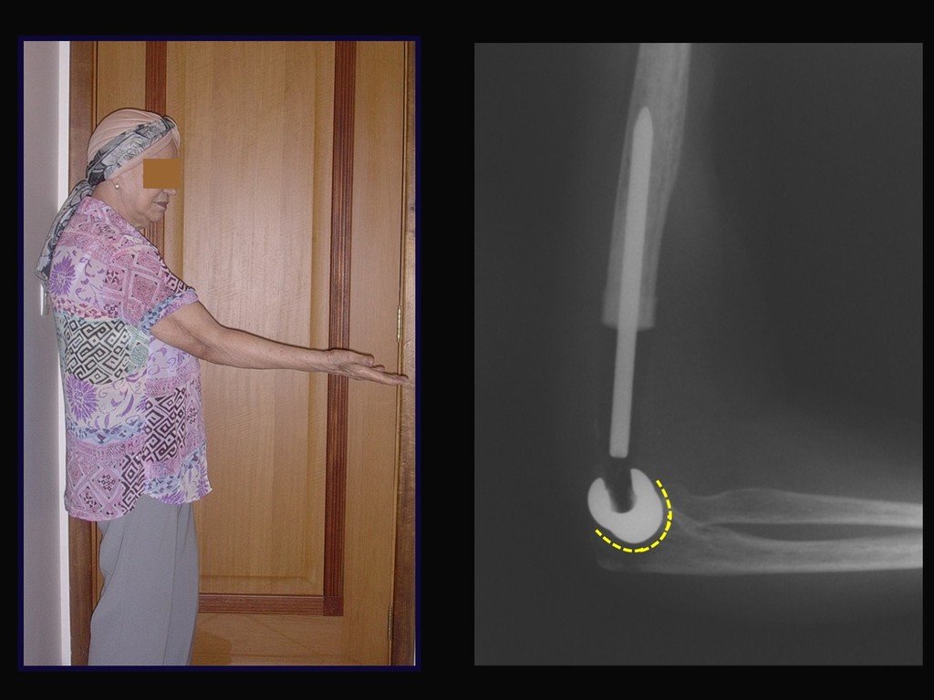
This is an example of an elbow prosthesis, custom-made, especially for this patient.
Figura 9: Metástase de carcinoma de mama.
Figure 9: Breast carcinoma metastasis.
Figura 10: Prótese fabricada sob medica. Feita com molde de radiografia do paciente.
Figure 10: Medically manufactured prosthesis. Made with a mold of the patient's x-ray.
Figura 11: Aspecto clínico. Prótese com curvatura adaptada ao olécrano.
Figure 11: Clinical appearance. Prosthesis with curvature adapted to the olecranon.
Figura 12: Boa função do cotovêlo. Radiografia evidenciando boa congruência com o cotovêlo da paciente.
Figure 12: Good elbow function. Radiograph showing good congruence with the patient's elbow.

Author: Prof. Dr. Pedro Péricles Ribeiro Baptista

 Orthopedic Oncosurgery at the Dr. Arnaldo Vieira de Carvalho Cancer Institute

Hello! How can we assist you?