This digital library houses the book on Oncology and Orthopedic Oncosurgery.

It includes academic lectures, presentations from national and international congresses, published papers, case discussions, performed surgical procedures, and proprietary techniques developed.

The digital format was chosen because the web allows the inclusion of texts with numerous visual resources, such as images and videos, which would not be possible in a printed book.

The content is intended for students, healthcare professionals, and the general public interested in the field.

Chondrosarcoma – Pelvectomy

1f5f6ad95cc908a20bb7e30ee28a5958 2

Total Internal Pelvectomy Technique

Chondrosarcoma – Pelvectomy. Female patient, 19 years old, complaining of discomfort and increased volume of the left pelvis. Clinically, there was mild pain upon palpation of the iliac wing, with a palpable, hard, adherent tumor. Responded to on December 18, 2002, with the following imaging exams: 11/13/1983 12/18/2002
Figura 1: Radiografia de 18/12/2002, bacia frente, com lesão condensante acometendo o ilíaco esquerdo
Figure 1: Radiograph of 12/18/2002, front pelvis, with condensing lesion affecting the left iliac
Figura 2: Radiografia da bacia in let, evidenciando inúmeros focos de calcificação.
Figure 2: Radiograph of the basin in let, showing numerous foci of calcification.
Figura 3: Radiografia em alar, grande tumor extracortical, intra e extrapélvico.
Figure 3: Alar radiograph, large extracortical, intra and extrapelvic tumor.
Figura 4: Radiografia da bacia em obturatriz, tumor obliterando o forame.
Figure 4: X-ray of the pelvis in obturatrix, tumor obliterating the foramen.
Figura 5: Radiografia do tórax, de 18/12/2002, sem alterações.
Figure 5: Chest X-ray, dated 12/18/2002, no changes.
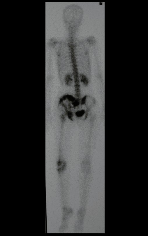
Figura 6: Cintilografia óssea, fase tardia, frente, com hipercaptação no ilíaco direito.
Figure 6: Bone scintigraphy, late phase, front, with high uptake in the right iliac.
Figura 7: Cintilografia óssea, fase tardia, posterior, com hipercaptação no ilíaco direito.
Figure 7: Bone scintigraphy, late, posterior phase, with high uptake in the right iliac.
Figura 8: Cintilografia localizada da bacia, detalhando o comprometimento do ilíaco direito.
Figure 8: Localized pelvis scintigraphy, detailing the involvement of the right iliac.
Figura 9: RM axial, com baixo sinal em T1, com grande tumor extracortical, comprometendo a tábua interna e a tábua externa do ilíaco direito.
Figure 9: Axial MRI, with low signal on T1, with a large extracortical tumor, affecting the internal and external tables of the right iliac.
Figura 10: RM coronal, com baixo sinal em T1, com grande tumor extracortical, comprometendo as tábuas internas e externa do ilíaco direito.
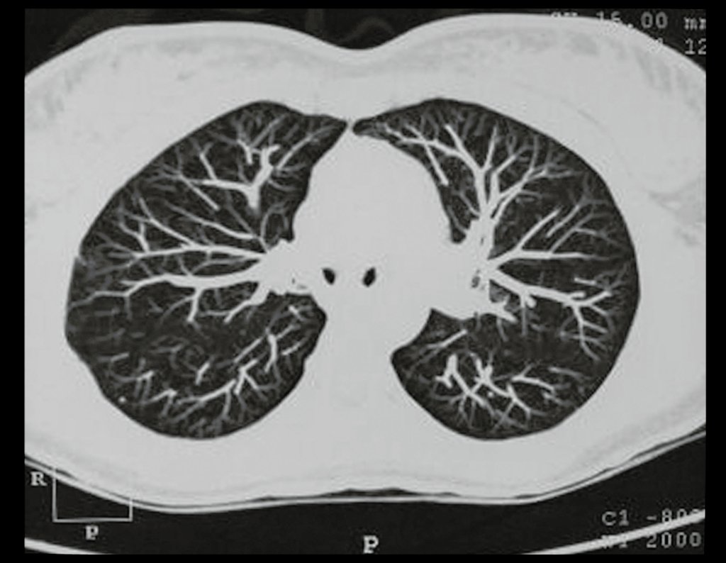
Figure 10: Coronal MRI, with low signal on T1, with a large extracortical tumor, affecting the internal and external tables of the right iliac.
Figura 11: RM axial alto sinal em T2, com grande tumor extracortical, comprometendo a tábua interna e a tábua externa do ilíaco direito.
Figure 11: Axial high-signal T2 MRI, with a large extracortical tumor, affecting the internal and external tables of the right iliac.
Figura 12: RM coronal, com alto sinal em T2, com grande tumor extracortical, comprometendo a tábua interna e a tábua externa do ilíaco direito.
Figure 12: Coronal MRI, with high signal on T2, with a large extracortical tumor, affecting the internal and external tables of the right iliac.
Figura 13: RM axial T1 spir, com supressão de gordura, evidenciando reação periosteal em espículas e focos de calcificação.
Figure 13: Axial T1 spir MRI, with fat suppression, showing periosteal reaction in spicules and foci of calcification.
Figura 14: RM axial T1, com saturação de gordura e contraste, evidenciando espessa área de captação (alto sinal) devido à capa de cartilagem neoplásica maligna, ao redor da lesão (com baixo sinal).
Figure 14: Axial T1 MRI, with fat saturation and contrast, showing a thick uptake area (high signal) due to the layer of malignant neoplastic cartilage around the lesion (low signal).
Figura 15: Outro corte de axial T1, com saturação de gordura e contraste, evidenciando espessa área de captação (alto sinal) devido à capa de cartilagem neoplásica maligna, ao redor da lesão (com baixo sinal).
Figure 15: Another T1 axial section, with fat saturation and contrast, showing a thick uptake area (high signal) due to the layer of malignant neoplastic cartilage around the lesion (low signal).
Figura 16: RM coronal T1, com saturação de gordura e contraste, evidenciando espessa área de captação (alto sinal) devido à capa de cartilagem neoplásica maligna, ao redor da lesão (com baixo sinal).
Figure 16: Coronal T1 MRI, with fat saturation and contrast, showing a thick uptake area (high signal) due to the layer of malignant neoplastic cartilage around the lesion (low signal).
Figura 17: Tomografia axial, densidade para osso, grande tumor acometendo o ilíaco esquerdo.
Figure 17: Axial tomography, bone density, large tumor affecting the left iliac bone.
Figura 18: Tomografia axial, densidade para tecidos moles, grande tumor acometendo o ilíaco esquerdo.
Figure 18: Axial tomography, soft tissue density, large tumor affecting the left iliac.
Figura 19: Tomografia coronal, densidade para osso, com grande tumor acometendo o ilíaco esquerdo.
Figure 19: Coronal tomography, bone density, with a large tumor affecting the left iliac bone.
Figura 20: Tomografia coronal, densidade para tecidos moles, com grande tumor acometendo o ilíaco esquerdo.
Figure 20: Coronal tomography, soft tissue density, with a large tumor affecting the left iliac bone.
Figura 21: Paciente em decúbito lateral direito, demarcação do retalho de pele que será ressecado em bloco, juntamente com o tumor. A seta aponta a biópsia realizada em outro serviço por cirurgião geral, em sentido perpendicular à incisão cirúrgica, complicando a abordagem.
Figure 21: Patient in right lateral decubitus, demarcation of the skin flap that will be resected en bloc, together with the tumor. The arrow points to the biopsy performed in another service by a general surgeon, in a perpendicular direction to the surgical incision, complicating the approach.
Figura 22: Incisão ampla para permitir uma ressecção com margem de segurança, para garantir a cura deste condrossarcoma, já que a cirurgia é o único recurso para o tratamento desta lesão. Observe o grande retalho de pele que precisa ser removido, devido à incisão errônea da biópsia.
Figure 22: Wide incision to allow a resection with a safety margin, to guarantee the cure of this chondrosarcoma, as surgery is the only resource for treating this lesion. Note the large flap of skin that needs to be removed due to the erroneous biopsy incision.
Figura 23: Dissecção com cuidadosa hemostasia, seccionando e amarrando os segmentos musculares seccionados.
Figure 23: Dissection with careful hemostasis, sectioning and tying the sectioned muscle segments.
Figura 24: Isolamento do nervo femoral.
Figure 24: Isolation of the femoral nerve.
Figura 25: Dissecção dos vasos ilíacos.
Figure 25: Dissection of the iliac vessels.
Figura 26: Exposição da veia ilíaca interna para ligadura.
Figure 26: Exposure of the internal iliac vein for ligation.
Figura 27: Ligadura da veia ilíaca interna.
Figure 27: Ligation of the internal iliac vein.
Figura 28: Ligadura da artéria ilíaca interna.
Figure 28: Ligation of the internal iliac artery.
Figura 29: Dissecção da região sacro ilíaca e preparação para osteotomia.
Figure 29: Dissection of the sacroiliac region and preparation for osteotomy.
Figura 30: Dissecção do tendão do músculo psoas, separando-o do músculo ilíaco, que será removido junto com a peça cirúrgica, como margem de segurança oncológica.
Figure 30: Dissection of the psoas muscle tendon, separating it from the iliac muscle, which will be removed along with the surgical piece, as an oncological safety margin.
Figura 31: Ressecção em bloco da hemipelve esquerda e revisão da hemostasia.
Figure 31: En bloc resection of the left hemipelvis and review of hemostasis.
Figura 32: Aspecto da reconstrução, com o retalho de pele para o fechamento da ferida operatória.
Figure 32: Aspect of the reconstruction, with the skin flap to close the surgical wound.
Figura 33: Aspecto da peça cirúrgica, ressecada em bloco, juntamente com a pele, o trajeto da biópsia e tecidos moles sadios ao redor, como margem.
Figure 33: Appearance of the surgical specimen, resected en bloc, together with the skin, the biopsy path and surrounding healthy soft tissues, as a margin.
Figura 34: Visão do anel obturatório e do segmento da sínfise púbica do lado esquerdo, superiormente.
Figure 34: View of the obturator ring and the pubic symphysis segment on the left side, superiorly.
Figura 35: Visão da face ventral da hemipelve ressecada, juntamente com o colo femoral e o complexo capsulo-ligamentar do quadril, evidenciando o grande volume tumoral.
Figure 35: View of the ventral surface of the resected hemipelvis, together with the femoral neck and the capsulo-ligamentous complex of the hip, showing the large tumor volume.
Figura 36: Radiografia do pós-operatório imediato, após a ressecção completa da hemipelve esquerda, juntamente com o colo femoral e o complexo capsulo-ligamentar da articulação do quadril esquerdo.
Figure 36: Radiograph of the immediate postoperative period, after complete resection of the left hemipelvis, together with the femoral neck and the capsulo-ligamentous complex of the left hip joint.
Figura 37: Radiografia da peça cirúrgica, mostrando o tumor extracortical, acometendo a tábua interna e a tábua externa do ilíaco esquerdo.
Figure 37: Radiograph of the surgical specimen, showing the extracortical tumor, affecting the internal table and the external table of the left iliac bone.
Figura 38: Radiografia da peça cirurgica, mostrando o acetábulo e a cabeça femoral ressecada em bloco.
Figure 38: Radiograph of the surgical specimen, showing the acetabulum and femoral head resected en bloc.
Figura 39: Radiografia da peça cirúrgica, em alar.
Figure 39: Radiograph of the surgical specimen, in alar.
Figura 40: Aspecto da peça cirurgia com tecidos moles macroscopicamente sadios, envolvendo o tumor.
Figure 40: Aspect of the surgical specimen with macroscopically healthy soft tissues surrounding the tumor.
Figura 41: Corte da peça cirúrgica mostrando a lesão circundada por tecido sadio.
Figure 41: Section of the surgical specimen showing the lesion surrounded by healthy tissue.
Figura 42: Avaliação da peça cirúrgica pelo departamento de Patologia.
Figure 42: Assessment of the surgical specimen by the Pathology department.
Figura 43: Vários cortes para o estuda das margens.
Figure 43: Various cuts to study the margins.
Figura 44: Em maior aumento, detalhando o aspecto macroscópico deste condrossarcoma.
Figure 44: In higher magnification, detailing the macroscopic appearance of this chondrosarcoma.
Figura 45: Os vários cortes permitem avaliar melhor a peça de ressecção.
Figure 45: The various cuts allow a better evaluation of the resection piece.
Figura 46: Corte evidenciando o revestimento pela pele e tecido celular subcutâneo, o tumor e a cabeça femoral ressecada.
Figure 46: Section showing the covering of the skin and subcutaneous cellular tissue, the tumor and the resected femoral head.
Figura 47: Paciente com 20 anos de idade, pós-operatório de um ano. Carga total com auxílio de muletas.
Figure 47: 20-year-old patient, one year postoperative. Full load with the aid of crutches.
Figura 48: Pós -operatório de um ano, carga total nos MMII, com flexão dos joelhos, visão de frente.
Figure 48: One year post-surgery, full load on the lower limbs, with knee flexion, frontal view.
Figura 49: Pós-operatório de um ano, carga total nos MMII, com flexão dos joelhos, visão de perfil esquerdo.
Figure 49: One year postoperatively, full load on the lower limbs, with knee flexion, left profile view.
Figura 50: Pós-operatório de um ano, carga total nos MMII, com flexão dos joelhos, visão de perfil direito, boa função.
Figure 50: One year post-surgery, full load on the lower limbs, with knee flexion, right profile view, good function.
Figura 51: Pós-operatório de um ano e seis meses, carga total nos MMII, ortostática, visão de frente.
Figure 51: One year and six months postoperative, full load on the lower limbs, standing, frontal view.
Figura 52: Pós-operatório de um ano e seis meses, carga total nos MMII, ortostática, visão posterior. Observem o encurtamento do lado esquerdo, pela ascensão do fêmur.
Figure 52: One year and six months postoperative, full load on the lower limbs, standing, posterior view. Note the shortening on the left side, due to the rise of the femur.
Figura 53: Pós-operatório de um ano e seis meses, carga total nos MMII, ortostática, visão de perfil.
Figure 53: One year and six months postoperative, full load on the lower limbs, standing, profile view.
Figura 54: Pós-operatório de um ano e seis meses, carga total nos MMII, ortostática, visão lateral.
Figure 54: One year and six months postoperative, full load on the lower limbs, standing, lateral view.
Figura 55: Após um ano e seis meses, carga total com flexão dos MMII.
Figure 55: After one year and six months, full load with lower limb flexion.
Figura 56: Carga total monopodal sobre o membro operado!!! Boa recuperação funcional.
Figure 56: Total single-leg load on the operated limb!!! Good functional recovery.
Figura 57: Radiografia do pós-operatório de um ano e seis meses. O membro inferior esquerdo parece flutuar, mas na realidade existe uma fibrose cicatricial que apoia o membro e impede maior ascensão.
Figure 57: One year and six months postoperative radiograph. The left lower limb appears to float, but in reality there is a scarring fibrosis that supports the limb and prevents further ascension.
Figura 58: RM coronal T1, após dois anos da cirurgia, sem sinais de recidiva.
Figure 58: Coronal T1 MRI, two years after surgery, with no signs of recurrence.
Figura 59: RM axial T1, após dois anos da cirurgia, sem sinais de recidiva.
Figure 59: Axial T1 MRI, two years after surgery, with no signs of recurrence.
Figura 60: RM coronal T2 Stir, após dois anos da cirurgia, sem sinais de recidiva.
Figure 60: Coronal T2 Stir MRI, two years after surgery, with no signs of recurrence.
Figura 61: RM axial T2 Stir, após dois anos da cirurgia, bom aspecto local.
Figure 61: Axial T2 Stir MRI, two years after surgery, good local appearance.
Figura 62: RM coronal T2, após dois anos da cirurgia.
Figure 62: Coronal T2 MRI, two years after surgery.
Figura 63: RM sagital T1 Spir, com contraste, após dois anos da cirurgia.
Figure 63: Sagittal T1 Spir MRI, with contrast, two years after surgery.
Figura 64: RM axial T1 Spir, com contraste, após dois anos da cirurgia.
Figure 64: Axial T1 Spir MRI, with contrast, two years after surgery.
Figura 65: RM coronal T1 Spir, com contraste, após dois anos da cirurgia, bom aspecto, sem recidiva.
Figure 65: Coronal T1 Spir MRI, with contrast, two years after surgery, good appearance, without recurrence.
Figura 66: RM coronal T1 Spir, com contraste, após dois anos da cirurgia.
Figure 66: Coronal T1 Spir MRI, with contrast, two years after surgery.
Figura 67: Tomografia do tórax normal, após três anos da cirurgia.
Figure 67: Normal chest tomography, three years after surgery.
Figura 68: RM do tórax normal, após três anos da cirurgia.
Figure 68: MRI of the normal chest, three years after surgery.
Figura 69: Pós-operatório de três anos, carga total nos MMII, ortostática, visão posterior.
Figure 69: Three years post-surgery, full load on the lower limbs, standing, posterior view.
Figura 70: Pós-operatório de três anos, visão em perfil. Observem o encurtamento do lado esquerdo, pela ascensão do fêmur.
Figure 70: Three years post-operative, profile view. Note the shortening on the left side, due to the rise of the femur.
Figura 71: Pós-operatório de três anos. O encurtamento do lado esquerdo, é facilmente compensado por um salto tipo Anabela.
Figure 71: Three-year postoperative period. The shortening on the left side is easily compensated by an Anabela-type heel.
Figura 72: Pós-operatório de três anos, visão de perfil. O encurtamento é compensado pelo salto tipo Anabela.
Figure 72: Three years post-operative, profile view. The shortening is compensated by the Anabela heel.
Figura 73: Quadris e joelhos em flexão, com carga total, sem auxílio de muletas, com compensação no calçado.
Figure 73: Hips and knees in flexion, with full load, without the aid of crutches, with compensation in the shoes.
Figura 74: Carga total, monopodal, sobre o membro operado, no pós-operatório de três anos.
Figure 74: Total single-leg load on the operated limb, three years postoperatively.
Figura 75: Aspecto clínico da paciente após quatro anos da cirurgia, 07/02/2006.
Figure 75: Clinical appearance of the patient four years after surgery, 02/07/2006.
Figura 76: Após quatro anos da cirurgia, compensação com calçado.
Figure 76: Four years after surgery, compensation with shoes.
Figura 77: Após 12 anos e três meses da cirurgia, Bom aspecto clínico, discrepância de MMII inalterada.
Figure 77: 12 years and three months after surgery, good clinical appearance, unchanged lower limb discrepancy.
Figura 78: Carga total monopodal, sem auxílio, apoiando no membro operado, após 12 anos e três meses da cirurgia.
Figure 78: Single-leg total load, without assistance, supporting the operated limb, 12 years and three months after surgery.
Figura 79: Flexão dos MMII com carga, muito boa função, Pelvectomia sem reconstrução.
Figure 79: Lower limb flexion with load, very good function, Pelvectomy without reconstruction.
Figura 80: Em 12/05/2015, após 12 anos e três meses da cirurgia. Aspecto estável da discrepância. Muito bom resultado cosmético e funcional.
Figure 80: On 05/12/2015, 12 years and three months after surgery. Stable aspect of the discrepancy. Very good cosmetic and functional result.
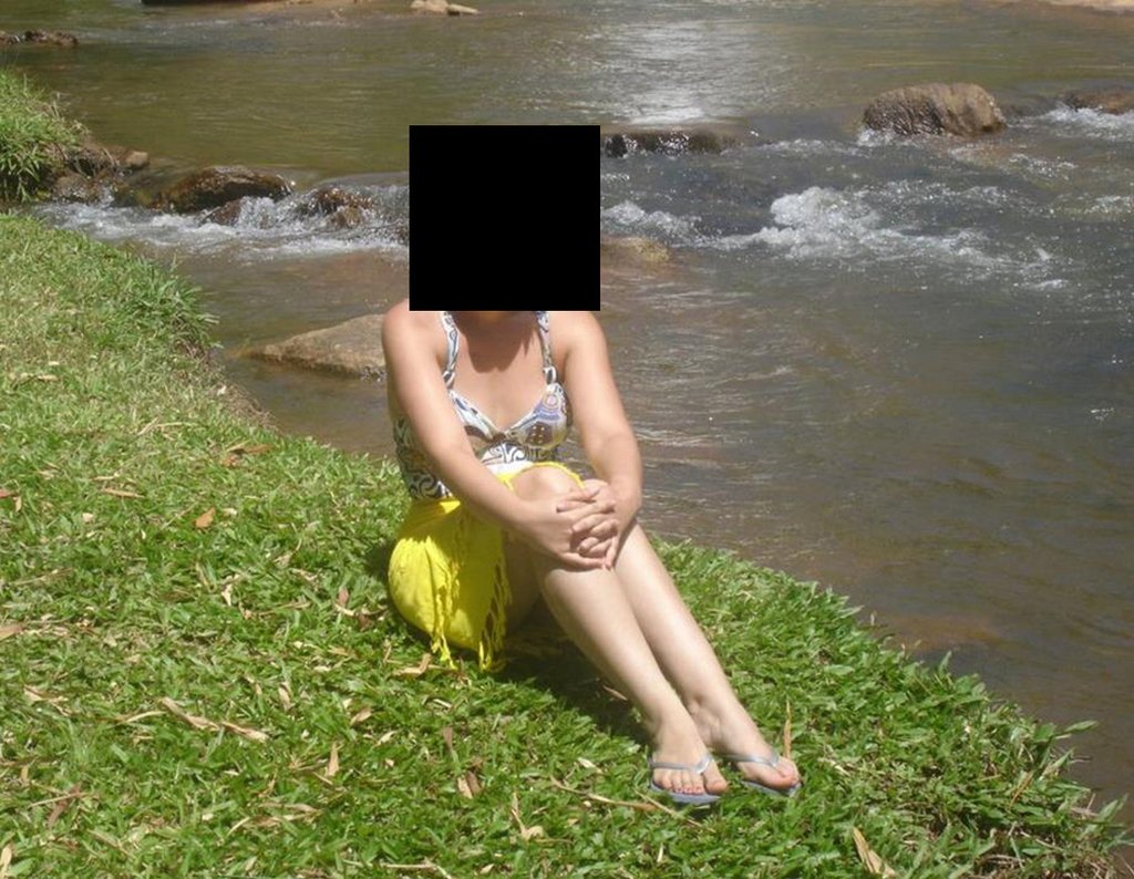
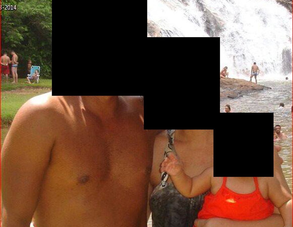
The patient happily displays her life without significant limitations. Shows complete overcoming of the functional deficit, carrying out activities of daily living with ease, figures 81 to 90.
Figura 81: Piquenique com amigos.
Figure 81: Picnic with friends.
Figura 82: Curtindo a Cachoeira.
Figure 82: Enjoying the Waterfall.
Figura 83: Namoro feliz.
Figure 83: Happy dating.
Figura 84: Jogando boliche!!!
Figure 84: Playing bowling!!!
Figura 85: Exibindo feliz a gravidez.
Figure 85: Happily displaying the pregnancy.
Figura 86: Batizando a filha.
Figure 86: Baptizing the daughter.
Figura 87: Passeio no parque.
Figure 87: Walk in the park.
Figura 88: Família completa. De volta à cachoeira.
Figure 88: Complete family. Back to the waterfall.
Figura 89: Opa!!! Torcendo na copa de 2014!
Figure 89: Oops!!! Supporting the 2014 World Cup!
Figura 90: Curtindo o campo.
Figure 90: Enjoying the countryside.
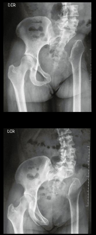
Figura 91: Radiografias da bacia, mostrando a pelvectomia interna à esquerda, em 12/05/2015.
Figure 91: X-rays of the pelvis, showing the internal pelvectomy on the left, on 05/12/2015.
Figura 92: Pós-operatório de 12 anos e três meses de pelvectomia à esquerda. Aspecto da adaptação do MIE.
Figure 92: Postoperative period 12 years and three months after left pelvectomy. Aspect of MIE adaptation.
Figura 93: Escanometria mostrando o encurtamento do MIE de 5,08 cm, em 12/05/2015.
Figure 93: Scanometry showing the shortening of the MIE of 5.08 cm, on 05/12/2015.
Figura 94: Radiografia da bacia evidenciando a ascensão proximal do MIE, apoiado em tecido fibroso cicatricial.
Figure 94: X-ray of the pelvis showing the proximal rise of the MIE, supported by fibrous scar tissue.
Figura 95: Radiografia com carga, em 12/05/2015.
Figure 95: X-ray with load, on 05/12/2015.
Figura 96: Radiografia da bacia em Lowenstein, em 16/06/2015.
Figure 96: Radiograph of the basin in Lowenstein, on 06/16/2015.
Figura 97: Laudo das radiografias de 12/05/2015.
Figure 97: X-ray report from 05/12/2015.
Figura 98: RM do tórax normal, em 16/06/2015.
Figure 98: Normal chest MRI, on 06/16/2015.
Figura 99: RM do tórax, sem alteração, 16/06/2015.
Figure 99: MRI of the chest, no changes, 06/16/2015.
Figura 100: RM da pelve evidenciando a acomodação do MIE, em 16/06/2015.
Figure 100: MRI of the pelvis showing accommodation of the MIE, on 06/16/2015.
Figura 101: RM da pelve, coronal com saturação de gordura, com as alterações adaptativas do MIE, em 16/06/2015.
Figure 101: MRI of the pelvis, coronal with fat saturation, with adaptive MIE changes, on 06/16/2015.
Figura 102: RM da pelve, em 16/06/2015.
Figure 102: MRI of the pelvis, on 06/16/2015.
Figura 103: RM da pelve, em 16/06/2015.
Figure 103: MRI of the pelvis, on 06/16/2015.
Figura 104: Laudo da RM da pelve, de 16/06/2015, parte a.
Figure 104: Pelvis MRI report, dated 06/16/2015, part a.
Figura 105: Laudo da RM da pelve, em 16/06/2015, parte b.
Figure 105: Pelvis MRI report, on 06/16/2015, part b.

Video 1 : Gait with full support on the operated limb, single-leg load with good balance, good flexion function of the hips and knees with load. Excellent functional result for a total internal hemipelvectomy, without reconstruction, 12 years and three months after surgery.

Author: Prof. Dr. Pedro Péricles Ribeiro Baptista

 Orthopedic Oncosurgery at the Dr. Arnaldo Vieira de Carvalho Cancer Institute

Hello! How can we assist you?