This digital library houses the book on Oncology and Orthopedic Oncosurgery.

It includes academic lectures, presentations from national and international congresses, published papers, case discussions, performed surgical procedures, and proprietary techniques developed.

The digital format was chosen because the web allows the inclusion of texts with numerous visual resources, such as images and videos, which would not be possible in a printed book.

The content is intended for students, healthcare professionals, and the general public interested in the field.

Chondrosarcoma of The Iliac Branch

7a70c831f7cd407750d00c839b23a496 2

Branch Iliac Chondrosarcoma

The right hip X-ray showed a lesion in the iliopubic branch (Figure 1) and the scintigraphy only captured this region (Figure 2).
Figura 1: Radiografia da pelve direita com lesão de rarefação óssea e focos de condensação.
Figure 1: Radiograph of the right pelvis with bone rarefaction lesion and condensation foci.
Figura 2: Cintilografia óssea com hiper captação apenas no ramo íleo púbico direito.
Figure 2: Bone scintigraphy with hyperuptake only in the right pubic ileum branch.
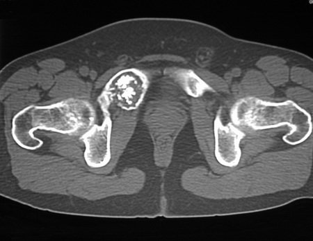
The tomography showed a lytic lesion, affecting part of the acetabular cavity and containing points of condensation suggestive of calcification foci (Figures 3 and 4). Detailed analysis of the radiographs (Figures 5 and 6) and tomographic sections (Figures 7 and 8) show an aggressive lesion, in the root of a limb, in a patient in his fourth decade.
Figura 3: Corte de tomografia evidenciando a lesão, com comprometimento do ramo íleo púbico direito e parte da parede do acetábulo.
Figure 3: Tomography section showing the injury, with involvement of the right pubic ileum branch and part of the acetabulum wall.
Figura 4: Podemos observar a ruptura da cortical que deve ter provocado a dor e início do sintoma do paciente. Portanto é uma lesão ativa.
Figure 4: We can observe the cortical rupture that must have caused the pain and the onset of the patient's symptoms. Therefore it is an active injury.
Figura 5: Radiografia da bacia frente, com a lesão do ramo íleo púbico direito.
Figure 5: Radiograph of the front pelvis, with the lesion of the right pubic ileum branch.
Figura 6: Em detalhe ampliado observamos o afilamento e a insuflação da cortical, com inúmeros focos de calcificação. O paciente tem dor, motivo pelo qual procurou o médico, e apresenta lesão ativa, localmente agressiva.
Figure 6: In enlarged detail we observe the thinning and inflation of the cortex, with numerous foci of calcification. The patient has pain, which is why he sought the doctor, and has an active, locally aggressive lesion.
Figura 7: Corte tomográfico com densidade para osso. Lesão de rarefação óssea com insuflação e erosão da cortical, com evidentes focos de condensação que sugerem fortemente tratar-se de focos de calcificação.
Figure 7: Tomographic section with density for bone. Bone rarefaction lesion with cortical inflation and erosion, with evident condensation foci that strongly suggest that these are calcification foci.
Figura 8: Corte tomográfico com densidade para tecidos moles. Observamos o comprometimento de parte do acetábulo e a evidente ruptura da cortical.
Figure 8: Tomographic section with density for soft tissues. We observed the involvement of part of the acetabulum and the evident rupture of the cortex.
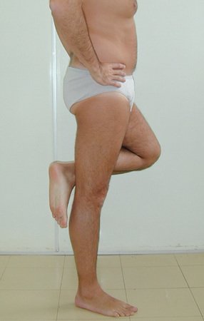
We perform monthly image control, without biopsy. In the third month, with the images revealing the evolution of the lesion, we treated it oncologically as the aggressive lesion it was, considering the most likely hypothesis of chondrosarcoma. We performed complete resection of the lesion, with margins (Figures 9 and 10). You can observe the incision and the patient’s function after ten years (Figures 11 to 13).
Figura 9: Radiografia da bacia, após a ressecção do ramo íleo púbico direito, com parte da parede do acetábulo.
Figure 9: X-ray of the pelvis, after resection of the right pubic ileum branch, with part of the acetabulum wall.
Figura 10: Radiografia da peça cirúrgica, em bloco. Observamos as áreas líticas e os focos de condensação.
Figure 10: Radiograph of the surgical specimen, en bloc. We observed the lithic areas and condensation foci.
Figura 12: Paciente com carga total e flexão completa dos MMII.
Figure 11: Surgery incision. Figure 12: Patient with full load and complete flexion of the lower limbs.
Figura 13: Carga monopodal no membro operado, sem nenhum déficit funcional. O paciente encontra-se curado do tumor e é praticante de tênis.
Figure 13: Single leg load on the operated limb, without any functional deficit. The patient is cured of the tumor and plays tennis.

Author: Prof. Dr. Pedro Péricles Ribeiro Baptista

 Orthopedic Oncosurgery at the Dr. Arnaldo Vieira de Carvalho Cancer Institute

Hello! How can we assist you?