This digital library houses the book on Oncology and Orthopedic Oncosurgery.

It includes academic lectures, presentations from national and international congresses, published papers, case discussions, performed surgical procedures, and proprietary techniques developed.

The digital format was chosen because the web allows the inclusion of texts with numerous visual resources, such as images and videos, which would not be possible in a printed book.

The content is intended for students, healthcare professionals, and the general public interested in the field.

Chondrosarcoma of The Femur

Condrosarcoma del Fémur

Chondrosarcoma of the Femur

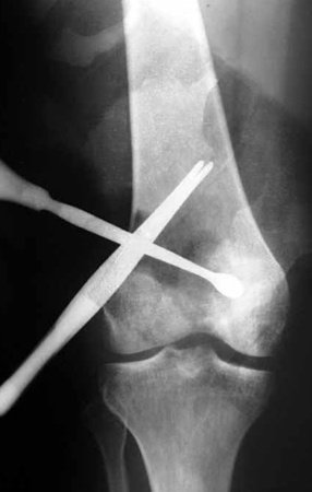
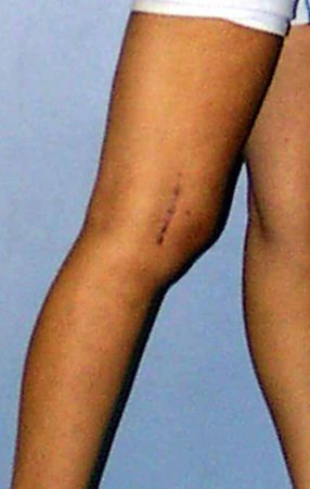
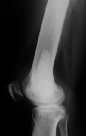
Chondrosarcoma of the Femur. Female patient, 33 years old, with pain in her right knee for 6 months. She was treated at the University Hospital, where she underwent imaging tests that revealed a bone rarefaction lesion and foci of calcification. She underwent biopsy and histopathological diagnosis was chondroma. The patient underwent surgery through a small lateral incision. They performed intralesional curettage and filled the cavity with cement. They documented the procedure with the argument that they could reach the entire cavity through this incision and performed a postoperative x-ray (Figures 1 – 4).
Figura 1: Radiografia do procedimento intralesional por pequena incisão!!!??? A cureta e a pinça apontam a profundidade, querendo o cirurgião inferir que por este acesso consegue atingir toda a lesão. Sabemos que isto não é correto, apenas espalha a lesão.
Figure 1: Radiograph of the intralesional procedure through a small incision!!!??? The curette and forceps point out the depth, the surgeon wanting to infer that through this access he can reach the entire lesion. We know this is not correct, it only spreads the injury.
Figura 2: Foto da cicatriz operatória, em 13/jun./1998. Esta incisão pequena é contraindicada no tratamento oncológico desta lesão.
Figure 2: Photo of the surgical scar, on June 13, 1998. This small incision is contraindicated in the oncological treatment of this lesion.
Figura 3: Radiografia do pós-operatório de curetagem intralesional indevida. Nesta radiografia já se pode observar áreas de rarefação ao redor de todo o cimento, indicando a presença de tumor.
Figure 3: Post-operative radiograph of improper intralesional curettage. In this radiograph, areas of rarefaction can be seen around the entire cement, indicating the presence of a tumor.
Figura 4: Radiografia, em perfil, com rarefação anterior, indicando a presença de tumor. Devemos assinalar que isto não se trata de recidiva e sim neoplasia que continuou na cavidade.
Figure 4: Radiograph, in profile, with anterior rarefaction, indicating the presence of a tumor. We must point out that this is not a recurrence but rather a neoplasia that continued in the cavity.

Intralesional curettage can be used in some cases of aggressive benign lesions, such as GCT (giant cellular tumor). However, a small incision cannot be used in any situation. When the curettage technique is indicated, we must have a broad view of the entire cavity and perform local adjuvant therapy, when necessary. 
    In this specific case, curettage is not indicated for the treatment of this lesion, much less with a small incision. The biopsy suggested the diagnosis of chondroma, but it was a grade II chondrosarcoma, which was the definitive diagnosis of the cured material. 
    With the diagnosis of malignant neoplasm and the “recurrence” condition, amputation of the limb was indicated. The patient does not accept the treatment and seeks us for a second opinion.

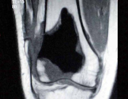
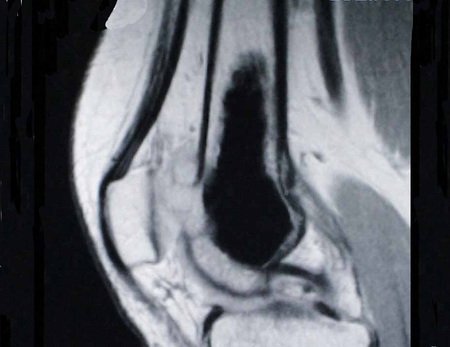
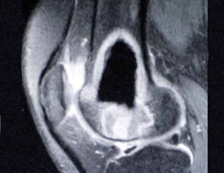
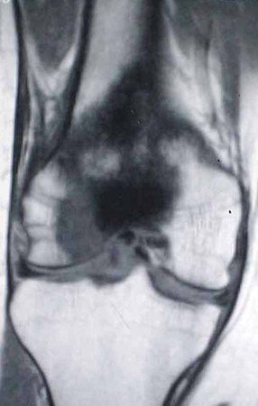
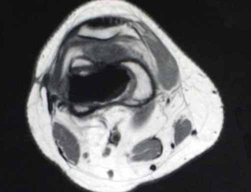
     We can observe the presence of a remaining tumor in the immediate postoperative radiographs. The resonance images detail this situation (Figures 5 – 10)

Figura 5: Ressonância magnética, em corte coronal T1, mostrando a permanência de tumor, sinal intermediário ao redor do cimento (baixo sinal). Nota-se ainda extravasamento da lesão pela abertura cirúrgica da cortical lateral e em tecidos moles.
Figure 5: Magnetic resonance imaging, in coronal T1 section, showing the presence of tumor, intermediate signal around the cement (low signal). Extravasation of the lesion is also noted through the surgical opening of the lateral cortex and into soft tissues.
Figura 6: Ressonância magnética, corte sagital T1, ressaltando extravasamento da lesão na face anterior do fêmur, sob a patela.
Figure 6: Magnetic resonance imaging, sagittal T1 view, highlighting extravasation of the lesion on the anterior surface of the femur, under the patella.
Figura 7: RM, sagital com contraste, demarcando a permanência de tumor e o extravasamento da lesão, em 22/fev./1999.
Figure 7: MRI, sagittal with contrast, demarcating the presence of the tumor and the extravasation of the lesion, on February 22, 1999.
Figura 8: RM, coronal T1. O condrossarcoma atinge a epífise femoral.
Figure 8: MRI, coronal T1. Chondrosarcoma affects the femoral epiphysis.
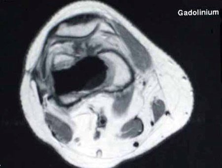
Figura 10: RM, axial T1, com lesão nos tecidos moles da face lateral da coxa.
Figure 9: MRI, ax T1, detailing the permanence and progression of the tumor, with extra-cortical tumor. Figure 10: MRI, axial T1, with lesion in the soft tissues of the lateral aspect of the thigh.

We analyzed that, despite inadequate manipulation, we were facing a slowly evolving neoplasm and that we could attempt a wide resection of the affected region and reconstruct it with a non-conventional endoprosthesis.
If the study of the specimen did not reveal dedifferentiation, we could perform post-operative clinical and imaging controls every three months initially, and only perform the alternative of amputation if there was a recurrence after this wide resection.
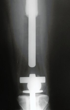
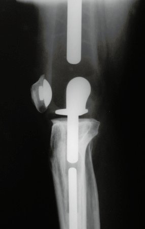
We resected the distal third of the femur that was compromised, through a wide incision, seen in Figures 11 and 12 and documented in the radiographs of the prosthesis, Figures 13 and 14.

Figura 10: Paciente com um ano e oito meses após a ressecção ampla e reconstrução com endoprótese não convencional. Incisão ampla lateral da coxa.
Figure 10: Patient one year and eight months after wide resection and reconstruction with non-conventional endoprosthesis. Wide lateral thigh incision.
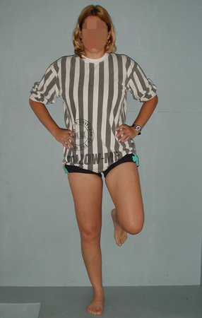
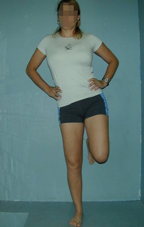
Figura 11: Carga monopodal após um ano e oito meses, em 17/Out/2000.
Figure 11: Single leg load after one year and eight months, on 17/Oct/2000.
Figura 12: Radiografia de controle, frente, após vinte meses.
Figure 12: Control x-ray, front, after twenty months.
Figura 13: Radiografia de controle, perfil. (17/Out/2000).
Figure 13: Control radiograph, profile. (17/Oct/2000).
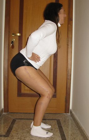
We can observe the patient’s function after three years, Figures 15 and 16 and evaluation after twelve years, Figures 17-20.
Figura 14: Paciente com boa função de flexão do joelho operado,17/Jul/2001.
Figure 14: Patient with good flexion function of the operated knee, 17/Jul/2001.
Figura 15: Carga monopodal após dois anos e meio. Nenhum sinal de recidiva do tumor, 17/Jul/2001.
Figure 15: Single-leg loading after two and a half years. No sign of tumor recurrence, 17/Jul/2001.
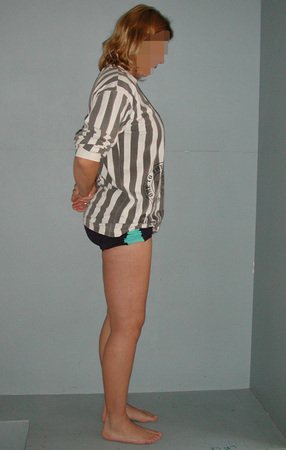
Figura 16: Paciente após 12 anos e meio da cirurgia de ressecção ampla, sem sinais de recorrência da lesão, 14/Jul/2011.
Figure 16: Patient after 12 and a half years of wide resection surgery, with no signs of recurrence of the lesion, 14/Jul/2011.
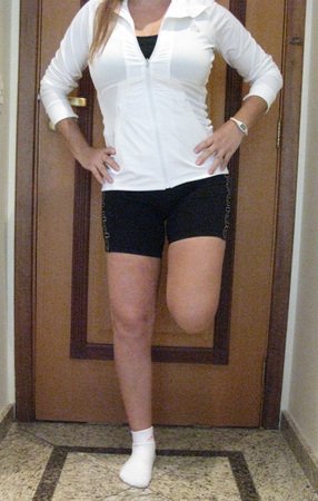
Figura 17: Carga monopodal, boa força muscular.
Figure 17: Single leg load, good muscle strength.
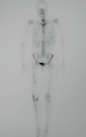
Figura 18: Cintilografia óssea sem sinais de recidiva.
Figure 18: Bone scintigraphy without signs of recurrence.
Figura 19: Paciente com boa função, após 12 anos da cirurgia de ressecção do 1/3 distal do fêmur e reconstrução com endoprótese não convencional de polietileno, sem recidiva, 02/03.2010.
Figure 19: Patient with good function, 12 years after resection of the distal 1/3 of the femur and reconstruction with non-conventional polyethylene endoprosthesis, without recurrence, 03/02/2010.

Author: Prof. Dr. Pedro Péricles Ribeiro Baptista

 Orthopedic Oncosurgery at the Dr. Arnaldo Vieira de Carvalho Cancer Institute

Hello! How can we assist you?