This digital library houses the book on Oncology and Orthopedic Oncosurgery.

It includes academic lectures, presentations from national and international congresses, published papers, case discussions, performed surgical procedures, and proprietary techniques developed.

The digital format was chosen because the web allows the inclusion of texts with numerous visual resources, such as images and videos, which would not be possible in a printed book.

The content is intended for students, healthcare professionals, and the general public interested in the field.

Chondrosarcoma of the femoral head

Condrosarcoma de la Cabeza Femoral

Femoral Head Chondrosarcoma

Chondrosarcoma of the femoral head. Male patient, 49 years old, reported pain in his hip for ten months. He performs an x-ray of the pelvis which reveals an injury to the head and right femoral neck (Figure 1). The tomography details the aspects of the injury (Figure 2).
Figura 1: Radiografia da pelve direita, com lesão de rarefação óssea no colo femoral e focos de maior densidade, sugerindo calcificações.
Figure 1: Radiograph of the right pelvis, with bone rarefaction lesion in the femoral neck and foci of greater density, suggesting calcifications.
Figura 2: Tomografia com lesão de rarefação óssea na cabeça femoral direito, erosão da cortical interna (em saca bocado) e focos de calcificação. Lesão agressiva localmente, ativa, destruindo a estrutura óssea.
Figure 2: Tomography showing bone rarefaction lesion in the right femoral head, erosion of the internal cortex (in a saccharine shape) and foci of calcification. Locally aggressive, active injury, destroying the bone structure.
Chondroma or chondrosarcoma? The doctor chooses to perform a percutaneous biopsy whose pathology concludes chondroma. Cartilaginous lesion, in a long bone, with pain, erosion of the internal cortex, foci of calcification in a patient over forty years of age should be treated as chondrosarcoma. It is treated as a chondroma, a conventional prosthesis (Figure 3).
After a few months, there was an increase in volume in the right thigh and pain, and a progressive increase in the hip (Figure 4). The radiograph shows an expansive lesion (Figure 5).
Figura 3: Radiografia da pelve direita, após reconstrução com prótese convencional. O paciente foi tratado como condroma, inadequadamente.
Figure 3: Radiograph of the right pelvis, after reconstruction with a conventional prosthesis. The patient was treated as chondroma, inappropriately.
Figura 4: Paciente com aumento de volume e dor no quadril direito devido a recidiva e desdiferenciação de condrossarcoma.
Figure 4: Patient with increased volume and pain in the right hip due to recurrence and dedifferentiation of chondrosarcoma.
Figura 5: Radiografia da pelve direita, após alguns meses da reconstrução com prótese convencional, A recidiva do condrossarcoma é evidente.
Figure 5: Radiograph of the right pelvis, a few months after reconstruction with a conventional prosthesis. The recurrence of chondrosarcoma is evident.
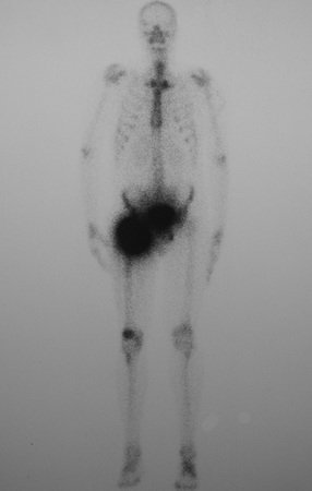
Figura 6: Cintilografia óssea com hiper captação acentuada da pelve direita e terço proximal do fêmur.
Figure 6: Bone scintigraphy with marked hyperuptake of the right pelvis and proximal third of the femur.
Figura 7: Paciente posicionado para a realização de hemipelvectomia devido a disseminação do condrossarcoma, operado indevidamente.
Figure 7: Patient positioned for hemipelvectomy due to dissemination of chondrosarcoma, improperly operated.
Figura 8: Radiografia da bacia, após a realização da hemipelvectomia em paciente portador de condrossarcoma, inicialmente grau I, tratado indevidamente como condroma, que se desdiferenciou. A cirurgia adequada poderia ter levado a cura do tumor com a preservação do membro.
Figure 8: X-ray of the pelvis, after performing hemipelvectomy in a patient with chondrosarcoma, initially grade I, improperly treated as a chondroma, which dedifferentiated. Appropriate surgery could have cured the tumor while preserving the limb.
Figura 9: Esta hemipelvectomia teria sido evitada se não tivesse havido supervalorização da patologia em detrimento do quadro clínico e da imagem.
Figure 9: This hemipelvectomy would have been avoided if there had not been an overvaluation of the pathology to the detriment of the clinical picture and image.
For this neoplasm, whose only treatment is surgery, it cannot be induced by anatomopathological examination of chondroma by biopsy. We should not perform another biopsy, as we already have a diagnosis of a cartilaginous lesion. The aggressive images, with foci of calcification and erosion of the internal cortex define the approach to treating this case as the chondrosarcoma it was (Figure 9).

Author: Prof. Dr. Pedro Péricles Ribeiro Baptista

 Orthopedic Oncosurgery at the Dr. Arnaldo Vieira de Carvalho Cancer Institute

Hello! How can we assist you?