This digital library houses the book on Oncology and Orthopedic Oncosurgery.

It includes academic lectures, presentations from national and international congresses, published papers, case discussions, performed surgical procedures, and proprietary techniques developed.

The digital format was chosen because the web allows the inclusion of texts with numerous visual resources, such as images and videos, which would not be possible in a printed book.

The content is intended for students, healthcare professionals, and the general public interested in the field.

Osteoma

Bdd1b613ee6fcac7694cf648430358ce 3
[auto_translate_button]
Osteoma Benign, slow-growing lesion, with mature bone tissue, with a lamellar structure, well differentiated. It is bone, dense , within the bone, whether in the cortical or medullary region .

Osteoma

It can manifest itself in three distinct clinical forms:

  • Exostoses  (dense, homogeneous bone, with an  ivory appearance ): this is the conventional osteoma, restricted to bones of intramembranous origin (facial bones, skullcap), figures 1 to 10.
Figura 1: Radiografia do crânio com lesão nodular, densa, homogênea, na calota.
Figure 1: X-ray of the skull with a nodular, dense, homogeneous lesion in the cap.
Figura 2: Tomografia axial do crânio, com lesão acometendo as duas tábuas, com abaulamento maior da cortical externa.
Figure 2: Axial tomography of the skull, with the lesion affecting both tables, with greater bulging of the external cortex.
Figura 3: Reconstrução tomográfica em três dimensões da lesão do crânio.
Figure 3: Three-dimensional tomographic reconstruction of the skull lesion.
Figura 4: Nódulo firme, saliente, na base de implantação do cabelo.
Figure 4: Firm, prominent nodule at the base of hair implantation.
Figura 6: Ostectomia com formão.
Figure 5: Asepsis and antisepsis, with plastic field placement. Figure 6: Ostectomy with chisel.
Figura 8: Fragmentos do osteoma ressecado.
Figure 7: Regularized surgical bed, without protrusions. Figure 8: Fragments of the resected osteoma.
Figura 9: Osteoma exofítico da região parietal, aspecto de marfin.
Figure 9: Exophytic osteoma of the parietal region, ivory appearance.
Figura 11: Segmento ressecado na base.
Figure 10: Ivory-looking lesion. Figure 11: Resected segment at the base.
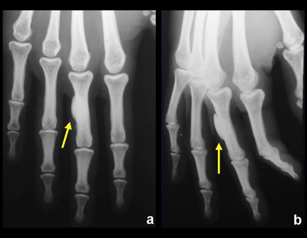
  • Parosteal  (juxtacortical) occurs on the external surface of long or short bones, figure 12.
  • Figura 12: Osteoma da falange proximal, justacortical.
    Figure 12: Osteoma of the proximal phalanx, juxtacortical.
    Figura 13 : Osteoma medular do colo femoral, enostose óssea.
    Figure 13: Medullary osteoma of the femoral neck, bone enostosis.
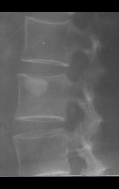
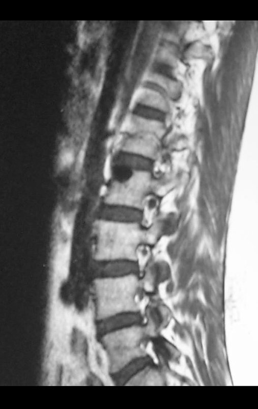
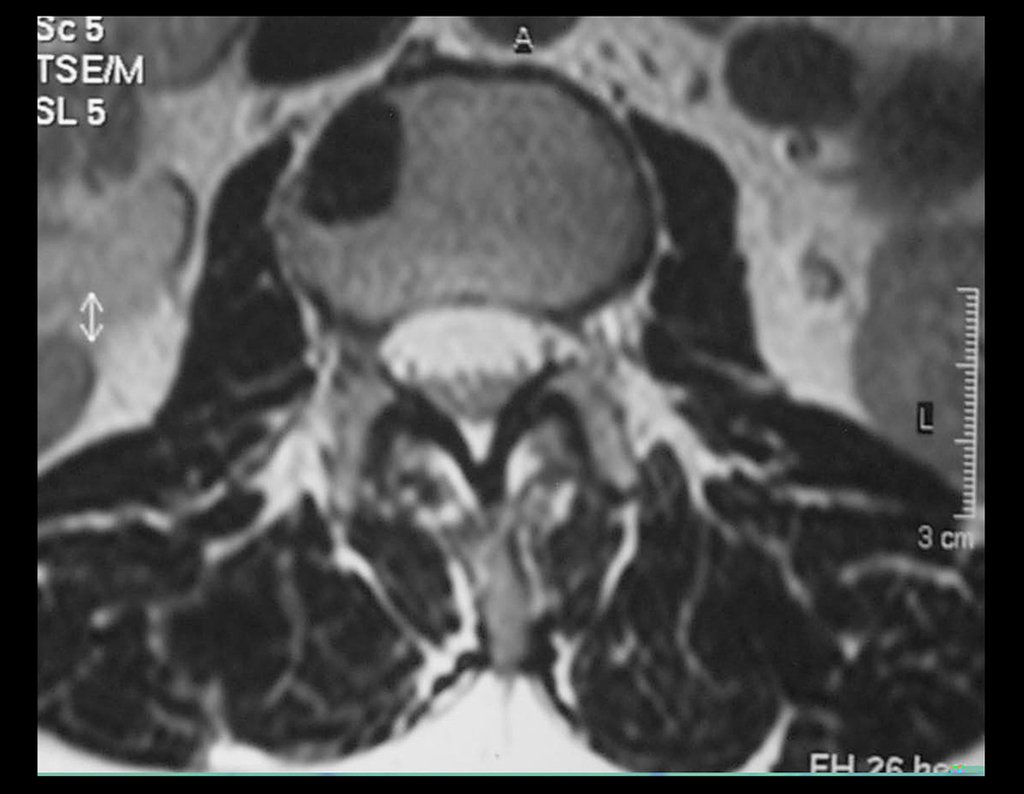
    • Medullary : known as enostosis or bone islet. Lesions histologically similar to mature bone, dense and homogeneous, without significant clinical signs and generally resulting from radiographic findings, figures 13 to 16.
    Figura 14 : Osteoma medular do corpo vertebral, enostose óssea.
    Figure 14: Medullary osteoma of the vertebral body, bone enostosis.
    Figura 15: Rm sagital de osteoma medular do corpo vertebral.
    Figure 15: Sagittal MRI of medullary osteoma of the vertebral body.
    Figura 16: Rm axial de osteoma medular do corpo vertebral.
    Figure 16: Axial MRI of medullary osteoma of the vertebral body.

    he differential diagnoses, both from a clinical, histological and radiographic point of view, include the following conditions:

    • Bone sclerosis  (inflammatory or post-traumatic cause)
    • Hyperostoses
    • Meningioma
    • Osteoid osteoma.

    Treatment consists of observation when asymptomatic or surgical resection when indicated due to aesthetic or functional changes (compression or obstruction – nasal fossa).

    Author: Prof. Dr. Pedro Péricles Ribeiro Baptista

     Orthopedic Oncosurgery at the Dr. Arnaldo Vieira de Carvalho Cancer Institute

    Leave a Reply

    Hello! How can we assist you?