Osteochondroma
Diagnosis and Treatment
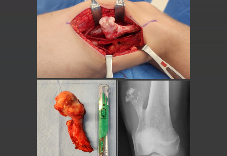
Osteochondroma, also known as osteocartilaginous exostosis, represents the most common benign bone lesion, although its incidence may be even higher than that reported in the literature due to many patients presenting asymptomatic osteochondromas.
This condition generally develops in the first and second decades of life, being located in the metaphyseal region of long bones. Radiographically, it is characterized by presenting a tumor composed of cartilage and bone. A distinctive feature is that the central cancellous bone of the exostosis continues with the medullary of the affected bone, while the dense cortical layer of the tumor continues with the normal cortical bone. On the surface of the lesion, there is a band of cartilage, through which the growth of the lesion occurs, hence the name “osteochondroma” – tumor forming cartilage and bone.
Osteochondromas can present as sessile bases (with an enlarged base) or pedunculated bases. They can be single or multiple, characterizing hereditary multiple osteochondromatosis.
The standard treatment for osteochondromas is surgery, generally involving resection, especially when the lesion compromises aesthetics, compresses vascular or nervous structures or limits function. It is important to note that these tumors usually continue to grow while the patient is in the growth phase.
When an osteochondroma increases in size after skeletal maturity, it may indicate post-traumatic bursitis or, more worryingly, malignancy to chondrosarcoma, which requires resection with oncological margins.
The risk of malignancy is low, with solitary osteochondromas having a malignancy rate of about 1%, while multiple osteochondromatosis can be as high as 10%.
In summary, a comprehensive understanding of the characteristics, diagnosis and treatment of osteochondroma is essential to ensure an effective and appropriate therapeutic approach, always aiming for the patient’s well-being and quality of life.
Author: Prof. Dr. Pedro Péricles Ribeiro Baptista
Orthopedic Oncosurgery at the Dr. Arnaldo Vieira de Carvalho Cancer Institute
Office : Rua General Jardim, 846 – Cj 41 – Cep: 01223-010 Higienópolis São Paulo – SP
Phone: +55 11 3231-4638 Cell:+55 11 99863-5577 Email: drpprb@gmail.com

