This digital library houses the book on Oncology and Orthopedic Oncosurgery.

It includes academic lectures, presentations from national and international congresses, published papers, case discussions, performed surgical procedures, and proprietary techniques developed.

The digital format was chosen because the web allows the inclusion of texts with numerous visual resources, such as images and videos, which would not be possible in a printed book.

The content is intended for students, healthcare professionals, and the general public interested in the field.

Growth Cartilage Autotransplantation Technique

D9bec6b996ff7a16fc57fb253e6255c8 3
Growth Cartilage Autotransplantation Technique. Patient aged twelve years and five months, presenting pain and tumor in the proximal third of the right leg in April 1999, figures 1 and 2, and tomography figures 3 to 8.

Growth Cartilage Autotransplantation Technique – Tibialization of the Proximal Fibula – Biological Reconstruction – Autologous Graft in Tibial Osteosarcoma
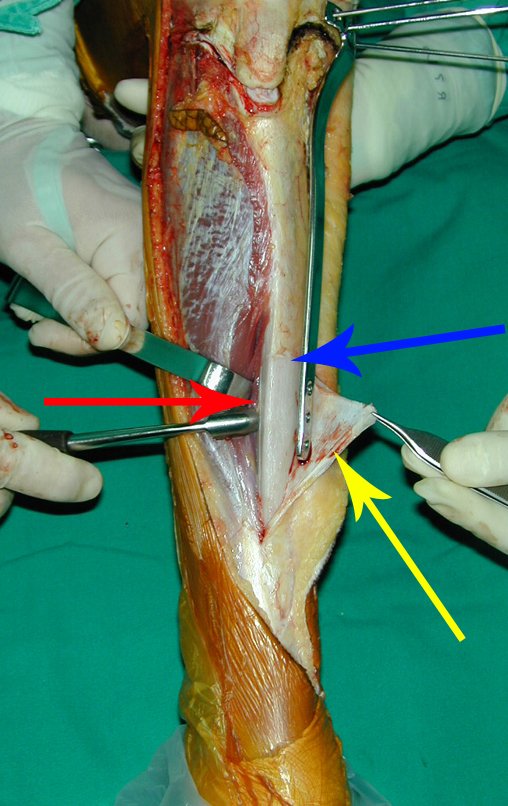
Figura 1: Radiografia do 1/3 proximal da tíbia direita, de 29/04/1999, com lesão na metáfise proximal, com áreas de rarefação e áreas de condensação óssea, com erosão da cortical lateral.
Figure 1: Radiograph of the proximal 1/3 of the right tibia, dated 04/29/1999, with injury to the proximal metaphysis, with areas of rarefaction and areas of bone condensation, with erosion of the lateral cortex.
Figura 2: Radiografia em perfil, com lesão na metáfise proximal da tíbia, com limites imprecisos, sem comprometimento da epífise tibial.
Figure 2: Profile x-ray, with injury to the proximal metaphysis of the tibia, with imprecise limits, without involvement of the tibial epiphysis.
Figura 3: Tomografia de 29/04/1999, lesão condensante na metáfise proximal da tíbia, formadora de osso, com tumor extracortical na região anterior, lateral e posterior, densidade para osso.
Figure 3: Tomography of 04/29/1999, condensing lesion in the proximal metaphysis of the tibia, bone forming, with extracortical tumor in the anterior, lateral and posterior region, density for bone.
Figura 4: Tomografia de 29/04/1999, lesão condensante na metáfise proximal da tíbia, formadora de osso, com tumor extracortical na região anterior, lateral e posterior, densidade para tecidos moles.
Figure 4: Tomography of 04/29/1999, condensing lesion in the proximal metaphysis of the tibia, forming bone, with extracortical tumor in the anterior, lateral and posterior region, density for soft tissues.
Figura 5: Tomografia de 29/04/1999, lesão condensante na medular do 1/3 proximal da tíbia, com lesão extracortical na região ântero-lateral, densidade para osso.
Figure 5: Tomography of 04/29/1999, condensing lesion in the spinal cord of the proximal 1/3 of the tibia, with extracortical lesion in the anterolateral region, density for bone.
Figura 6: Tomografia de 29/04/1999, lesão condensante na medular do 1/3 proximal da tíbia, com lesão extracortical na região ântero-lateral, densidade para tecidos moles.
Figure 6: Tomography of 04/29/1999, condensing lesion in the spinal cord of the proximal 1/3 of the tibia, with extracortical lesion in the anterolateral region, density for soft tissues.
Figura 7: Tomografia de 29/04/1999, corte mais inferior do 1/3 proximal da tíbia, com lesão extracortical na região ântero-lateral, densidade para osso.
Figure 7: Tomography of 04/29/1999, lower section of the proximal 1/3 of the tibia, with extracortical lesion in the anterolateral region, density for bone.
Figura 8: Tomografia de 29/04/1999, corte mais inferior do 1/3 proximal da tíbia, sem evidência de lesão na medular, porém com lesão extracortical na região ântero-lateral, densidade para tecidos moles.
Figure 8: Tomography of 04/29/1999, lower section of the proximal 1/3 of the tibia, without evidence of spinal cord injury, but with extracortical injury in the anterolateral region, density for soft tissues.
After staging the lesion, a biopsy of the leg was performed via an anterolateral approach. The anatomical pathological examination confirmed that it was osteosarcoma. In October 1999, the post-neoadjuvant chemotherapy MRI study showed a metaphyseal lesion that affected the anterolateral portion of the growth plate, figures 9 to 16.
Figura 9: RM em T1, de 02/10/1999, pós quimioterapia, com lesão de baixo sinal, na região lateral da metáfise proximal da tíbia direita e diminuição do volume tumoral extracortical.
Figure 9: T1 MRI, on 10/02/1999, after chemotherapy, with a low signal lesion, in the lateral region of the proximal metaphysis of the right tibia and a decrease in the extracortical tumor volume.
Figura 10: RM, coronal, de 02/10/1999, pós quimioterapia, com lesão na região lateral da metáfise proximal da tíbia direita e diminuição do volume tumoral extracortical.
Figure 10: MRI, coronal, from 10/02/1999, post chemotherapy, with lesion in the lateral region of the proximal metaphysis of the right tibia and reduction in extracortical tumor volume.
Figura 11: Ressonância magnética em corte sagital, com saturação de gordura, de 02/10/1999, pós quimioterapia, com lesão na região lateral da metáfise proximal da tíbia direita, logo abaixo da cartilagem de crescimento.
Figure 11: Magnetic resonance imaging in sagittal section, with fat saturation, from 10/02/1999, post chemotherapy, with lesion in the lateral region of the proximal metaphysis of the right tibia, just below the growth cartilage.
Figura 12: RM, T2, Stir, Dp, corte sagital e mais no centro da tíbia, com lesão metafisária distante da placa de crescimento.
Figure 12: MRI, T2, Stir, Dp, sagittal section and more in the center of the tibia, with a metaphyseal lesion far from the growth plate.
The lesion touches the growth cartilage, but does not invade the epiphysis, figures 9 and 10. In figure 11, we can see the lesion under the growth line, and in figure 12, at some distance from the tumor. In sagittal MRI scans, we observed that the lesion touches the growth cartilage in some sections, but does not invade the epiphysis in any of them.
Figura 13: RM pós Qt, de outubro de 1999, com boa resposta ao tratamento quimioterápico. Diminuição de edema e mineralização da lesão.
Figure 13: MRI after Qt, in October 1999, with good response to chemotherapy treatment. Decrease in edema and mineralization of the lesion.
Figura 14: RM axial pós Qt com ossificação do tumor extracortical.
Figure 14: Post-Qt axial MRI with ossification of the extracortical tumor.
Figura 15: RM axial T1 Spir, boa resposta ao tratamento neoadjuvante.
Figure 15: Axial T1 Spir MRI, good response to neoadjuvant treatment.
Figura 16: RM axial T1 Spir, corte mais inferior, boa resposta ao tratamento neoadjuvante.
Figure 16: Axial T1 Spir MRI, lower section, good response to neoadjuvant treatment.
Figura 17: Radiografia pré-operatória, pós quimioterapia, com boa resposta ao tratamento neoadjuvante.
Figure 17: Preoperative radiograph, post chemotherapy, with good response to neoadjuvant treatment.
Figura 18: Radiografia pré-operatória, perfil, de outubro de 1999, pós quimioterapia, com boa resposta ao tratamento neoadjuvante.
Figure 18: Preoperative radiograph, profile, from October 1999, post chemotherapy, with good response to neoadjuvant treatment.
Figura 19: Cintilografia de outubro de 1999, pré-operatória.
Figure 19: Scintigraphy from October 1999, pre-operative.
Figura 20: Mapeamento ósseo, anterior, de outubro de 1999.
Figure 20: Bone mapping, previous, from October 1999.
Figura 21: Mapeamento ósseo, posterior, de outubro de 1999.
Figure 21: Bone mapping, later, from October 1999.
Figura 22: Mapeamento ósseo, fase tardia, localizado, de outubro de 1999.
Figure 22: Bone mapping, late phase, localized, from October 1999.
Figura 23: Cirurgia realizada em 25/10/1999, marcação da incisão cirúrgica, ressecando o trajeto da biópsia ântero-lateral da tíbia e órtese confeccionada especialmente para o paciente, que será utilizada no pós-operatório.
Figure 23: Surgery performed on 10/25/1999, marking of the surgical incision, resecting the path of the anterolateral biopsy of the tibia and orthosis made especially for the patient, which will be used post-operatively.
Figura 24: Incisão cirúrgica, ressecando o trajeto da biópsia.
Figure 24: Surgical incision, resecting the biopsy path.
Figura 25: Dissecção do subcutâneo e desinserção dos tendões da pata de ganso.
Figure 25: Dissection of the subcutaneous tissue and disinsertion of the pes anserinus tendons.
Figura 26: Dissecção do ligamento patelar com eletrocautério, que permanecerá inserido na epífise femoral.
Figure 26: Dissection of the patellar ligament with electrocautery, which will remain inserted into the femoral epiphysis.
Figura 27: Delimitação do contorno do ligamento patelar - seta azul, abertura do perimísio do músculo tibial anterior - seta vermelha e posicionamento da placa, feita sob medida, que vai fixar a epífise tibial à diáfise - seta amarela, quando da reconstrução.
Figure 27: Delimitation of the contour of the patellar ligament - blue arrow, opening of the perimysium of the anterior tibial muscle - red arrow and positioning of the custom-made plate, which will fix the tibial epiphysis to the diaphysis - yellow arrow, during reconstruction.
Figura 28: Detalhe da ressecção em bloco da lesão incluindo o trajeto da biópsia - seta azul.
Figure 28: Detail of the en bloc resection of the lesion including the biopsy path - blue arrow.
Figura 29: Rebatimento dos tendões da pata de ganso e exposição do ligamento colateral medial, superficial e profundo.
Figure 29: Reflection of the pes ansus tendons and exposure of the medial, superficial and deep collateral ligament.
Figura 30: Após a marcação do trajeto dos parafusos da epífise tibial com fios de Kirschner, procede-se à dissecção distalmente, deixando o periósteo da tíbia como margem medial - seta azul, o perimísio do musculo tibial anterior como margem lateral - seta vermelha e rebate-se o periósteo abaixo do nível da osteotomia - seta amarela, preservando-o.
Figure 30: After marking the path of the tibial epiphysis screws with Kirschner wires, dissection proceeds distally, leaving the periosteum of the tibia as the medial margin - blue arrow, the perimysium of the anterior tibial muscle as the lateral margin - red arrow and the periosteum is reflected below the level of the osteotomy - yellow arrow, preserving it.
Figura 31: Secção do ligamento colateral medial e marcação da osteotomia proximal da tíbia do lado medial, curvilínea para preservar a epífise.
Figure 31: Section of the medial collateral ligament and marking of the proximal osteotomy of the tibia on the medial side, curvilinear to preserve the epiphysis.
Figura 32: Dissecção do músculo tibial anterior, deixando algumas fibras musculares, o perimísio e o periósteo como margem lateral.
Figure 32: Dissection of the anterior tibialis muscle, leaving some muscle fibers, the perimysium and the periosteum as the lateral margin.
Figura 33: Conferência da posição da placa e da orientação dos fios de Kirschner, antes da osteotomia.
Figure 33: Checking the position of the plate and the orientation of the Kirschner wires, before osteotomy.
Figura 34: Osteotomia da diáfise da tíbia, distalmente.
Figure 34: Osteotomy of the tibial shaft, distally.
Figura 35: Dissecção da porção proximal da fíbula, que vai ser transplantada para a tíbia, juntamente com a placa de crescimento.
Figure 35: Dissection of the proximal portion of the fibula, which will be transplanted to the tibia, together with the growth plate.
Figura 36: Remoção da cartilagem articular da epífise fibular proximal, expondo-se o osso epifisário - seta amarela, facilitando a consolidação com a epífise tibial.
Figure 36: Removal of the articular cartilage of the proximal fibular epiphysis, exposing the epiphyseal bone - yellow arrow, facilitating consolidation with the tibial epiphysis.
Figura 37: Segmento da metáfise tibial proximal ressecado, epífise da tíbia com os três fios guias. Boa hemostasia.
Figure 37: Resected segment of the proximal tibial metaphysis, tibial epiphysis with the three guide wires. Good hemostasis.
Figura 38: Dissecção e exposição da diáfise fibular distal que vai ser encavilhada na diáfise da tíbia.
Figure 38: Dissection and exposure of the distal fibular diaphysis that will be nailed into the tibial diaphysis.
Figura 39: Osteotomia da tíbia para o encavilhamento.
Figure 39: Tibial osteotomy for nailing.
Figura 40: Para viabilizar o posicionamento da fíbula, dentro do canal medular tibial, é necessário realizar uma janela na diáfise da tábua posterior da tíbia, para não lesar a artéria nutrícia.
Figure 40: To enable the positioning of the fibula, within the tibial medullary canal, it is necessary to create a window in the diaphysis of the posterior table of the tibia, so as not to damage the nutrient artery.
Figura 41: Janela realizada, com a retirada de um segmento da cortical posterior.
Figure 41: Window created, with the removal of a segment of the posterior cortex.
Figura 42: Fíbula encavilhada na tíbia, distalmente.
Figure 42: Fibula embedded in the tibia, distally.
Figura 43: Dispositivo de fixação interna extensível, feito sob moldagem especial para este paciente. Segmento proximal em T, com três orifícios - seta amarela, haste confeccionada já curvelínea e pré modelada - seta azul e dispositivo de deslizamento triangular, para adaptação à tíbia , com encaixe - seta vermelha.
Figure 43: Extensible internal fixation device, made under a special mold for this patient. Proximal segment in T, with three holes - yellow arrow, rod already curved and pre-shaped - blue arrow and triangular sliding device, to adapt to the tibia, with fitting - red arrow.
Figura 44: Placa especial desenhada e modelada para este paciente, encaixada no diapositivo de fixação interna extensível.
Figure 44: Special plate designed and modeled for this patient, fitted to the extendable internal fixation slide.
Figura 45: Colocação dos três parafusos proximais no "T" da placa especial e fixação à epífise da tíbia.
Figure 45: Placement of the three proximal screws in the “T” of the special plate and fixation to the tibial epiphysis.
Figura 46: Osteossíntese terminada, estabilizando a reconstrução do autotransplante da fíbula, com a sua cartilagem de crescimento, para a tíbia.
Figure 46: Osteosynthesis completed, stabilizing the reconstruction of the fibula autotransplant, with its growth cartilage, to the tibia.
Figura 47: Detalhe do dispositivo fixado distalmente. Ele permite que a haste da placa deslize, com o crescimento da epífise fibular.
Figure 47: Detail of the device fixed distally. It allows the stem of the plate to slide, with the growth of the fibular epiphysis.
Figura 48: Fechamento do plano muscular e revisão da hemostasia.
Figure 48: Closure of the muscular plane and review of hemostasis.
Figura 49: Sutura da pele.
Figure 49: Skin suture.
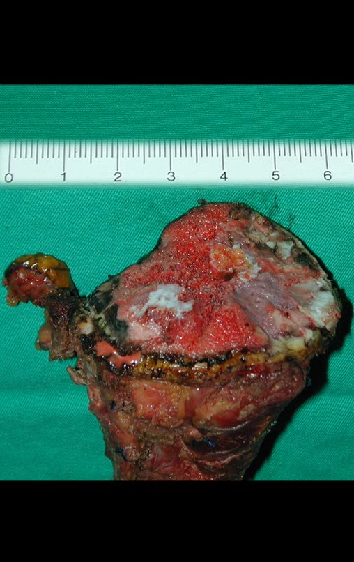
Figura 50: Peça ressecada, envolvida pelo periósteo, medialmente, e pelo periósteo e perimísio lateralmente.
Figure 50: Resected piece, surrounded by the periosteum, medially, and by the periosteum and perimysium laterally.
Figura 51: Detalhe do corte superior da tíbia, intraepifisário e curvilíneo, removendo toda a cartilagem de crescimento.
Figure 51: Detail of the upper tibia cut, intraepiphyseal and curvilinear, removing all growth cartilage.
Figura 52: Peça cortada ao meio, evidenciando a margem proximal e o tumor extracortical, com o triângulo de Codman.
Figure 52: Piece cut in half, showing the proximal margin and the extracortical tumor, with Codman's triangle.
Figura 53: Detalhe do corte da peça cirúrgica, boa margem de ressecção.
Figure 53: Detail of the cut of the surgical specimen, good resection margin.
Figura 54: Fatia preparada para descalcificação e estudo histopatológico.
Figure 54: Slice prepared for decalcification and histopathological study.
Figura 55: Radiografia da peça cirúrgica, frente.
Figure 55: Radiograph of the surgical specimen, front.
Figura 56: Radiografia da peça cirúrgica, perfil.
Figure 56: Radiograph of the surgical specimen, profile.
Figura 57: Radiografia de controle intraoperatório, frente.
Figure 57: Intraoperative control radiograph, front.
F9e70773c750b0b4ef0b7b2a0b07fe67 3
Figure 58: Intraoperative control radiograph, profile.
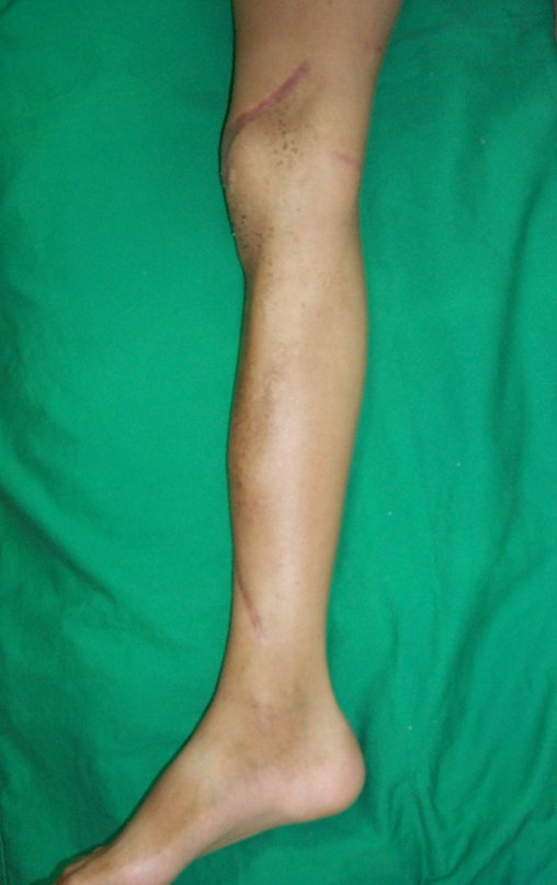
Figura 59: Após três dias, em 28/10/1999, ferida operatória em cicatrização, com sinal de sofrimento da pele, na região do joelho.
Figure 59: After three days, on 10/28/1999, surgical wound healing, with signs of skin distress, in the knee region.
Figura 60: Detalhe do sofrimento de pele, na região do joelho. Provável compressão pelo curativo (gaze com sangue seco, endurecida e enfaixamento sob pressão).
Figure 60: Detail of skin damage in the knee region. Probable compression by the dressing (gauze with dried blood, hardened and bandaging under pressure).
Figura 61: Aspecto em 10/11/1999, após duas semanas da cirurgia, delimitação da área de necrose.
Figure 61: Appearance on 11/10/1999, two weeks after surgery, delimitation of the area of ​​necrosis.
Figura 62: Aspecto em 10/11/1999, após desbridamento da área de necrose. Pontos retirados, boa cicatrização no restante da incisão.
Figure 62: Appearance on 11/10/1999, after debridement of the area of ​​necrosis. Stitches removed, good healing in the rest of the incision.
Figura 63: Aspecto clínico após quatro meses, em 29/02/2000.
Figure 63: Clinical appearance after four months, on 02/29/2000.
Figura 64: Aspecto clínico em 29/02/2000, perfil.
Figure 64: Clinical appearance on 02/29/2000, profile.
Figura 65: Aspecto clínico após sete meses, com valgismo do membro operado, em 22/05/2000. Deambulando com discreto apoio.
Figure 65: Clinical appearance after seven months, with valgus in the operated limb, on 05/22/2000. Walking with discreet support.
Figura 66: Aspecto clínico após sete meses, sem carga, em 22/05/2000.
Figure 66: Clinical appearance after seven months, without load, on 05/22/2000.
Figura 67: Flexão do joelho 90 graus, sem carga, após sete meses, em 22/05/2000.
Figure 67: Knee flexion 90 degrees, without load, after seven months, on 05/22/2000.
Figura 68: Detalhe da ferida operatória e do valgismo, após sete meses da cirurgia.
Figure 68: Detail of the surgical wound and valgus, seven months after surgery.
Figura 69: Melhora do valgismo!!!...??? Após nove meses, em 08/08/2000.
Figure 69: Valgus improvement!!!...??? After nine months, on 08/08/2000.
Figura 70: Melhora da flexão, 135 graus, sem carga, após nove meses, em 08/08/2000.
Figure 70: Improvement in flexion, 135 degrees, without load, after nine months, on 08/08/2000.
Figura 71: Aspecto da cicatrização após nove meses, em 08/08/2000.
Figure 71: Appearance of healing after nine months, on 08/08/2000.
Figura 72: Aspecto clínico após nove meses, em 08/08/2000.
Figure 72: Clinical appearance after nine months, on 08/08/2000.
Figura 73: Valgismo corrigido!!! ... Como??? Após um ano, em 07/11/2000.
Figure 73: Valgus corrected!!! ... As??? After one year, on 11/07/2000.
Figura 74: Vista posterior, membro alinhado, após um ano da cirurgia.
Figure 74: Posterior view, limb aligned, one year after surgery.
Figura 75: Boa função do joelho, após um ano da cirurgia.
Figure 75: Good knee function, one year after surgery.
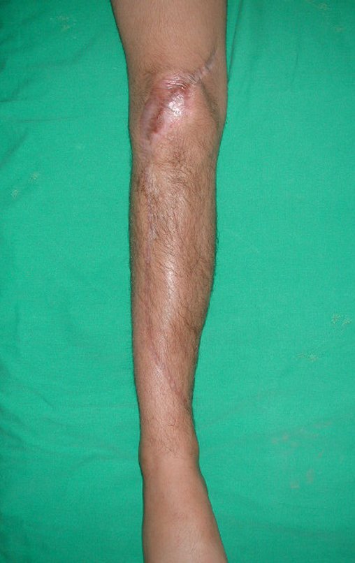
Figura 76: Aspecto cosmético da cicatriz, após um ano da cirurgia.
Figure 76: Cosmetic appearance of the scar, one year after surgery.
Figura 77: Radiografia de 01/03/2000, após cinco meses da cirurgia, com valgismo.
Figure 77: Radiograph from 03/01/2000, five months after surgery, with valgus.
Figura 78: Radiografia de 01/03/2000, perfil, após cinco meses da cirurgia.
Figure 78: Radiograph from 03/01/2000, profile, five months after surgery.
Figura 79: Radiografia de 24/07/2000, após oito meses da cirurgia. Melhora do valgismo pois a físe da fíbula está crescendo e empurrando a epífise da tíbia horizontalizando os parafusos e corrigindo a deformidade.
Figure 79: Radiograph from 07/24/2000, eight months after surgery. Improvement of valgusism as the fibular physis is growing and pushing the tibial epiphysis, horizontalizing the screws and correcting the deformity.
Figura 80: Escanograma corroborando este crescimento.
Figure 80: Scanogram corroborating this growth.
Figura 81: Escanograma de 07/11/2000, após um ano da cirurgia, evidenciando a horizontalização dos parafusos e o alinhamento do joelho.
Figure 81: Scanogram of 11/07/2000, one year after surgery, showing the horizontalization of the screws and the alignment of the knee.
Figura 82: Correção total do valgismo. Parafusos epifisários horizontais, corrigidos pelo crescimento da epífise fibular transplantada.
Figure 82: Total correction of valgusism. Horizontal epiphyseal screws, corrected by the growth of the transplanted fibular epiphysis.
Figura 83: Paciente após um ano e três meses. Membro alinhado, valgismo totalmente corrigido, carga total e boa função do joelho, em 06/02/2001.
Figure 83: Patient after one year and three months. Limb aligned, valgusism fully corrected, full load and good knee function, on 02/06/2001.
Figura 84: Carga total monopodal e boa função do joelho, após um ano e três meses, em 06/02/2001.
Figure 84: Total single-leg load and good knee function, after one year and three months, on 02/06/2001.
Figura 85: Cintilografia de 06/02/2001, após um ano e três meses, mostra a vitalidade da fíbula transplantada e a vitalidade da sua placa de crescimento.
Figure 85: Scintigraphy on 02/06/2001, after one year and three months, shows the vitality of the transplanted fibula and the vitality of its growth plate.
Figura 86: Cintilografia localizada, lateral direita, mostra a vitalidade da fíbula transplantada e a vitalidade da sua placa de crescimento.
Figure 86: Localized scintigraphy, right side, shows the vitality of the transplanted fibula and the vitality of its growth plate.
Figura 87: Cintilografia localizada, lateral esquerda, mostra a vitalidade da fíbula transplantada e a vitalidade da sua placa de crescimento.
Figure 87: Localized scintigraphy, left side, shows the vitality of the transplanted fibula and the vitality of its growth plate.

Autotransplantation of the vascularized fibula, with its growth plate, is proving to be an effective biological solution. The fibula is being “tibialized” and its growth plate continues to grow, replacing the tibial growth plate, which was resected along with the tumor.

Figura 88: Paciente após um ano e sete meses da cirurgia, em 08/05/2001. Trabalhando em fazenda, lidando com gado no dia a dia.
Figure 88: Patient one year and seven months after surgery, on 05/08/2001. Working on a farm, dealing with livestock on a daily basis.
Figura 89: Carga total, monopodal, em 08/05/2001.
Figure 89: Total load, single leg, on 05/08/2001.
Figura 90: Flexão com carga total, em 08/05/2001.
Figure 90: Flexion with full load, on 05/08/2001.
Figura 91: Carga total monopodal, em 08/05/2001.
Figure 91: Total single-leg load, on 05/08/2001.
Figura 92: Bom trofismo e boa função do membro operado, em 08/05/2001.
Figure 92: Good trophism and good function of the operated limb, on 05/08/2001.
Figura 93: Membro inferior alinhado e com a perna operada mais curta.
Figure 93: Lower limb aligned and with the operated leg shorter.
Figura 94: A fíbula continua aumentando a sua espessura, "tibializando-se". Radiografia de 08/05/2001.
Figure 94: The fibula continues to increase in thickness, "tibializing". Radiograph of 05/08/2001.
Figura 95: A fíbula com o aumento da sua espessura, "tibializando-se". Radiografia em perfil de 08/05/2001.
Figure 95: The fibula increases in thickness, "tibializing". Profile x-ray dated 05/08/2001.

Lower limb aligned and clinically with the shorter leg, as shortening occurred right at the beginning, due to the inclination of the epiphysis, causing inferior sliding of the plate stem, already demonstrated with the inclination of the epiphyseal screws.

Figura 96: Paciente em 07/08/2001, bem.
Figure 96: Patient on 08/07/2001, well.
Figura 97: Membros alinhados, paciente bem, em 07/08/2001.
Figure 97: Limbs aligned, patient well, on 08/07/2001.
Figura 98: Carga monopodal, bom trofismo, em 07/08/2001.
Figure 98: Single-legged load, good trophism, on 08/07/2001.
Figura 99: Flexão com carga, paciente ganhando altura e massa muscular, em 07/08/2001.
Figure 99: Flexion with load, patient gaining height and muscle mass, on 07/08/2001.
Figura 100: Escanograma de 07/08/2001, com a fise fibular encerrando o seu crescimento e a perna operada ligeiramente mais curta, devido à perda no pós-operatório precoce, já comentado.
Figure 100: Scanogram of 08/07/2001, with the fibular physis ending its growth and the operated leg slightly shorter, due to the loss in the early postoperative period, already mentioned.
Figura 101: Escanograma de 07/08/2001, perfil, com a fise fibular encerrando o seu crescimento e a perna operada ligeiramente mais curta, devido à perda no pós-operatório precoce, já comentado.
Figure 101: Scanogram of 07/08/2001, profile, with the fibular physis ending its growth and the operated leg slightly shorter, due to the loss in the early postoperative period, already mentioned.
Figura 102: Paciente após dois anos da cirurgia, boa função do joelho em 29/10/2001.
Figure 102: Patient two years after surgery, good knee function on 10/29/2001.
Figura 103: Flexão com carga total após dois anos, em 29/10/2001.
Figure 103: Flexion with full load after two years, on 10/29/2001.
Figura 104: Carga total monopodal após dois anos da cirurgia.
Figure 104: Total single-leg weightlifting two years after surgery.
Figura 105: Carga total monopodal, perfil, após dois anos da cirurgia.
Figure 105: Total single-leg load, profile, two years after surgery.
Figura 106: Pós-operatório de dois anos e seis meses, em 28/05/2002.
Figure 106: Two years and six months after surgery, on 05/28/2002.
Figura 107: Flexão com carga após dois anos e seis meses, em 28/05/2002.
Figure 107: Flexion with load after two years and six months, on 05/28/2002.
Figura 108: Carga monopodal após dois anos e seis meses, em 28/05/2002.
Figure 108: Single leg load after two years and six months, on 05/28/2002.
Figura 109: Boa função e trofismo após dois anos e seis meses, em 28/05/2002.
Figure 109: Good function and trophism after two years and six months, on 05/28/2002.
Figura 110: Paciente após dois anos e sete meses, em 30/07/2002.
Figure 110: Patient after two years and seven months, on 07/30/2002.
Figura 111: Carga total, bom alinhamento após dois anos e sete meses, em 30/07/2002.
Figure 111: Full load, good alignment after two years and seven months, on 07/30/2002.
Figura 112: Flexão com carga total, após dois anos e sete meses, em 30/07/2002.
Figure 112: Flexion with full load, after two years and seven months, on 07/30/2002.
Figura 113: Flexão de 145º, após dois anos e sete meses, em 30/07/2002.
Figure 113: 145º flexion, after two years and seven months, on 07/30/2002.
Figura 114: Flexão do joelho e rotação externa, boa função com solução biológica, autóloga e definitiva, sem necessidade de revisões. Paciente curado, após dois anos e sete meses, em 30/07/2002.
Figure 114: Knee flexion and external rotation, good function with biological, autologous and definitive solution, without the need for revisions. Patient cured, after two years and seven months, on 07/30/2002.
Figura 115: Escanograma de 30/07/2002 confirmando a correção do valgismo e a eficácia do método.
Figure 115: Scanogram of 07/30/2002 confirming the correction of valgusism and the effectiveness of the method.
Figura 116: Escanograma de 30/07/2002, perfil, evidenciando a hipertrofia da fíbula.
Figure 116: Scanogram of 07/30/2002, profile, showing hypertrophy of the fibula.
Figura 117: Após três anos, função normal do membro operado, em 21/10/2002.
Figure 117: After three years, normal function of the operated limb, on 10/21/2002.
F2b6806d6ed60d2d87b0dd5ae62e6f20 3
Figure 118: Full load, single leg, after three years, normal function of the operated limb, on 10/21/2002.
087bc7552322045ad94868390dce5b37 3
Figure 119: Symmetric function, after three years, on 10/21/2002.
Figura 120: Flexão com carga total de 150º, após três anos, em 21/10/2002.
Figure 120: Flexion with full load of 150º, after three years, on 10/21/2002.
Figura 121: Extensão com carga total normal, após três anos, em 21/10/2002
Figure 121: Extension with normal full load, after three years, on 10/21/2002
Figura 122: Boa função, após três anos, em 21/10/2002.
Figure 122: Good function, after three years, on 10/21/2002.
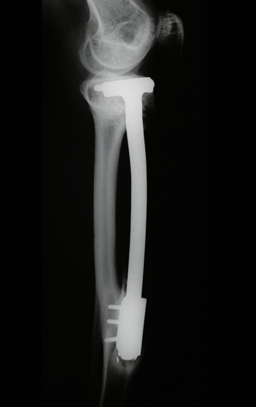
Figura 123: Radiografia de 21/10/2002, após três anos, com boa hipertrofia da fíbula e bom alinhamento do membro operado.
Figure 123: Radiograph of 10/21/2002, after three years, with good hypertrophy of the fibula and good alignment of the operated limb.
Figura 124: Radiografia perfil, de 21/10/2002, após três anos, boa hipertrofia da fíbula e alinhamento do membro operado.
Figure 124: Profile x-ray, dated 10/21/2002, after three years, good hypertrophy of the fibula and alignment of the operated limb.
Video 01: Normal function of the operated limb after three years, on 10/21/2002.
Figura 125: Carga total, monopodal, após três anos e dois meses, em 31/12/2002.
Figure 125: Total load, single leg, after three years and two months, on 12/31/2002.
Figura 126: Flexão com carga total, após três anos e dois meses, em 31/12/2002.
Figure 126: Flexion with full load, after three years and two months, on 12/31/2002.
Figura 127: Radiografia de 31/12/2002, após três anos e dois meses, com boa hipertrofia da fíbula e bom alinhamento do membro operado.
Figure 127: Radiograph from 12/31/2002, after three years and two months, with good hypertrophy of the fibula and good alignment of the operated limb.
Figura 128: Radiografia perfil, de 31/12/2002, após três anos e dois meses.
Figure 128: Profile x-ray, dated 12/31/2002, after three years and two months.
Figura 129: Após três anos e oito meses, função normal do membro operado, em 03/06/2003.
Figure 129: After three years and eight months, normal function of the operated limb, on 06/03/2003.
Figura 130: Função e estética após três anos e oito meses, em 03/06/2003.
Figure 130: Function and aesthetics after three years and eight months, on 06/03/2003.
Figura 131: Carga total monopodal, após três anos e oito meses, em 03/06/2003.
Figure 131: Total single-leg load, after three years and eight months, on 06/03/2003.
Figura 132: Flexão com carga total, aspecto frontal, após três anos e oito meses, em 03/06/2003.
Figure 132: Flexion with full load, frontal aspect, after three years and eight months, on 06/03/2003.
Figura 133: Radiografia de 03/06/2003, após três anos e oito meses, com boa hipertrofia da fíbula e bom alinhamento do membro operado.
Figure 133: Radiograph from 06/03/2003, after three years and eight months, with good hypertrophy of the fibula and good alignment of the operated limb.
Figura 134: Radiografia perfil, com excelente hipertrofia da fíbula, em 03/06/2003, após três anos e oito meses.
Figure 134: Profile x-ray, with excellent hypertrophy of the fibula, on 06/03/2003, after three years and eight months.
Figura 135: Radiografia perfil, com excelente hipertrofia da fíbula, em 03/06/2003, após três anos e oito meses.
Figure 135: Profile x-ray, with excellent hypertrophy of the fibula, on 06/03/2003, after three years and eight months.
Figura 136: Após quatro anos, função normal do membro operado, em 07/10/2003.
Figure 136: After four years, normal function of the operated limb, on 10/07/2003.
Figura 137: Extensão com carga total normal, após quatro anos, em 07/10/2003.
Figure 137: Extension with normal full load, after four years, on 10/07/2003.
Figura 138: Função simétrica, após quatro anos, em 07/10/2003.
Figure 138: Symmetric function, after four years, on 10/07/2003.
Figura 139: Flexão com carga total, aspecto frontal, após quatro anos, em 07/10/2003.
Figure 139: Flexion with full load, frontal aspect, after four years, on 10/07/2003.
Figura 140: Radiografia de 30/03/2004, após quatro anos e cinco meses, com boa hipertrofia da fíbula e bom alinhamento do membro operado.
Figure 140: Radiograph from 03/30/2004, after four years and five months, with good hypertrophy of the fibula and good alignment of the operated limb.
Figura 141: Radiografia perfil, com excelente hipertrofia da fíbula, em 30/03/2004, após quatro anos e cinco meses.
Figure 141: Profile x-ray, with excellent hypertrophy of the fibula, on 03/30/2004, after four years and five months.
Figura 142: Flexão com carga total, aspecto no perfil, função simétrica e normal, após quatro anos e cinco meses, em 30/03/2004.
Figure 142: Flexion with full load, profile appearance, symmetrical and normal function, after four years and five months, on 03/30/2004.
Video 2: Symmetrical and normal function of the lower limbs, after four years and five months, on 03/30/2004.
Figura 143: Após seis anos e dois meses, função normal do membro operado, em 13/12/2005.
Figure 143: After six years and two months, normal function of the operated limb, on 12/13/2005.
Figura 144: Carga total, monopodal, após seis anos e dois meses, função normal do membro operado, em 13/12/2005.
Figure 144: Full load, single leg, after six years and two months, normal function of the operated limb, on 12/13/2005.
Figura 145: Extensão com carga total normal, após seis anos e dois meses, em 13/12/2005.
Figure 145: Extension with normal full load, after six years and two months, on 12/13/2005.
Figura 146: Boa função, após três anos, em 13/12/2005.
Figure 146: Good function, after three years, on 12/13/2005.
Figura 147: Radiografia de 13/12/2005, após seis anos e dois meses, com boa hipertrofia da fíbula e bom alinhamento do membro operado.
Figure 147: Radiograph from 12/13/2005, after six years and two months, with good hypertrophy of the fibula and good alignment of the operated limb.
Figura 148: Radiografia perfil, de 13/12/2005, após seis anos e dois meses, boa hipertrofia da fíbula e alinhamento do membro operado.
Figure 148: Profile x-ray, dated 12/13/2005, after six years and two months, good hypertrophy of the fibula and alignment of the operated limb.
Figura 149: Flexão com carga total de 160º, após seis anos e dois meses, em 13/12/2005.
Figure 149: Flexion with full load of 160º, after six years and two months, on 12/13/2005.
Figura 150: Amanhecer no litoral paulista - novembro/2015.
Figure 150: Dawn on the coast of São Paulo - November/2015.
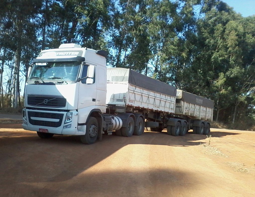
Figura 151: Modelo de caminhão trucado.
Figure 151: Truck model.

After the evaluation in 2005, ten years have passed. The patient, now 28 years old, returns on November 15, 2015. He currently works as a driver, transporting bulk sugar from the state of Minas Gerais to the port of Santos. He travels 800 km driving a truck, using his right leg to accelerate and brake. The reconstruction of the leg, carried out with autotransplantation of growth cartilage, proves to be an excellent alternative to endoprostheses for growing children, with good functional results. A biological, autologous and definitive solution.

Figura 152: Após 16 anos, função normal do membro operado, em 15/11/2015.
Figure 152: After 16 years, normal function of the operated limb, on 11/15/2015.
Figura 153: Extensão com carga total normal, após 16 anos, em 15/11/2015.
Figure 153: Extension with normal full load, after 16 years, on 11/15/2015.
Figura 154: Carga total, monopodal, após 16 anos, em 15/11/2015.
Figure 154: Total load, single leg, after 16 years, on 11/15/2015.
Figura 155: Flexão com carga total, após 16 anos, em 15/11/2015.
Figure 155: Flexion with full load, after 16 years, on 11/15/2015.
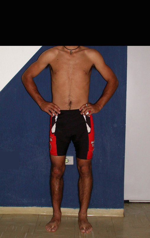
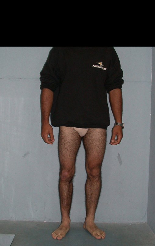
Figura 156: Paciente com os membros alinhados, em 15/11/2015, após 16 anos da cirurgia.
Figure 156: Patient with limbs aligned, on 11/15/2015, 16 years after surgery.
Figura 157: Aspecto clínico, frente, após 16 anos, em 15/11/2015.
Figure 157: Clinical appearance, front, after 16 years, on 11/15/2015.
Figura 158: Aspecto clínico, perfil, após 16 anos, em 15/11/2015.
Figure 158: Clinical appearance, profile, after 16 years, on 11/15/2015.
Video 3: Normal flexion and extension, patient well after 16 years.
Figura 159: Radiografia de 15/11/2015, após 16 anos, com excelente hipertrofia da fíbula e bom alinhamento do membro operado.
Figure 159: Radiograph from 11/15/2015, after 16 years, with excellent hypertrophy of the fibula and good alignment of the operated limb.
Figura 160: Radiografia perfil, com excelente hipertrofia da fíbula, já tibializada, em 15/11/2015, após 16 anos da cirurgia.
Figure 160: Profile x-ray, with excellent hypertrophy of the fibula, already tibialized, on 11/15/2015, 16 years after surgery.
Figura 161: Radiografia do tórax normal, em 15/11/2015.
Figure 161: Normal chest X-ray, on 11/15/2015.
Figura 162: Bom aspecto da cicatriz cirúrgica, membros alinhados, excelente trofismo muscular, após 16 anos.
Figure 162: Good appearance of the surgical scar, aligned limbs, excellent muscle trophism, after 16 years.
Figura 163: Carga total, com extensão e alinhamento normais, após 16 anos de tratamento.
Figure 163: Full load, with normal extension and alignment, after 16 years of treatment.
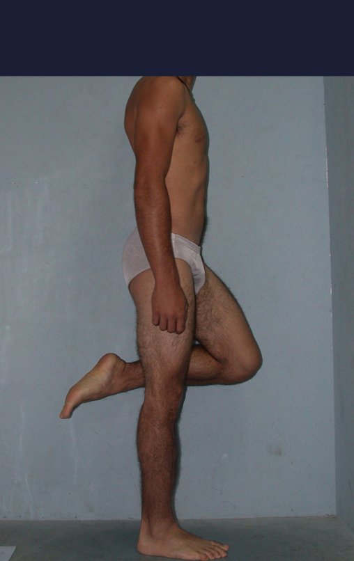
Figura 164: Carga total, monopodal, com membro alinhado, após 16 anos.
Figure 164: Full load, single leg, with limb aligned, after 16 years.
Figura 165: Flexão com carga total simétrica, após 16 anos. em 15/11/2015.
Figure 165: Flexion with symmetrical full load, after 16 years. on 11/15/2015.
Video 4: Patient does not report any functional limitations. Play sports and drive your truck without any difficulty. Assessment on 11/15/2015, 16 years after surgery.

See the article on this extensible internal fixation device, which we developed, as well as the use of this technique in the first two cases, which was published in the Revista Brasileira de Ortopedia – Vol. 36, No. 7 – July 2001, Figure 166. This complete article can be accessed and downloaded in PDF directly from the link below:

https://www.oncocirurgia.com.br/2015/08/19/dispósito-de-fixacao-interna-extensivel/

This technique, of growth cartilage autotransplantation, has been publicized at several national and international congresses in recent years.

We carried out research on Poodle dogs, in partnership with the Botucatu Veterinary College. This work resulted in a Master’s Thesis, in the Area of ​​Veterinary Medicine, in which we acted as Co-supervisor. This thesis was later published in an international journal, in ZEITSCHRIFTEN – VETERINARY AND COMPARATIVE ORTHOPAEDICS AND TRAUMATOLOGY – ARCHIVE – ISSUE 2 2008, Figure 167. This article can be accessed at the link below:

https://www.oncocirurgia.com.br/2015/11/07/use-of-extensible-internal-device-in-the-femur-of-young-dogs/

Figura 166: Artigo publicado na Revista Brasileira de Ortopedia - Vol. 36, Nº 7 - Julho de 2001.
Figure 166: Article published in the Brazilian Journal of Orthopedics - Vol. 36, No. 7 - July 2001.
Figura 167: Artigo publicado na VCOT - Veterinary and Comparative Orthopaedics and Traumatology 2008 21 2:133-139
Figure 167: Article published in VCOT - Veterinary and Comparative Orthopedics and Traumatology 2008 21 2:133-139

In 2016, we published an updated article with the evolution of this case, with sixteen years of follow-up,  see:

http://springerplus.springeropen.com/articles/10.1186/s40064-016-2042-7

Author: Prof. Dr. Pedro Péricles Ribeiro Baptista

 Orthopedic Oncosurgery at the Dr. Arnaldo Vieira de Carvalho Cancer Institute

Leave a Reply

Hello! How can we assist you?