This digital library houses the book on Oncology and Orthopedic Oncosurgery.

It includes academic lectures, presentations from national and international congresses, published papers, case discussions, performed surgical procedures, and proprietary techniques developed.

The digital format was chosen because the web allows the inclusion of texts with numerous visual resources, such as images and videos, which would not be possible in a printed book.

The content is intended for students, healthcare professionals, and the general public interested in the field.

Extendable internal fixation device

F591ed4b09492933c2de77c78c9d9a66 3
[auto_translate_button]
Extendable internal fixation device. The authors present a dynamic internal fixation method that consists of a stainless steel piece that has a flap adapted to the bone that will be stabilized and a channel that will contain one of the ends of the plate to be used in osteosynthesis. This fixation prevents rotational deviations, valgus, varus, antecurvatum and retrocurvatum, but does not block bone growth of the epiphysis that is fixed. This device, called “device for extensible internal fixation”, was developed at the “Pavilhão Fernandinho Simonsen”, by the Oncological Orthopedics Group of the Department of Orthopedics and Traumatology of Santa Casa de Misericórdia de São Paulo (DOT-SCMSP-SP) and is indicated for the surgical treatment of selected cases of aggressive tumor lesions, and can also be used in the treatment of other conditions, such as congenital malformations and sequelae of trauma or infections that require reconstructions with dynamic stabilization, without blocking bone growth.

Extendable internal fixation device

INTRODUCTION

The search for biological solutions that make it possible to resolve bone defects, caused by locally aggressive tumor or pseudotumor lesions, congenital malformations, traumas and infections, has increasingly aroused the interest of orthopedists(1).

Advances in therapeutic resources in the treatment of malignant bone tumors have provided greater survival and even the prospect of a cure for patients. On the other hand, there is the inherent complication of endoprostheses over time. The correlation of these factors therefore requires the improvement of biological reconstruction methods that aim to be definitive(1-4). Reviewing the literature regarding bone reconstructions in the developing skeleton, it can be seen that this subject is of current interest(1,2,4-6).

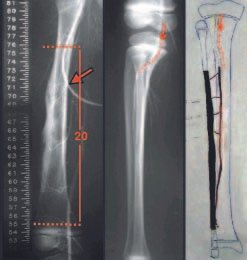
Fig. 1 – Arteriografia do fêmur, mensuração de 20cm de ressecção. Arteriografia da fíbula e esquema do enxerto vascularizado.
Fig. 1 – Arteriography of the femur, measurement of 20cm of resection. Arteriography of the fibula and diagram of the vascularized graft.
However, we did not find any publication that made reference to any form of internal fixation of long bones that would allow stabilizing the epiphysis with the diaphyseal segment (fixing the epiphyseal plate), but at the same time would not block its growth. Such a mechanism should somehow allow the osteosynthesis system to slide, so as not to impede bone growth in the stabilized segment. As a result of treating a patient with Ewing’s sarcoma, we developed an internal fixation device that maintains the stabilization of the reconstruction and, at the same time, allows the bone to grow, either through its own epiphyseal plate or through the epiphyseal plate of the bone graft. transported or transplanted by microsurgical technique. This device prevents rotational deviations, valgus, varus, antecurvatum and retrocurvatum, but does not block bone growth of the epiphyseal plate that has been stabilized. For better understanding, we will present a description of the first case treated with this device, as well as exemplifying its use and possibility of adaptation to other bone segments.
Fig. 2 – Acesso medial, facilitando as anastomoses. Detalhes da peça ressecada, da placa angulada e das anastomoses.
Fig. 2 – Medial access, facilitating anastomoses. Details of the resected piece, the angled plate and the anastomoses.
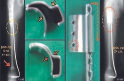
Fig. 3 – Após 8 meses da 1ª cirurgia – peças de aço inoxidável – encaixe da placa permitindo o deslizamento – RX após a 2ª cirurgia.
Fig. 3 – 8 months after the 1st surgery – stainless steel parts – plate fitting allowing sliding – RX after the 2nd surgery.

DESCRIPTION OF THE TECHNIQUE

In February 1999, the nine-year-old male LCAA patient was undergoing preoperative chemotherapy treatment for diaphyseal Ewing sarcoma of the right femur at the Pediatric Hematology and Oncology Service of the Department of Pediatrics and Child Care of Santa Catarina. Casa Misericórida de São Paulo.
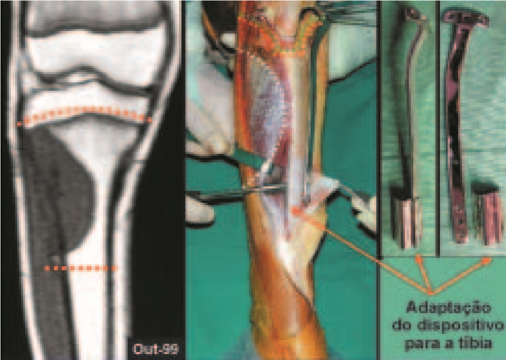
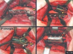
Fig. 4 – Acesso medial para retirada dos parafusos da placa angulada. Colocação da lâmina curva entre o fêmur e a placa. Aposição da segunda lâmina por sobre a primeira e a placa. Fixação do dispositivo com parafusos de anterior para posterior.
Fig. 4 – Medial access to remove the angled plate screws. Placement of the curved blade between the femur and the plate. Place the second blade over the first and the plate. Fixing the device with screws from anterior to posterior.
With the favorable response to neo-adjuvant chemotherapy, we decided to resect the affected segment and perform a biological reconstruction solution with transplantation of a vascularized contralateral fibula, using a microsurgical technique. To assess the extent of spinal involvement, we performed radiographs, bone scintigraphy, tomography and magnetic resonance imaging, pre- and post-chemotherapy, which safely showed us the possibility of resection of the diaphyseal segment, with preservation of the growth plate of the affected femur. In surgical planning, we calculated 20cm of resection, performed arteriography, chose the vessels for the anastomoses (fig. 1) and opted for a medial approach to facilitate resection, the placement of an angled plate fixing the femoral epiphysis and diaphysis and the vascular anastomoses through microsurgery (fig. 2). Eight months later, the control radiograph showed the bone transplant was fully integrated, both in the proximal and distal segments (fig. 3). However, the fibula had not yet obtained the necessary thickening to dispense with the protection of the angled plate. This osteosynthesis with an angled plate, fixed to the diaphysis and distal epiphysis of the femur, acted to block the growth of the bone provided by the distal epiphyseal plate of the femur, in addition to preventing the transported fibula from receiving the appropriate load request, in order to react and thicken it. faster.
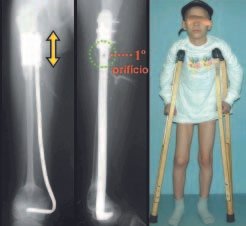
Fig. 5 – L.C.C.A., masculino, 9 anos. Dispositivo estabilizando a osteossíntese. Início do deslizamento da placa (aparece o primeiro orifício).
Fig. 5 – LCCA, male, 9 years old. Device stabilizing osteosynthesis. Start of plate sliding (first hole appears).
We are afraid to replace the stabilization method with external fixators, given its complications, both due to degenerative muscle injuries, trophic and functional changes they cause, and the risk of infection in patients undergoing chemotherapy. To resolve that situation, we requested the manufacture of a device consisting of two pieces of stainless steel that could be adapted to the proximal segment of the osteosynthesis, in order to maintain the support provided by the angled plate and at the same time allow it to slide and not block the bone growth (fig. 3). We performed a small medial surgical access, at the proximal end, removed the screws fixing the plate stem to the bone (directed from medial to lateral), and placed the curved blade between the femur and the plate, affixed the second blade shaped to adapt We placed it over the rod of the angled plate and screwed it in the anteroposterior direction (fig. 4).
Fig. 6 –Evidência de crescimento do osso e deslizamento da placa. Aparece o segundo “espaço de parafuso” – espessamento do enxerto – membros equalizados
Fig. 6 – Evidence of bone growth and plate slippage. The second “screw space” appears – thickening of the graft – equalized limbs
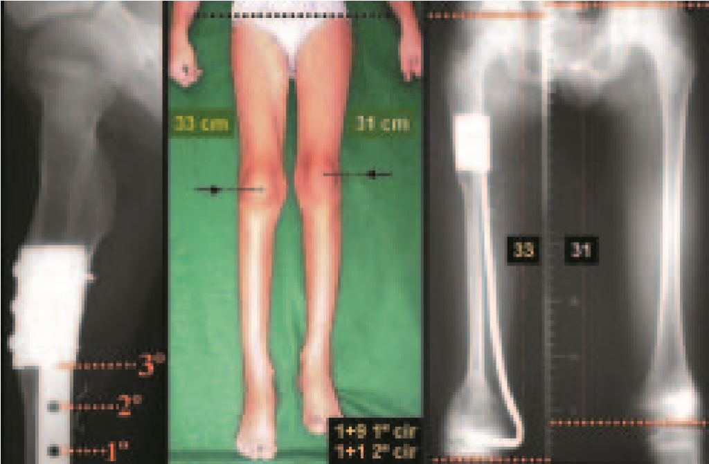
Fig. 7 – Continua o crescimento (aparece o terceiro orifício de parafuso). O fêmur operado cresceu mais que o outro lado. Escanograma confirmando.
Fig. 7 – Growth continues (third screw hole appears). The operated femur grew more than the other side. Scanogram confirming.
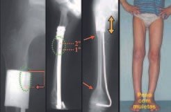
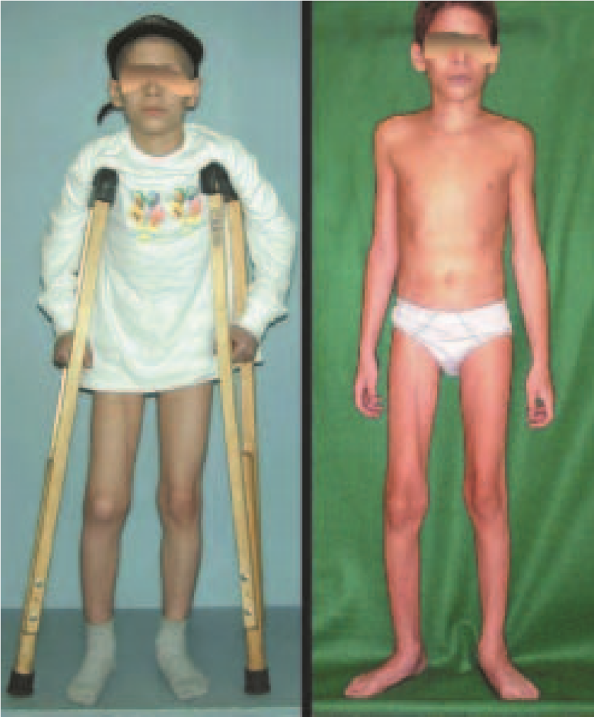
Fig. 8 – Pós-operatório de oito meses da primeira cirurgia (carga parcial) – joelhos desnivelados (maior à direita), carga total (1 ano e 1 mês da 2ª cirurgia).
Fig. 8 – Eight months post-operative period after the first surgery (partial weight-bearing) – unlevel knees (larger on the right), full weight-bearing (1 year and 1 month after the 2nd surgery).
In this way, we obtained good stability in the sense of blocking the efforts of rotational, varus, valgus, retrocurvatum or antecurvatum movements, but allowing the plate stem to slide as bone growth occurred (fig. 5).
Fig. 9 – R.N.M. determinando nível da ressecção, sacrificando a placa de crescimento da tíbia. Detalhe do periósteo recobrindo a lesão, dissecção do tendão patelar e músculo tibial anterior. Placa especial confeccionada e modelada para o paciente e dispositivo ocluído no extremo distal, com a aba de fixação angulada para adaptar-se ao formato triangular da tíbia.
Fig. 9 – MRI determining the level of resection, sacrificing the tibial growth plate. Detail of the periosteum covering the injury, dissection of the patellar tendon and anterior tibialis muscle. Special plate made and shaped for the patient and device occluded at the distal end, with the fixation flap angled to adapt to the triangular shape of the tibia.
In the fourth month postoperatively, after the placement of the extensible device (second surgery), we were able to verify the growth spurt and the sliding of the angled plate rod (fig. 6), at a distance of approximately one “screw space” ( fig. 5). The patient began partial weight bearing, walking with the aid of axillary crutches (fig. 5). In the control radiograph one year after placement of the device for extensible internal fixation, we were able to observe the continued sliding (fig. 6) of the angled plate rod, in which we visualized the advancement of yet another “screw space” (fig. 6). The fibula becomes thicker (fig. 6) and the patient increases weight on the operated limb (fig. 6). In the thirteenth month the third hole in the plate begins to appear (fig. 7). During the clinical examination of the patient (fig. 7), we were able to observe that the operated side grew two centimeters more than the non-operated side, confirmed by scanometry (fig. 7). This greater growth was due to the stimuli caused by the first surgery, the vascularized graft and the second surgery (placement of the extensible device). We observed that there has been an equalization in the size of members and we believe that at the end of the growth the members will be the same size or the difference will be minimal. The patient began to walk with full load (and 2cm compensation), and the longer operated side can be seen, where it can be seen that the knee level is lower on the operated side (fig. 8).
Fig. 10 – Emprego da fíbula proximal, com sua placa de crescimento, detalhe da inclinação em valgo do planalto tibial. Paciente com carga parcial, detalhe clínico do joelho em valgo. RX após 14 meses, com crescimento de 0,75cm pela placa fisária da fíbula e correção da angulação do planalto tibial. Paciente com carga e com correção clínica do valgo.
Fig. 10 – Use of the proximal fibula, with its growth plate, detail of the valgus inclination of the tibial plateau. Patient with partial weight bearing, clinical detail of the knee in valgus. X-ray after 14 months, with growth of 0.75cm through the fibular physeal plate and correction of the angulation of the tibial plateau. Patient with weight bearing and clinical valgus correction.
Fig. 11 – W.R.C., 15 anos, PO 18 meses. Paciente em crescimento, membros equalizados e alinhados. Boa função do joelho.
Fig. 11 – WRC, 15 years, PO 18 months. Patient growing, limbs equalized and aligned. Good knee function.
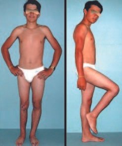
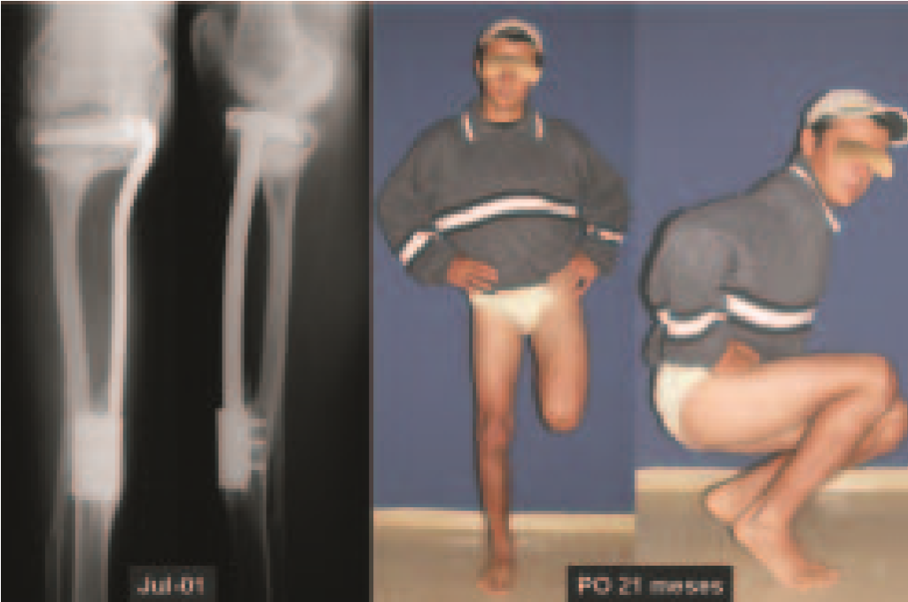
Fig. 12 – W.R.C., 16 anos. Pós-operatório 21 meses. Radiografia mostrando o fechamento da linha epifisária. Patiente com carga total e flexão do joelho.
Fig. 12 – WRC, 16 years old. Post-operative 21 months. Radiograph showing closure of the epiphyseal line. Patient with full load and knee flexion.

We currently use this device for immediately extensible internal fixation, and it currently consists of a single piece that has a curved side flap to adapt to the femur and humerus or it can be flat with an angle to adapt to the shape. triangular shape of the tibia (fig. 9). Another patient, WRC, 14 years old, with osteosarcoma of the right tibia, is a relatively recent example of reconstruction of the proximal metaphyseal segment of the tibia, with resection that also included the growth plate; We performed reconstruction with the fibula, including its epiphysis and using the epiphyseal plate of this fibula to provide growth (fig. 9). We can observe a slip of 0.75 cm by comparing the distances between the physeal plate of the transported fibula and the limit of the extensible device. Radiographic correction of the valgus inclination of the tibial plateau and clinical realignment of the knee can be seen (fig. 10). The patient is currently in the final phase of growth and has good knee function (Figs. 11 and 12).

COMMENTS

We believe that this device for extensible internal fixation, which we developed, can be used both for the treatment of selected cases of aggressive tumor lesions and also for other conditions, such as congenital malformations and sequelae of trauma or infections, which may require reconstructions that require a stabilization mechanism that allows the epiphysis to be fixed, but without blocking bone growth.

REFERENCES

1. Manfrini M., Gasbarrini A., Malaguti C., et al: Intraepiphyseal resection of the proximal tibia and its impact on lower limb growth. Clin Orthop 358: 111-119, 1999. 2. Eckardt JJ, Kabo JM, Kelley CM, et al: Expandable endoprosthesis reconstruction in skeletally immature patients with tumors. Clin Orthop 373: 51-61, 2000. 3. Capanna R., Bufalini C., Campanacci M.: A new technique for reconstructions of large metadiaphyseal bone defects. Orthop Traumatol 2:159-177, 1993. 4. Cool WP, Carter SR, Grimer RJ, Tillman RM, Walker PS: Growth after extendible endoprosthetic replacement of the distal femur. J Bone Joint Surg [Br] 79: 938-942, 1997. 5. Baptista PPR, Guedes A., Reggiani R., Lavieri RF, Lopes JAS: Tibialization of the distal fibula with preservation of the epiphyseal plate: preliminary report. Rev Bras Ortop 33: 841-846, 1998. 6. Baptista PPR, Guedes A., Reggiani R., Lavieri RF, Pires CEF: Tibialization of the fibula: description of the surgical approach. Rev Bras Ortop 33: 861-866, 1998.

Click here to download the PDF 

Author: Prof. Dr. Pedro Péricles Ribeiro Baptista

 Orthopedic Oncosurgery at the Dr. Arnaldo Vieira de Carvalho Cancer Institute

Leave a Reply

Hello! How can we assist you?