This digital library houses the book on Oncology and Orthopedic Oncosurgery.

It includes academic lectures, presentations from national and international congresses, published papers, case discussions, performed surgical procedures, and proprietary techniques developed.

The digital format was chosen because the web allows the inclusion of texts with numerous visual resources, such as images and videos, which would not be possible in a printed book.

The content is intended for students, healthcare professionals, and the general public interested in the field.

Ewing sarcoma of the humerus in children

54e0e46b6647aa736c13ef9d09eab432 3
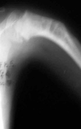
Ewing sarcoma of the humerus in children. A four-year-old and five-month-old patient presented with pain and a tumor in the left humerus in January 1991. The biopsy revealed Ewing’s sarcoma. Staging did not reveal another focus. He underwent treatment with neoadjuvant chemotherapy, showing a good radiographic response to treatment, with mineralization of the lesion and angular deformity due to neoplastic plasticity, figures 1 to 4.

Ewing sarcoma of the humerus in a child – Management – ​​Resection and reconstruction techniques with special plate – Combined autologous fibula and iliac graft

Figura 1: Radiografia do úmero, frente, com lesão de rarefação óssea metafisária, comprometendo a placa de crescimento, em 10/02/1991.
Figure 1: Radiograph of the humerus, front, with metaphyseal bone rarefaction lesion, compromising the growth plate, on 02/10/1991.
Figura 2: Radiografia oblíqua, mostrando que a lesão estende-se até o meio da diáfise.
Figure 2: Oblique radiograph, showing that the lesion extends to the middle of the diaphysis.
Figura 3: Radiografia frente, após a quimioterapia neoadjuvante, mostrando boa resposta radiográfica, em 15/05/1991.
Figure 3: Front radiograph, after neoadjuvant chemotherapy, showing good radiographic response, on 05/15/1991.
Figura 4: Radiografia em perfil, após a QT neoadjuvante, com deformidade angular, em 15/05/1991.
Figure 4: Profile x-ray, after neoadjuvant CT, with angular deformity, on 05/15/1991.
Figura 5: Cirurgia em 20/05/1991, dissecção de pele e tecido celular subcutâneo e exposição do segmento de úmero a ser ressecado. Nervo radial isolado e estudo da colocação da placa.
Figure 5: Surgery on 05/20/1991, dissection of skin and subcutaneous cellular tissue and exposure of the humerus segment to be resected. Isolated radial nerve and plate placement study.
Figura 6: Ressecção do segmento metadiafisário, incluindo a placa de crescimento e posicionamento da lâmina da placa, na fenda feita na cabeça umeral, previamente à retirada do tumor.
Figure 6: Resection of the metadiaphyseal segment, including the growth plate and positioning of the plate blade, in the gap made in the humeral head, prior to tumor removal.
Figura 7: Enxerto autólogo de fíbula para a reconstrução.
Figure 7: Autologous fibula graft for reconstruction.
Figura 8: Reconstrução da falha óssea com enxertos autólogos de segmento de fíbula, (seta azul) e tricortical do ilíaco (seta amarela).
Figure 8: Reconstruction of the bone defect with autologous fibula segment (blue arrow) and tricortical iliac (yellow arrow) grafts.
Figura 9: Foto do segmento ósseo ressecado, com margem oncológica.
Figure 9: Photo of the resected bone segment, with oncological margin.
Figura 10: Corte da peça, fixada em formol, sendo preparado para descalcificação.
Figure 10: Section of the piece, fixed in formaldehyde, being prepared for descaling.
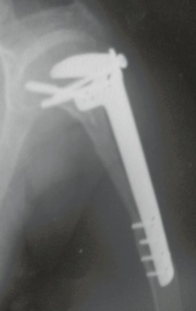
The control radiograph one month postoperatively and the function of the operated limb are shown in figures 11 to 14.
Figura 11: Radiografia do primeiro mês pós-operatório.
Figure 11: Radiograph of the first month after surgery.
Figura 12: Radiografia do pós-operatório de um mês, reconstrução com enxerto autólogo de fíbula (seta azul), e de crista ilíaca (seta amarela).
Figure 12: One-month postoperative radiograph, reconstruction with autologous fibula graft (blue arrow) and iliac crest graft (yellow arrow).
Figura 13: Paciente em quimioterapia adjuvante, no segundo mês após a cirurgia, boa cicatrização da ferida operatória, boa rotação interna do MSE.
Figure 13: Patient undergoing adjuvant chemotherapy, in the second month after surgery, good healing of the surgical wound, good internal rotation of the MSE.
Figura 14: Pós-operatório de dois meses, elevação ativa do MSE.
Figure 14: Two months postoperatively, active elevation of the MSE.
Figura 15: Radiografia de controle em 15/07/1991, frente.
Figure 15: Control radiograph on 07/15/1991, front.
Figura 16: Radiografia de controle em 15/07/1991, perfil.
Figure 16: Control radiograph on 07/15/1991, profile.
Figura 17: Paciente após três anos e dez meses da cirurgia, boa rotação externa.
Figure 17: Patient three years and ten months after surgery, good external rotation.
Figura 18: Paciente após três anos e dez meses da cirurgia, observe o encurtamento do lado operado, em 19/06/1995.
Figure 18: Patient three years and ten months after surgery, observe the shortening of the operated side, on 06/19/1995.
Figura 19: Paciente após três anos e dez meses da cirurgia, elevação ativa do membro operado.
Figure 19: Patient three years and ten months after surgery, active elevation of the operated limb.
Figura 20: Paciente após 21 anos da cirurgia, observe o encurtamento do MSE, em 11/11/2011.
Figure 20: Patient 21 years after surgery, observe the shortening of the MSE, on 11/11/2011.
Figura 21: Radiografia de controle em 11/11/2011, oblíqua.
Figure 21: Control radiograph on 11/11/2011, oblique.
Figura 22: Radiografia de controle em 11/11/2011, frente.
Figure 22: Control x-ray on 11/11/2011, front.
Figura 23: Após 22 anos da cirurgia. Observem a estética e a discrepância dos MMS.
Figure 23: 22 years after surgery. Note the aesthetics and discrepancy of MMS.
Figura 24: Rotação interna normal do membro operado, após 22 anos da cirurgia, em 11/01/2012.
Figure 24: Normal internal rotation of the operated limb, 22 years after surgery, on 01/11/2012.
Figura 25: Boa função do MSE, após 22 anos da cirurgia.
Figure 25: Good function of the MSE, 22 years after surgery.
Figura 26: Elevação ativa dos MMSS, encurtamento do úmero, porém com boa função.
Figure 26: Active elevation of the upper limbs, shortening of the humerus, but with good function.
Figura 27: Aspecto no perfil, após 22 anos da cirurgia.
Figure 27: Profile appearance, 22 years after surgery.
Figura 28: Aspecto da cicatriz da perna esquerda, local de retirada do segmento de fíbula, enxerto autólogo.
Figure 28: Appearance of the scar on the left leg, site of removal of the fibula segment, autologous graft.
Video 1: Good aesthetics, despite shortening, good function, 22 years after surgery, on 01/11/2012.
Figura 29: Radiografia de controle, oblíqua, após 22 anos.
Figure 29: Control radiograph, oblique, after 22 years.
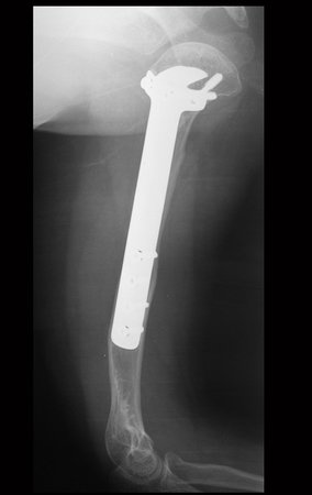
Figura 30: Radiografia de controle, perfil, após 22 anos.
Figure 30: Control X-ray, profile, after 22 years.
Figura 31: Radiografia da bacia, frente, local da retirada de enxerto da crista ilíaca esquerda, após 22 anos.
Figure 31: X-ray of the pelvis, front, site of graft removal from the left iliac crest, after 22 years.
Figura 32: Radiografia da perna esquerda, mostrando a reconstituição da fíbula, no sítio de retirada do enxerto.
Figure 32: Radiograph of the left leg, showing the reconstruction of the fibula, at the graft removal site.
Figura 33: Cintilografia óssea de controle, frente, após 22 anos, em 12/02/2012.
Figure 33: Control bone scintigraphy, front, after 22 years, on 02/12/2012.
Figura 34: Cintilografia óssea de controle, posterior, após 22 anos, em 12/02/2012.
Figure 34: Control bone scintigraphy, later, after 22 years, on 02/12/2012.
Figura 35: Boa função e estética, em outubro de 2012, após 22 anos de pós-operatório.
Figure 35: Good function and aesthetics, in October 2012, after 22 years post-surgery.
Figura 36: Paciente em julho de 2014. Esteticamente e funcionalmente bem.
Figure 36: Patient in July 2014. Aesthetically and functionally well.

As figuras 37 a 42, ilustram etapas da evolução deste caso de Sarcoma de Ewing, tratado cirurgicamente com uma solução biológica.

Figura 37: Em 15/07/1991, com cinco anos de idade, realizando quimioterapia adjuvante, no pós-operatório de dois meses.
Figure 37: On 07/15/1991, at the age of five, undergoing adjuvant chemotherapy, two months post-operatively.
Figura 38: Em 19/06/1995, no pós-operatório de três anos e dez meses.
Figure 38: On 06/19/1995, three years and ten months after surgery.
Figura 39: Adolescente com encurtamento do lado esquerdo, devido à ressecção da placa de crescimento proximal do úmero. Esteticamente e funcionalmente bem.
Figure 39: Adolescent with shortening on the left side, due to resection of the proximal growth plate of the humerus. Aesthetically and functionally well.
Figura 40: Paciente bem, sem queixas, aspecto estético em 2013, pós-operatório de 23 anos.
Figure 40: Patient well, no complaints, aesthetic appearance in 2013, 23 years post-operative.
Figura 41: Aspecto da cicatriz cirúrgica no úmero esquerdo, funcionalmente bem, em 2013.
Figure 41: Appearance of the surgical scar on the left humerus, functionally well, in 2013.
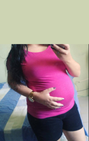
Figura 42: Após 24 anos de pós-operatório, paciente grávida, quinto mês de gestação, boa função do MSE, em fevereiro de 2015.
Figure 42: After 24 years post-surgery, pregnant patient, fifth month of pregnancy, good MSE function, in February 2015.
In May 2015, the patient had her first child, giving birth to a boy. In 1991, we had not yet performed autotransplantation of growth cartilage, reconstructing this segment with a vascularized fibula with the growth plate, to replace the humerus plate, which when resected. However, the upper limb better accepts the length discrepancy, crowning the alternative we used at the time.

Author: Prof. Dr. Pedro Péricles Ribeiro Baptista

 Orthopedic Oncosurgery at the Dr. Arnaldo Vieira de Carvalho Cancer Institute

Leave a Reply

Hello! How can we assist you?