This digital library houses the book on Oncology and Orthopedic Oncosurgery.

It includes academic lectures, presentations from national and international congresses, published papers, case discussions, performed surgical procedures, and proprietary techniques developed.

The digital format was chosen because the web allows the inclusion of texts with numerous visual resources, such as images and videos, which would not be possible in a printed book.

The content is intended for students, healthcare professionals, and the general public interested in the field.

Chapters

Ad92350e4633462fd6eb5956da8876f2 3

Gigantocellular tumor

[auto_translate_button]

Giant Cell Tumor

Giant Cell Tumor.

Gigantocellular tumor is a neoplasm of mesenchymal nature, characterized by the proliferation of giant multinucleated cells – gigantocytes – that resemble osteoclasts, amidst a stroma of mononucleated cells.

Due to this histological aspect present in several other processes, the gigantocellular tumor could be confused, often requiring analysis of the clinical and radiographic appearance to confirm its diagnosis.

The giant cell tumor is also known by the acronym TGC, by the names giant cell tumor and osteoclastoma.

The main manifestation is intermittent local pain, accompanied or not by an increase in volume in the affected region and limitation of movement in the neighboring joint. The length of history, on average 6 to 12 months, varies from case to case and depends on the affected bone.

Some patients seek treatment due to pain, others because of the perception of the tumor or a pathological fracture. It is not uncommon to refer to more or less intense trauma as the beginning of the clinical history.

As the tumor is most often epiphyseal, clinical involvement of the neighboring joint is frequent, with progressive functional impotence.

Joint effusion and clinical symptoms simulating meniscal or arthritis processes may occur.

In tumors located in the spine and sacrum, in addition to pain and increased volume, neurological manifestations may occur. The volume is sometimes enormous, predominating over the other symptoms.

The gigantocellular tumor generally affects a single bone. When there are lesions in several bones, the possibility of it being a “brown tumor of hyperparathyroidism” must be checked, whose lesions have similar radiographic aspects, but are multiple and the patient presents alterations in calcium and phosphorus.

GCT is classically known as a tumor of the epiphysis of long bones, most common in the knee region, that is, in the distal epiphysis of the femur and proximal epiphysis of the tibia and, then, in order of frequency, in the proximal region of the humerus and distal region of the radius. . It is rare in the axial skeleton and when it occurs, it predominates in the sacrum.

It occurs in the third and fourth decade, affecting both sexes equally.

Radiographically, it is described as an epiphyseal lesion characterized by bone rarefaction, initially eccentric, initially respecting the cortical limits. As it progresses, cortical rupture and joint involvement may occur (fig. 27).

Histologically, giant cells and the stroma are the most important elements of this tumor. It is characterized by having numerous giant cells that resemble osteoclasts in a stroma of spindle cells.

The main clinical, radiographic and anatomopathological differential diagnoses are aneurysmal bone cyst, teleangectatic osteosarcoma and chondroblastoma.

         The treatment of giant cell tumor is currently well established. Whenever possible, segmental resection of the lesion should be chosen, with an oncological safety margin in both bone and soft tissues. This way the surgeon will provide the greatest opportunity for cure, without risk of recurrence.

         However, in regions where segmental resection is not feasible, cervical spine for example, the most judicious endocavitary curettage possible must be performed and complemented with adjuvant therapy such as the CO 2 laser , phenol diluted in 4% alcohol, the liquid nitrogen and electrothermia. Methylmethacrylate has a low adjuvant effect and, when used to fill the cavity, it must be preceded by one of the therapies mentioned.

         In the past, curettage had high recurrence rates due to the lack of large openings for effective cleaning and the lack of use of local adjuvants. Today, when endocavitary curettage is indicated, it is recommended that a large bone window be created to allow the surgeon a broad view of the cavity. We also complement this curettage with the use of an electric scalpel (electrothermia).

         This electrothermal technique is very effective because with the curved tip of the scalpel we can reach areas that are more difficult to access. This scalpel, in addition to cauterization, also performs a complement to curettage, as those tumor cells, remaining in the small “cavities” of the bone wall, are destroyed and become detached, allowing for easier removal.

         We complement our curettage, after using electrothermia, with milling the cavity. For this we use the  Lentodril , with a spherical dental milling cutter.

         In the knee region (distal end of the femur and proximal end of the tibia), the site with the highest incidence of TGC, we frequently recommend endocavitary curettage, followed by electrothermia and milling with  Lentodril . This is because segmental resection of this region would imply arthrodesis or replacement with endoprostheses or homologous grafts. Arthrodesis of the knee joint causes the greatest limitation for the patient and should be avoided. Replacements in young patients can result in problems in the near future, and their indication must be judicious.

         Therefore, we first recommend the most conservative therapy: – curettage followed by adjuvant therapy for this region, except in advanced cases, with significant destruction of the bone structure, in which both the function and local control of the disease may be compromised.

         A brief comment remains regarding filling the treated cavity. This can be done with autologous or heterologous bone graft or with methyl methacrylate, each of them has its advantages and disadvantages.

         Methyl methacrylate makes it possible to visualize possible recurrences easily, is easy to use and allows for earlier loading, however it is not a biological solution and fractures may occur in the region.

The bone graft is a biological and definitive solution, however it makes it difficult to visualize possible early recurrence, which can be confused with reabsorption/integration of the graft and still requires six months on average for full load. The homologous graft has a longer integration period, is not always available, but on the other hand it shortens surgical time. The autologous graft has the advantage of biocompatibility and faster integration, but it prolongs surgical time.

Author: Prof. Dr. Pedro Péricles Ribeiro Baptista

 Orthopedic Oncosurgery at the Dr. Arnaldo Vieira de Carvalho Cancer Institute

7cf68b210274ef46d38b0cd76e059af6 3

Soft Tissue Sarcoma

[auto_translate_button]
Soft Tissue Sarcoma. Orthopedic oncological surgery covers  the treatment of musculoskeletal injuries, including benign and malignant bone neoplasms, pseudotumor lesions and benign and malignant neoplasms of soft tissues.

Soft Tissue Sarcoma

    Soft tissue sarcoma is a malignant neoplasm, derived from the  mesenchyme  that occurs in soft tissues, such as muscles, fascia, tendons, etc. It is not called carcinoma because this name refers to malignant neoplasms that have their embryonic origin in the ectoderm, such as the skin, nerves, viscera or glands.

            This neoplasm arises from a mesenchymal cell that undergoes mutation and begins to have an anomalous cellular reproduction, producing polymorphic cells, with varying sizes, hyperchromatic, resulting in an atypical tissue, therefore malignant.

            Their nomenclature is given by their similarity to the tissue they try to reproduce. Thus, for example, when atypical neoformed tissue presents an appearance similar to fatty tissue, it is called liposarcoma.

Etiology:

           Most soft tissue sarcomas do not have a defined etiology, but some risk factors are well described, such as previous radiotherapy, lymphedema, Li-Fraumeni syndrome, type I neurofibromatosis, individual genetic propensity and HIV infection.

Incidence:

            It is a rare tumor, as the group of all these sarcomas comprises approximately 12 percent of pediatric neoplasms, with the “small round cell” pattern predominating, such as embryonal rhabdomyosarcoma and primitive peripheral neuroectodermal tumor, and only one percent of all adult malignant tumors  1,2 , Table 1.

            It is estimated that there are 12 thousand new cases of soft tissue sarcomas per year in the USA, causing around 4700 deaths  1 .

            About 60 percent of soft tissue sarcomas arise in the limbs, with the majority of them occurring in the thigh. It also occurs in the chest wall and retroperitoneum. In order of frequency, soft tissue sarcomas appear in the lower limb (46%), back (18%), upper limb (13%), retroperitoneum (13%) and head and neck (9%) 1 .

Quadro 1: Sarcomas de tecidos moles mais frequentes na população adulta e pediátrica
Table 1: Most common soft tissue sarcomas in the adult and pediatric population

Classification:

           The World Health Organization (WHO) classifies soft tissue sarcoma according to the histological subtype that the neoplasm resembles, such as liposarcoma, synovial sarcoma, rhabdomyosarcoma, etc. 1

            In some cases, the histology is uncertain and the morphological aspect is used to classify them as alveolar sarcoma or clear cell sarcoma.

          The most common soft tissue sarcomas in adults are: undifferentiated, liposarcoma, synovial sarcoma, leiomyosarcoma and malignant tumor of the peripheral nerve sheath, which is included in this chapter, despite originating in the ectoderm, as it presents biological behavior , treatment and prognosis similar to soft tissue sarcomas  1,2 .

            Histological grade is also used to classify soft tissue sarcomas, being classically divided into  Grade 1 ,  well differentiated  with a low degree of histological malignancy;  Grade 2 ,  moderately differentiated  and  Grade 3 ,  poorly differentiated  with a high degree of malignancy  1 .

 Clinical condition:

           The initial clinical picture is palpable tumor bulging, often painless, with progressive growth that can reach considerable size, especially in the thigh.

            Some patients may eventually experience pain and paresthesia due to the compressive effect of the tumor. They are clinically eutrophic in most cases, with fever or weight loss being exceptional symptoms.

          Soft tissue sarcomas tend to grow between adjacent muscles, pushing and compressing the surrounding structures, rarely extending beyond the anatomical compartments. Growth speed varies between histological subtypes.

          There are no absolute clinical criteria to differentiate benign from malignant soft tissue tumors. However, all deep tumors, that is, below the plane of the muscular fascia, and superficial tumors greater than 5 cm, have a high probability of being malignant  2 .

Staging:

           At the time of diagnosis, soft tissue sarcoma rarely metastasizes, not exceeding 10 percent of cases. This occurs more frequently in large volume tumors, deep to the muscular fascia and of high grade  2 .

            The dissemination pattern for most sarcomas is hematogenous and the main site of metastasis is the lung. For this reason, all patients with soft tissue sarcoma must undergo chest staging. Chest tomography to search for lung metastasis should always be indicated, especially for high-risk patients, such as superficial lesions larger than 5 cm, deep tumors and those with a high degree of malignancy.

            Regional lymph nodes are the site of metastases in 2.6% of cases, however, in some histological subtypes these may be more frequent and should be investigated, such as rhabdomyosarcoma, synovial sarcoma, epithelioid sarcoma, clear cell sarcoma and angiosarcoma  2 .

            Tomography of the abdomen and pelvis is recommended, especially in the staging of round cell soft tissue sarcoma and myxoid liposarcoma, due to the high rate of extrapulmonary metastasis to the abdomen and retroperitoneum. In the case of angiosarcoma, evaluation of the central nervous system, through skull tomography, is necessary due to the propensity for metastasis to the brain.

 Imaging exams:

 a)  Radiography:

            Radiography is the cheapest and most accessible imaging test. It can be used as the first line of tumor evaluation, to differentiate a tumor of skeletal origin from a soft tissue sarcoma. Furthermore, it may show calcifications inside.

b) Magnetic resonance imaging:

          Magnetic resonance imaging is still the test of choice to evaluate soft tissue sarcomas when there are no metallic elements, such as an orthopedic prosthesis. It can detail the limits of the tumor and its relationship with neighboring structures. The use of contrast can also differentiate solid lesions from lesions filled with liquid, and the examination using contrast should be requested.

c) Tomography:

          Tomography is the exam of choice in retroperitoneal tumors and in the search for pulmonary metastasis, as resonance is not adequate due to respiratory movement and intestinal peristalsis. In the study of MTS of extremities, resonance is superior in delineating neoplastic extension  2 .

d) Pet-Cet:

          Fluorine deoxyglucose positron emission tomography (FDG-PET/CT) allows, with high sensitivity, to differentiate high-grade soft tissue sarcoma from benign soft tissue tumors. However, it loses value in differentiating between low-grade and low-grade tumors. intermediaries  2 .

            FDG-PET/CET should not be used routinely in the initial evaluation of a patient with tissue tumor, however its use is indicated in determining prognosis and response to chemotherapy treatment  , 3 .

            The  standaruptakevalue  (SUV) is a quantification of fluorine-labeled glucose consumed by the tumor and demonstrates tumor metabolism. Soft tissue sarcoma with SUV equal to or greater than 6, which after chemotherapy shows a reduction in this value of less than 40 percent, presents a higher risk of systemic recurrence of the disease  2 .

e) Scintigraphy:

            Bone scintigraphy is not always performed in the initial staging of soft tissue sarcoma, as bone metastases in adults are infrequent in the initial stage of the disease. The exception is round cell tumors and myxoid liposarcoma, but skeletal mapping can be false negative and evaluation using magnetic resonance imaging is recommended  2 .  

Biopsy:

           Biopsy is indicated for the histological diagnosis of MTS, since imaging tests do not present characteristics suggestive of the histological subtype, as we can infer in some bone neoplasms, such as osteosarcoma.

            A biopsy is not always necessary for treatment, as the treatment of MTS is still primarily surgical and, in some cases, the biopsy can be replaced by excisional resection or also called excisional biopsy.

            Superficial tumors, above the muscular fascia, smaller than 5 cm, have a low probability of being malignant. In these cases, they can be resected without histological diagnosis if they are subject to oncological surgery  (wide resection with an oncological margin, without compromising surrounding noble structures) , as this would be the correct treatment in the event that we are dealing with a high-quality soft tissue sarcoma. degree.

            The biopsy must be performed by the surgeon who will carry out the definitive treatment or be guided by him. The biopsy path must be in line with the incision that will be used for resection and this entire biopsy path must be removed en bloc, along with the tumor.

            Incisional biopsy, performed through a surgical incision, exposing part of the tumor, was the most used for the diagnosis of soft tissue sarcoma. Currently, percutaneous biopsies have gained ground with the development of appropriate needles, such as the Trucut®, and imaging methods such as ultrasound and tomography to guide them.

Pathology:

            The pathologist must always be present to perform the frozen section examination, in order to confirm and ensure that the material collected is representative of the lesion, avoiding the need to repeat the procedure. The definitive histological result must await paraffin histology and possibly immunohistochemistry.

            Percutaneous needle biopsy, performed with these precautions, generally makes it possible to differentiate malignant tumors from benign ones and experienced pathologists are able to correctly determine the histological grade of the tumors in 97.6 and 86.3 percent respectively, in addition to this technique causing less local dissemination of the tumor. injury during the procedure  3 .

Soft tissue sarcoma – Treatment:

           The treatment of soft tissue tumors is generally surgical, however, as with most neoplasms, monitoring of cancer patients must always be multidisciplinary.

            From diagnosis to staging and treatment, a team of several professionals interacts in managing the case, such as the orthopedist who will perform the  oncological surgery , the pathologist, the radiologist, the clinical oncologist, the radiotherapist, the psychologist, the social worker, etc.

          The objective of treatment is to preserve the patient’s life, avoiding local recurrence, maximizing the function of the affected limb and minimizing treatment morbidity.

Chemotherapy:

          Soft tissue sarcomas, in the pediatric population, respond to systemic chemotherapy by presenting a greater response to drugs, providing an improvement in survival, as the child is able to withstand the doses of chemotherapy necessary to control the disease, unlike adults.

            In children, the most common soft tissue sarcomas are rhabdomyosarcoma, the extraosseous form of osteosarcoma, and the extraosseous form of Ewing’s sarcoma. All of these with neoadjuvant and adjuvant chemotherapy protocols.

            In the rare cases of these neoplasms occurring in adults, the pediatric chemotherapy protocol is chosen, at doses supported for adults and their possible comorbidities.

          In adult soft tissue sarcomas, there is great controversy regarding the use of chemotherapy. The meta-analysis published in 1997 by the  Sarcoma Meta-analysiscollaboration  showed that the use of doxorubicin in these patients increased the time free from local recurrence and distant metastasis, but there was no statistically significant benefit in the patients’ overall survival  3 .

            A new updated meta-analysis was published in 2008 demonstrating that the use of doxorubicin associated with ifosfamide promoted an improvement in patient survival  3 . However, chemotherapy is extremely toxic, especially in the adult population where the presence of clinical comorbidities can make effective treatment unfeasible. The current recommendation is to individualize each case, remembering that systemic treatment does not correct an inadequate surgical resection.

            In general, adult MTS are poor responders to chemotherapy. Treatment, therefore, relies on surgical resection.

Oncological surgery:

 Soft tissue sarcoma resection surgery must be performed with wide margins, so that the entire tumor is removed en bloc and covered by healthy tissue, aiming to ensure that there are no residual neoplastic cells after resection.

          Soft tissue sarcomas have a surrounding pseudocapsule. This structure is represented by an inflammatory process produced by tumor aggression and is not always a sufficient barrier for neoplastic cells, which permeate the surrounding tissues. Adjuvant radiotherapy may be indicated to act on the margins when they need to be small, due to the need to preserve noble structures, such as vessels and nerves.

            Tumor resection through the pseudocapsule can leave microscopic neoplastic tissue in the patient and may be a risk factor for local recurrence and worse prognosis.

          Soft tissue sarcomas tend to grow by expanding and pushing against surrounding tissue, but rarely by infiltrating it. Therefore, invasion of bone or adjacent muscle tissues is uncommon.

            Most of the time when the tumor approaches the bone, it is possible to dissect it by removing the periosteum that surrounds the bone in question, along with the tumor. This deperiostization associated with radiotherapy, which is generally used as adjuvant, increases the risk of fracture, especially in the femur  4 .

Recurrence:

          The thickness of the wide margins around the tumor is questionable, with 1 cm being recommended, but this rarely happens homogeneously throughout its circumference.

            To preserve noble tissues, such as large nerves or vessels, the surgeon eventually reduces this margin to avoid resection of such a structure, which can put both the patient and the limb itself at risk in order to preserve the best function.

            On the other hand, anatomical tissue planes represent different barriers to the tumor, such as muscular fascia where 1 to 2 millimeter margins may be adequate for safe resection.

            Other tissues such as fat or muscle belly require wider margins.

          Large nerves can be preserved by dissecting and removing the outer nerve sheath as a margin, since soft tissue sarcomas do not usually infiltrate nerves. When the tumor surrounds the nerve, there is greater technical difficulty and this nerve may need to be sectioned and reconstructed with microsurgical sutures.

          Nerve grafts for reconstruction are rarely useful in the lower limbs of adults, especially in a site that will need to be irradiated. Children, however, may have better results.

            Ablative surgery is generally avoided, as even limited function of the lower limb may be viable, even in cases requiring complete resection of the sciatic nerve.

Radiotherapy:

          The use of  adjuvant radiotherapy  promotes the destruction of tumor cells around the main lesion. When associated with limb-preserving surgical resection, it promotes increased local control, minimizing recurrence to 10 to 15%  4 . However, the association of this therapeutic modality does not increase overall survival.

          Radiotherapy is indicated for almost all cases of soft tissue sarcoma.

            Exclusive surgical resection may be sufficient for cases of low-grade superficial tumors or small-volume, totally intramuscular tumors, as long as wide margins are guaranteed in the  oncological surgery  performed.

          With the improvement in local control, radiotherapy as an adjuvant in soft tissue sarcomas has reduced the rate of amputation of extremities from 50% in the 1970s to 1% today, without compromising survival  5 .

          The indication for amputation in the presence of MTS may be necessary in excessively extensive tumors, such that the residual limb has no function.

            An example is the need to resect a main nerve trunk, such as the brachial plexus, with the impossibility of obtaining wide margins. In this case, the loss of sensation results in a non-viable remaining limb.

             The involvement of large vessels by the tumor was already an indication for amputation in the past, however with the vascular reconstruction technique it became possible to resect the tumor en bloc with the vessels and reconstruct them with a vascular graft, allowing a viable limb.

Surgical margins:

          The main variable in local control of the disease is the surgical margins of tumor resection  5 .

            The presence of margins coinciding with the tumor increases the risk of local recurrence and recurrence increases the risk of distant metastasis, which can lead to shorter survival.

          The presence of compromised margins, however, does not represent certainty of local recurrence, since only 30 percent of cases undergoing incomplete resection and undergoing adjuvant radiotherapy suffered recurrence.

            Wide margins, on the other hand, are also no guarantee of local control, since, even when associated with radiotherapy, recurrence in this situation is around 5 to 10 percent.

          The presence of positive margins in the resection may require a new approach to the surgical bed, seeking to expand the previously obtained margins.

            This new approach will depend on the analysis of which anatomical structure will need to be resected for this expansion.

            In reference centers for oncological surgery, the presence of compromised margins is usually related to a possible residual tumor in large nerve bundles that would require amputation for resection. If this is the case, a higher dose of radiotherapy (66-68 Gy) can be used or amputation can be chosen as a last resort.

Benign soft tissue tumors:

            Benign soft tissue tumors are one hundred times more common than malignant tumors. Among them, subcutaneous lipoma is among the most diagnosed in clinical practice. 

            These lesions do not cause pain and are diagnosed when they are superficial and protrude into the skin or when they are deep and reach a large volume. The exception is schwannoma that arises from a peripheral nerve and can present pain, paresthesia and, less frequently, motor changes.

            Despite being benign, as these tumors grow, compression of adjacent structures may occur and only then will symptoms appear.

            The treatment of most benign soft tissue tumors depends on the symptoms they eventually produce. As most of them are asymptomatic, surgical resection is indicated when they increase in size, causing discomfort or aesthetic changes.

            In superficial, small-sized, asymptomatic tumors without signs suggestive of malignancy on imaging exams, it is possible to choose to observe the clinical evolution.

            Desmoid tumor, despite being benign, is a locally aggressive soft tissue neoplasm, which produces symptoms by attacking neighboring tissues.

            Unlike most benign soft tissue neoplasms, desmoid resection should be indicated with wide margins, an oncological surgery similar to the treatment for malignant tumors.

            Desmoid tumor is highly recurrent, even after adequate resection. In some cases, the patient may require amputation of the limb due to numerous recurrences, or when experiencing intractable pain or dysfunction and a new resection becomes impossible.

Discussion:

  • One consideration is to perform a  prior biopsy  or  excisional biopsy  ( biopsy resection ) in cases of small, superficial soft tissue sarcomas, or in places where oncological surgery is possible, with wide margins around the entire circumference of the tumor?
  • Another consideration is regarding adult soft tissue sarcomas, in which neoadjuvant chemotherapy does not improve locally nor interfere with survival. There is controversy regarding the use of neoadjuvant radiotherapy:
  1. A) Radiotherapy + surgery + radiotherapy or
  2. B) Surgery + radiotherapy?

Table 2 below summarizes parameters that we must consider.

Quadro 2: Os tumores de tecidos moles são raros, representam 5 % das neoplasias, ocorrendo em todas as idades e localizações, sendo 50 % nos membros inferiores. Representam a quinta neoplasia maligna na infância e sua disseminação é hematogênica. A biópsia é apenas uma amostra da lesão!
Table 2: Soft tissue tumors are rare, representing 5% of neoplasms, occurring at all ages and locations, 50% in the lower limbs. They represent the fifth malignant neoplasm in childhood and their dissemination is hematogenous. The biopsy is just a sample of the lesion!

 To define  oncological surgery , we must analyze the relationship between the tumor and the skin, aponeuroses, vessels and nerves. It is also necessary to define whether or not a prior biopsy is necessary or the possibility of performing a biopsy resection, excising the lesion in a single step, as long as it is possible to operate on it with an oncological margin without compromising function.

            For this purpose, imaging studies will define this possibility in tumors that are sufficiently distant from noble structures.

            The possibility of resection without biopsy is advantageous as it facilitates surgery and avoids the need to repeat biopsies with inappropriate tumor samples and review surgeries performed with inadequate diagnoses. 

            Many soft tissue sarcomas are initially operated on as benign tumors ( including after an anatomopathological study of the biopsy, which is just a sample of the lesion ), but in reality they were malignant neoplasms, which were later diagnosed with a complete study of the surgical material that was resected. .

            We will discuss the definition of treatment by discussing the following cases:

Case 1 – Soft tissue tumor of the shoulder , figure 1.

            What surgical approach will be recommended? Previous biopsy? Surgical resection only? In this case, a biopsy was performed prior to surgery.

            The first diagnosis of this biopsy sample was indeterminate spindle cell sarcoma.

            The pathology review carried out by other pathologists from Brazil and the USA reported: “…  unclassified spindle cell sarcoma, of intermediate grade, with some possibility that it could be dedifferentiated liposarcoma  …”

            Oncological surgery needs to be performed with a safety margin. This case must be operated on as a malignant neoplasm, as it is a heterogeneous lesion.

Figura 1: Neste exemplo era possível que tivéssemos realizado de imediato a ressecção biópsia com margem oncológica, removendo a neoplasia com bom resultado estético e funcional, pois a biópsia não foi suficiente para definir o diagnóstico nem para sugerir terapia adjuvante.
Figure 1: In this example, it was possible that we would have immediately performed the biopsy resection with oncological margin, removing the neoplasm with good aesthetic and functional results, as the biopsy was not sufficient to define the diagnosis nor to suggest adjuvant therapy.

Case 2 – High-grade soft tissue sarcoma of the lower limb , figures 2 to 4.

What is the treatment for this injury in the anterior thigh region?

Figura 2: Neoplasia sólida, de grande volume, profunda, heterogênea, intensa captação de contraste, intimamente relacionada aos feixes vasculares e nervosos e ainda em contato com mais da metade circunferencial do fêmur?
Figure 2: Solid, large-volume, deep, heterogeneous neoplasm, intense contrast uptake, closely related to the vascular and nervous bundles and still in contact with more than the circumferential half of the femur?

 This lesion is certainly a malignant soft tissue neoplasm.

            Even if the biopsy infers that it is a benign lesion, the surgical approach must necessarily be performed with the technique indicated for malignant neoplasms, an oncological surgery, due to the heterogeneity of the lesion.

            The biopsy is just a sample of the tumor and can obtain material from a benign area and not help at all. The procedure in these cases must be performed by an orthopedist specialized in oncosurgery, to perform the biopsy during the future surgery. A biopsy was performed, figure 3.

Figura 3: Sarcoma indiferenciado de alto grau. … ?
Figure 3: High-grade undifferentiated sarcoma. …?

This type of lesion should be resected with as much margin as possible. In this case we know that the limits will be tight.

            However, there is no indication for ablative surgery, since amputation does not improve survival.

  • Neoadjuvant chemotherapy ? Low probability of response. It delays surgery and, as the injury increases, the possibility of preserving the limb decreases.
  • Neoadjuvant radiotherapy ? Intense fibrosis with greater difficulty releasing vessels and nerves and complications with the surgical wound.

            As both neoadjuvant chemotherapy and/or radiotherapy do not change the survival prognosis, we do not recommend neoadjuvant treatment in these cases and perform the surgery first, using a dual approach.

            First medial access, for safe isolation of the vascular and nerve bundle, medial release of the periosteum and later lateral access, complementing local control with adjuvant radiotherapy, figure 4.

Figura 4: Para uma adequada ressecção com segurança realizamos dois acessos cirúrgicos. Primeiramente o acesso medial da coxa, para isolamento do feixe femoral vascular e nervoso e descolamento periosteal medial, complementando com a via lateral, para a dissecção e descolamento periosteal lateral e anterior.
Figure 4: For an adequate and safe resection, we perform two surgical approaches. Firstly, the medial approach to the thigh, for isolation of the femoral vascular and nerve bundle and medial periosteal detachment, complementing with the lateral approach, for dissection and lateral and anterior periosteal detachment.

 Case 3 – High-grade STM of the popliteal cavity , figures 5 to 11 and video 1.

             What is the recommended approach for this case?

Figura 5: Tumor do cavo poplíteo, de evolução rápida, com íntima relação com o feixe vascular, envolvendo, dividindo e afastando os ramos do nervo ciático. Qual será o diagnóstico?
Figure 5: Tumor of the popliteal cavus, of rapid evolution, with an intimate relationship with the vascular bundle, involving, dividing and separating the branches of the sciatic nerve. What will be the diagnosis?

  The choice of biopsy route must be determined by  oncology orthopedics . The point of greatest heterogeneity should be chosen, outside the area of ​​possible necrosis and along the path of the future surgical incision.

Figura 6: A Ressonância revela neoplasia profunda, de grande volume, heterogênea e com intensa captação de contraste. Todas as características clínicas e de imagem indicam tratar-se de lesão agressiva e de natureza maligna. O procedimento cirúrgico deverá ser de ressecção com a maior margem possível, independentemente do que revelar a biópsia. Esta pode ser realizada, para atender aspectos legais e acadêmicos, mas a cirurgia deverá ser sempre uma cirurgia oncológica.
Figure 6: The MRI reveals a deep, large-volume, heterogeneous neoplasm with intense contrast uptake. All clinical and imaging characteristics indicate that it is an aggressive and malignant lesion. The surgical procedure should involve resection with the largest possible margin, regardless of what the biopsy reveals. This can be performed to meet legal and academic aspects, but the surgery must always be an oncological surgery.
Figura 7: Se optarmos por realizar a biópsia, ela deverá estar no trajeto da futura ressecção cirúrgica e o seu trajeto deverá ser removido, juntamente com a lesão. Neste caso a biópsia foi realizada no trajeto medial, com agulha fina e auxílio da ultrassonografia, realizada no ponto de maior heterogeneidade, visando objetivar o diagnóstico preciso.
Figure 7: If we choose to perform the biopsy, it must be in the path of the future surgical resection and its path must be removed, along with the lesion. In this case, the biopsy was performed in the medial path, with a fine needle and the aid of ultrasound, carried out at the point of greatest heterogeneity, aiming to obtain an accurate diagnosis.
Figura 8: Incisão ampla póstero medial e curvando-se sobre a prega poplítea, com exérese do trajeto da biópsia. Operamos sem garrotear o membro, realizando cuidadosa hemostasia e expondo os ramos do nervo ciático deslocados pelo tumor.
Figure 8: Wide posteromedial incision curving over the popliteal fold, with excision of the biopsy path. We operated without garroting the limb, performing careful hemostasis and exposing the branches of the sciatic nerve displaced by the tumor.
Figura 9: Liberação cuidadosa dos ramos nervosos, o que poderia não ser viável com a realização de radioterapia neoadjuvante.
Figure 9: Careful release of the nerve branches, which might not be feasible with neoadjuvant radiotherapy.
Figura 10: Cirurgia oncológica com total preservação da função. Tumor ressecado com margens exíguas. Realizamos radioterapia adjuvante.
Figure 10: Oncological surgery with total preservation of function. Resected tumor with small margins. We perform adjuvant radiotherapy.
Figura 11: Função normal, após a cirurgia oncológica, possível neste paciente e radioterapia pós-operatória (adjuvante).
Figure 11: Normal function, after cancer surgery, possible in this patient and postoperative radiotherapy (adjuvant).

      In these three cases presented, we verified that a biopsy was performed. Surgical management, in all of them, was performed without neoadjuvant therapy.

            Therefore, in the majority of cases of neoplasms in which the clinic and imaging tests suggest malignancy and which are subject to resection with an oncological margin, preserving both aesthetics and function, we prefer to opt for a frozen section biopsy, performed during surgery. , with the presence of the pathologist, who confirmed that it was a malignant neoplasm and we performed the biopsy resection ( excisional biopsy )  first, as was performed in case 4, described below.

Case 4 – TMS of the thigh with metastasis to the sternum bone , figures 12 to 20.

            Patient seen in 1997, with pain and a rapidly growing tumor on the medial aspect of the right thigh, affecting the soft tissues.

            The best imaging test to evaluate soft tissue injuries is magnetic resonance imaging (MRI). This examination revealed the presence of a solid lesion measuring approximately 5.5 cm by 7.0 cm, within the adductor muscles of the thigh, figure 12.

Figura 12: a- Rm axial T1, com lesão de baixo sinal, heterogênea, junto ao plano da musculatura adutora da coxa. b- Rm axial, captação de contraste, heterogênea, vascularizada e com aparente delimitação por pseudocapsula. c- RM axial T1 com saturação de gordura, evidencia o vaso nutrício da lesão. d- Rm sagital com captação de contraste e evidente vascularização.
Figure 12: a- Axial T1 MRI, with low-signal, heterogeneous lesion, close to the plane of the adductor muscles of the thigh. b- Axial MRI, contrast uptake, heterogeneous, vascularized and with apparent delimitation by a pseudocapsule. c- Axial T1 MRI with fat saturation, showing the nutrient vessel of the lesion. d- Sagittal MRI with contrast uptake and evident vascularization.

   With this clinical and imaging picture we have to answer the following question:

               1- Perform a biopsy, or biopsy resection, since in this case it is possible to excise the lesion with an oncological margin, without changing its function?

               2- If the biopsy result reports a lesion without atypia, fibroxanthoma for example, would the surgical resection be changed? Would a resection be more economical? Even with the characteristics of these images, since the biopsy is just a small sample of the lesion?

            In our guidance, surgical management cannot change. Resection must be performed with an oncological margin in this case. The biopsy must be performed with the purpose of guiding the indication or not of neoadjuvant therapy, if it is confirmed that it is a malignant neoplasm that responds, such as embryonal rhabdomyosarcoma, in the child.

                3- What type of biopsy to perform? With a tru-cut needle? Incisional? With frozen section diagnosis and oncological surgery at the same time?

Guidance:

            It is best to always perform a biopsy of soft tissue lesions with a tru-cut needle, guided by ultrasound and pathological frozen section anatomy, to validate the material collected.

            The resection procedure will depend on each case and can be performed in cases where the oncological margin is possible, without functional loss.

            Otherwise, you must wait for the results of the anatomopathological examination in paraffin and even immunohistochemistry, when necessary.

            In this case, the diagnosis was high-grade sarcoma, on frozen section and as resection with a margin was possible, the surgery was performed using the same anesthetic procedure, figure 14, where we can observe the anaplastic histological aspect of this tumor.

            Adjuvant radiotherapy was initiated in this surgery, with the placement of catheters for brachytherapy, which was still a highly recommended procedure at that time.

            These catheters are passed through a needle with sufficient tubular diameter for subsequent placement of the iridium wires. The catheters are passed and fixed with a plastic clip, figures 14  a  and  b .

            Subsequently, the catheters are removed and supplemented with external radiotherapy. Figure 14  c  shows the local appearance after adjuvant radiotherapy treatment. 

Figura 14: a- Cateteres para a braquiterapia, posicionados no leito operatório onde se encontrava o tecido tumoral. b- Pele suturada e cateteres posicionados paralelamente, distantes 1,3 cm entre si e fixados por presilha plástica. c- Aspecto da cicatrização da ferida operatória, após a cirurgia e o tratamento radioterápico adjuvante.
Figure 14: a- Catheters for brachytherapy, positioned in the operating bed where the tumor tissue was located. b- Skin sutured and catheters positioned parallel, 1.3 cm apart and fixed with a plastic clip. c- Aspect of surgical wound healing after surgery and adjuvant radiotherapy treatment.

The case was discussed in a multidisciplinary team that chose not to perform adjuvant chemotherapy, as the relationship between possible benefits and side effects was not favorable, taking into account the fact that this chemotherapy does not increase overall survival, Lancet 1997 20 .

Evolution:

            In the control evaluation, after four years and three months, the patient reported bulging and pain in the sternum. The x-ray highlights the presence of a lesion in the distal portion of the sternum. Figures 15  a  and  b . Bone scintigraphy performed for skeletal staging revealed a single lesion, with intense uptake in this region.

Figura 15: a- Radiografia de tórax, em perfil, evidencia lesão expansiva no esterno. b- A cintilografia apresenta intensa captação na metade inferior do esterno.
Figure 15: a- Chest X-ray, in profile, shows an expansive lesion in the sternum. b- The scintigraphy shows intense uptake in the lower half of the sternum.

Figure 15 :  a – Chest X-ray, in profile, shows an expansive lesion in the sternum. b- The scintigraphy shows intense uptake in the lower half of the sternum.

            The MRI study showed the presence of a solid, heterogeneous lesion, with cortical erosion and destruction of the bone tissue of the sternum, with intense contrast uptake, characterizing an aggressive tumor, in a patient with a history of high-grade pleomorphic sarcoma, figures 16  a  and  b .

Figura 16: a- Ressonância magnética sagital T1, evidenciando a erosão e abaulamento da região. b- Ressonância magnética sagital T1 com contraste mostrando uma lesão heterogenia de alto e baixo sinal.
Figure 16: a- T1 sagittal magnetic resonance imaging, showing erosion and bulging of the region. b- Sagittal T1 magnetic resonance imaging with contrast showing a heterogeneous lesion of high and low signal.

With this patient’s past clinical history and these images, we asked the following questions:

1- Perform a biopsy of this lesion?

2- What should we do if we receive a diagnosis of a benign tumor, such as fibrohistiocytoma for example? Or an aneurysmal bone cyst? What would be the surgical approach given the history of this patient who had already undergone surgery for high-grade sarcoma? Curettage? Biopsy repeats?

              We must remember that the biopsy is just a sample, therefore it is questionable to repeat biopsies until we obtain a diagnosis of a malignant lesion for this patient.

              We must assume the need for resection with a margin of this lesion, since oncological surgery is indicated and resection of the sternum does not cause aesthetic or functional damage.

              We still have to consider that neoadjuvant chemotherapy would not add any advantage to the case nor would prior radiotherapy change the need for resection of the compromised area.

Conduct:

            If we are going to resect the sternum, what access route should we use? What is the incision that allows the widest and most aesthetic resection?

            We chose the infra-mammary, horizontal access route, figure 18.

Figura 17: a- Radiografia evidenciando a lesão. b- Incisão realizada na região infra mamária. c- Ressecção do esterno. d- Imagem do local onde foi ressecado o esterno.
Figure 17: a- Radiograph showing the injury. b- Incision made in the infra-mammary region. c- Resection of the sternum. d- Image of the place where the sternum was resected.

Figure 17 :   a – Radiograph showing the injury.   b – Incision made in the infra-mammary region.  c – Resection of the sternum.  d – Image of the place where the sternum was resected.

We can observe the good hemostasis achieved, which must be careful to eliminate the need for drain placement and allow for better aesthetics.

4- How to reconstruct this resection? In this case, there is no need for a sternum prosthesis or the placement of a rigid cement plate, which, with respiratory mobility, can cause deformity and pain. No need for rigid structure, no sagging chest occurs

The aesthetic appearance of the scar can be observed three years and five months after the sternum operation, figure 18  d .

Figura 18: a- Visualização da peça ressecada. b- Radiografia da peça evidenciando a lesão. c- Reconstrução da cavidade com Tela de Marlex. d- Aspecto estético da cicatriz operatória, após 3 anos.
Figure 18: a- Visualization of the dried piece. b- Radiograph of the piece showing the injury. c- Reconstruction of the cavity with Marlex Mesh. d- Aesthetic appearance of the surgical scar, after 3 years.

 This case was discussed again in a multidisciplinary meeting.

            Postoperative radiotherapy was not indicated after the type of en bloc resection in this case, in this location.

            The literature review at the time did not indicate the benefit of chemotherapy versus toxicity and the evidence of overall survival was equal, Cancer 2008 10 . The patient did not receive adjuvant treatment.

            After seven years and eight months of treatment for the thigh tumor and another four years and two months of resection of the sternum, the patient presented metastasis in the thoracic vertebra, T11, figure 19.

Figura 19: a- Pet-Cet. com imagem evidenciando a lesão na vértebra torácica T11. b- Tomografia com a lesão na vértebra torácica T11.
Figure 19: a- Pet-Cet. with an image showing the injury to the T11 thoracic vertebra. b- Tomography with the lesion in the T11 thoracic vertebra.
The patient underwent adjuvant palliative chemotherapy and after two years the lesion spread, dying due to lung metastases, figure 20.
Figura 20: Tomografia de tórax, com inúmeras metástases, em ambos os pulmões, em 2011, quatorze anos após a primeira cirurgia, sendo doze deles em boa saúde e sem desconfortos.
Figure 20: Chest tomography, with numerous metastases, in both lungs, in 2011, fourteen years after the first surgery, twelve of which were in good health and without discomfort.

Video 1 : Walking with full support on the operated limb, single-leg load with good balance, good hip and knee flexion function with load. Excellent functional result for a total internal hemipelvectomy, without reconstruction, 12 years and three months after surgery.

Comments:

            A randomized study in 2012 of adjuvant chemotherapy with doxorubicin and ifosfamide did not show any benefit in relapse-free survival or overall survival, Lancet 28 .

In the literature in 2013, UpToDate 29             still reported the benefits of neoadjuvant and adjuvant chemotherapy as uncertain  .

            Chemotherapy for soft tissue sarcoma still needs to evolve a lot. It is necessary to individualize drug therapy for each particular histology.

            It is not possible to place all histological diagnoses of “Soft Tissue Sarcoma” in the same package and treat with the same protocol and, on top of that, carry out “scientific work” with prognostic conclusions, without particularizing each diagnosis.

            The vast majority of MTS treatments are not individualized. We know that chemotherapy for Ewing sarcoma is different from chemotherapy for osteosarcoma, which is why we have favorable results in the treatment of these conditions.

            Therefore, the large pool of sarcomas cannot be placed in the same treatment protocol, just as work on the results of treatment for this pool of tumors does not allow conclusions.

            Cases that respond are mixed with the majority that do not respond and the oncologist is left without parameters.

            In adults, toxicity is further aggravated by comorbidities and the cost/benefit ratio to date has been unfavorable.

            Surgery is still the primary treatment for soft tissue sarcomas.

            Radiotherapy has its role in controlling high-grade sarcomas, but it does not rescue an inadequate resection. Neoadjuvant radiotherapy has fewer complications for the surgical wound than brachytherapy. Adjuvant radiotherapy is recommended in practically all cases, with occasional exceptions being made for superficial lesions, resectable tumors smaller than five centimeters and lesions of low histological grade.

______________________________

Bibliography

  • Miller RW, Young JL Jr, Novakovic B. Childhood cancer. Cancer 1995; 75:395.
  • Fletcher CDM, Bridge JA, Hogendoorn PCW, Mertens F. World Health Organization Classification of tumors of soft tissue and bone, 4th, IARC Press, Lyon 2013.
  • Siegel R, Ma J, Zou Z, Jemal A. Cancer statistics, 2014. CA Cancer J Clin 2014; 64:9.
  • Fletcher CDM, Bridge JA, Hogendoorn PCW, Mertens F. World Health Organization Classification of tumors of soft tissue and bone, 4th, IARC Press, Lyon 2013. (2)
  • Zagars GK, Ballo MT, Pisters PW, et al. Prognostic factors for patients with localized soft-tissue sarcoma treated with conservation surgery and radiation therapy: an analysis of 1225 patients. Cancer 2003; 97:2530.
  • Coindre JM, Terrier P, Guillou L, et al. Predictive value of grade for metastasis development in the main histological types of adult soft tissue sarcomas: a study of 1240 patients from the French Federation of Cancer Centers Sarcoma Group. Cancer 2001; 91:1914.
  • Deyrup AT, Weiss SW. Grading of soft tissue sarcomas: the challenge of providing precise information in an imprecise world. Histopathology 2006; 48:42.
  • Lawrence W Jr, Donegan WL, Natarajan N, et al. Adult soft tissue sarcomas. A pattern of care survey of the American College of Surgeons. Ann Surg 1987; 205:349.
  • NCCN (2011). NCCN clinical practice guidelines in oncology: soft tissue sarcoma, version 2.2011. National Comprehensive cancer network Fort Washington, PA:National comprehensive Cancer Network Inc. (http://www.nccn.org/professionals/physician_gls/pdf/sarcoma.pdf)
  • Christie-Large M, James SL, Tiessen L, et al. Imaging strategy for detecting lung metastases at presentation in patients with soft tissue sarcomas. Eur J Cancer 2008; 44:1841.
  • Fong Y, Coit DG, Woodruff JM, Brennan MF. Lymph node metastasis from soft tissue sarcoma in adults.Analysis of data from a prospective database of 1772 sarcoma patients. Ann Surg 1993; 217:72.
  • Demas BE, Heelan RT, Lane J, et al. Soft-tissue sarcomas of the extremities: comparison of MR and CT in determining the extent of disease. AJR Am J Roentgenol 1988; 15:615.
  • Bastiaannet E, Groen H, Jager PL, et al. The value of FDG-PET in the detection, grading and response to therapy of soft tissue and bone sarcomas; a systematic review and meta-analysis. Cancer Treat Rev 2004; 30:83.
  • Dimitrakopoulou-Strauss A, Strauss LG, Schwarzbach M, et al. Dynamic PET 18F-FDG studies in patients with primary and recurrent soft-tissue sarcomas: impact on diagnosis and correlation with grading. J Nucl Med 2001; 42:713.
  • Folpe AL, Lyles RH, Sprouse JT, et al. (F-18) fluorodeoxyglucose positron emission tomography as a predictor of pathologic grade and other prognostic variables in bone and soft tissue sarcoma. Clin Cancer Res 2000; 6:1279.
  • National Comprehensive Cancer Network (NCCN) guidelines www.nccn.org (Accessed on May 18, 2011).
  • Schuetze SM, Rubin BP, Vernon C, et al. Use of positron emission tomography in localized extremity soft tissue sarcoma treated with neoadjuvant chemotherapy. Cancer 2005; 103:339.
  • Noble JL, Moskovic E, Fisher C, Judson I. Imaging of skeletal metastases in myxoidliposarcoma. Sarcoma 2010; 2010:262361.
  • Strauss DC, Qureshi YA, Hayes AJ, et al. The role of core needle biopsy in the diagnosis of suspected soft tissue tumors. J SurgOncol 2010; 102:523.
  • Adjuvant chemotherapy for localized resectable soft-tissue sarcoma of adults: meta-analysis of individual data. Sarcoma Meta-analysis Collaboration.Lancet. 1997 Dec 6;350(9092):1647-54.
  • A systematic meta-analysis of randomized controlled trials of adjuvant chemotherapy for localized resectable soft-tissue sarcoma. 2008 Aug 1;113(3):573-81
  • Gortzak Y, Lockwood GA, Mahendra A, et al. Prediction of pathological fracture risk of the femur after combined modality treatment of soft tissue sarcoma of the thigh. Cancer 2010; 116:1553.
  • Rosenberg SA, Tepper J, Glatstein E, et al. The treatment of soft-tissue sarcomas of the extremities: prospective randomized evaluations of (1) limb-sparing surgery plus radiation therapy compared with amputation and (2) the role of adjuvant chemotherapy. Ann Surg 1982; 196:305.
  • Canter RJ, Beal S, Borys D, et al. Interaction of histologic subtype and histologic grade in predicting survival for soft-tissue sarcomas. J Am CollSurg 2010; 210:191.
  • Zagars GK, Ballo MT, Pisters PW, et al. Prognostic factors for patients with localized soft-tissue sarcoma treated with conservation surgery and radiation therapy: an analysis of 1225 patients. Cancer 2003; 97:2530.

Author: Prof. Dr. Pedro Péricles Ribeiro Baptista

 Orthopedic Oncosurgery at the Dr. Arnaldo Vieira de Carvalho Cancer Institute

366c0196895c9eeaa75992bacac60426 3

Ewing’s sarcoma

[auto_translate_button]
Ewing’s sarcoma is a malignant tumor composed of small, round, undifferentiated cells.

Ewing's sarcoma

It has a maximum incidence in the first and second decades of life, being rare after the third, with a 2:1 preponderance of males over females.

There is much controversy regarding the cell of origin of Ewing’s sarcoma, however more recent studies support a neuroectodermal origin.

Ewing’s tumor mainly affects the metaphyseal region of the long tubular bones and the pelvis.

Macroscopically, the bone lesion is characterized by a whitish gray color, with a soft consistency. This tumor goes beyond the cortex early, causing elevation of the periosteum with subperiosteal formation of new bone, characteristic of this disease, providing a radiographic image with an “onion skin” appearance.

Histologically, it is composed of numerous uniformly distributed, small, round cells, similar to lymphocytes, but larger in size. The argent impregnation technique in the histological study reveals a scarcity of reticulin fibers, which are only found around vessels, unlike lymphomas in which a rich network of reticulin is observed. An abundant amount of glycogen can be identified in the cytoplasm of Ewing sarcoma cells by PAS staining.

The clinical manifestations of Ewing’s sarcoma are pain, swelling, hypersensitivity of the affected part, collateral circulation, high local temperature and increased erythrocyte sedimentation rate, which can masquerade as osteomyelitis. At first it is possible that there are no radiographic changes or that the changes are small and poorly defined. As the disease progresses, the tumor induces the formation of large areas of bone lysis that corrode the spongy trabeculae and then the cortex from the inside out, giving the radiograph a “moth-eaten” appearance. Very early on, an extra-cortical tumor mass appears, in large volume, containing more tumor outside the bone than inside it.

Differential diagnosis is made with osteosarcoma, eosinophilic granuloma, rhabdomyosarcoma and osteomyelitis

Current treatment consists of preoperative multidrug therapy, lesion resection surgery followed by postoperative multidrug therapy. In surgical treatment, after resection of the lesion, reconstruction of the segment can be performed with endoprostheses, bank bone graft and autologous graft.

Assessment of the response to preoperative chemotherapy guides treatment, has prognostic value and is carried out by studying the material resected during surgery. This assessment is classified into degrees, namely:

 – up to 50% tumor necrosis

II  – between 50% and 90% of tumor necrosis

III  – above 90% tumor necrosis

IV  – absence of histologically viable neoplasia

         Currently, with excellent responses to chemotherapy treatment and expectations of a “cure” for the disease (patients with more than fifteen years of treatment, alive and clinically well), surgical treatment with biological solutions is sought whenever possible, as they are definitive and they avoid complications arising from endoprostheses or bank grafts, whose durability is limited.

Click here to see case of Ewing Sarcoma of the pelvis.

Click here to see case of Ewing Sarcoma of the humerus

Author: Prof. Dr. Pedro Péricles Ribeiro Baptista

 Orthopedic Oncosurgery at the Dr. Arnaldo Vieira de Carvalho Cancer Institute

4c32296316cec19d27f62e4e8db15430 3

Surface osteosarcoma

[auto_translate_button]

Surface osteosarcoma, also known as  parosteal  or juxta-cortical, is one that begins on the surface of the bone and grows externally. There is no lesion in the bone marrow and on its surface we have the tumor growth zone represented, therefore the most immature region. It has two histological representations: -a)  low grade : there are practically no atypical mitoses and it resembles mature bone. Your diagnosis is confirmed by clinical and imaging.

Surface osteosarcoma

-b)  high grade  : presents the same clinical characteristics but with atypia and immature neoplasm polymorphism.

Its clinical manifestation is characterized by a  slow evolution , affecting the  third and fourth decade,  occurring in the posterior and distal metaphyseal region of the femur, popliteal cavus, proximal humerus and distal radius.

Radiologically, it is characterized by a dense lesion, of tumoral bone neoformation, whose base is in continuity with the cortex of the affected bone. The surface may have a cartilaginous layer and represents the immature zone.

The main differential diagnosis is  myositis ossificans  (which presents immaturity in the center of the lesion and whose periphery is more mature, calcified).

The treatment of low-grade surface osteosarcoma consists of oncological resection of the lesion, which may be partial parieltal or segmental, and reconstruction with a bone graft or endoprosthesis.

High grade is treated like central osteosarcoma, that is, chemotherapy – surgery – chemotherapy.

Author: Prof. Dr. Pedro Péricles Ribeiro Baptista

 Orthopedic Oncosurgery at the Dr. Arnaldo Vieira de Carvalho Cancer Institute

Fd4eeaf6abdc74d8c3a37fbfbb055cd9 3

Osteosarcoma

[auto_translate_button]
Osteosarcoma is an immature neoplasm that forms osteoid and neoplastic bone produced by malignant osteoblasts, with a strongly sarcomatous stroma. Histologically, it may appear as osteoblastic, chondroblastic, fibroblastic or teleangectatic, depending on the similarity of its cells. After myeloma, it is the most common primitive bone neoplasm.

Osteosarcoma

It affects young people in the first and second decade, affecting the metaphyseal region of long bones, close to the growth plate. Clinically, it presents  pain  in the affected region,  heat  due to hyperemia,  tumor  due to increased local volume plus some  functional limitation .

Radiographically, it presents a lesion in the metaphyseal region characterized by areas of new bone formation interspersed with areas of bone rarefaction, with imprecise limits, with cortical erosion and a thin lamellar periosteal reaction, forming Codman’s triangle. The presence of Codman’s triangle denotes the existence of an extra-cortical tumor.

Para o diagnóstico dos tumores ósseos é preciso fazer a correlação entre a clinica, a radiologia e a anatomia patológica.
To diagnose bone tumors, it is necessary to make a correlation between the clinic, radiology and pathological anatomy.

Codman’s triangle (figure 2) is a radiographic sign that occurs in other conditions such as osteomyelitis, eosinophilic granuloma, Ewing’s sarcoma and other tumors. It can appear whenever a lesion occurs inside a bone and grows outside of it. When the lesion  (or pus in osteomyelitis)  surpasses the cortical bone, it causes detachment of the periosteum that was attached to the cortical bone. The lesion therefore grows and this detachment stimulates the periosteum to produce bone. In this way, a layer of mineralization occurs as if a neocortical was formed. This process can occur successively, forming true layers (lamellae) (figures 3a and 3b). These layers can be thin  (thin lamellar periosteal reaction – characterizing the growth speed of the lesion)  or thick   (thick lamellar periosteal reaction – characterizing slow growth) . As the lesion progresses, this attempted “barrier” ruptures and a triangle appears radiographically delimited  externally by the periosteum  (which was successively detached and removed from the bone cortex),  internally by the  bone cortex and at the  base by the  expanding tumor.

         The imaging study of the lesion can be more detailed using tomography (figure 3c), which allows the evaluation of erosion of the bone cortex and extra-cortical involvement. Magnetic resonance imaging is important to define the extent of the tumor, both in the medullary and extracortical areas (figures 4d, 4e and 4f). This exam also allows you to evaluate the relationship between the tumor and the soft parts (muscles, vessels and nerves) and is essential for surgical planning.

Figura 2: Triangulo de Codman (presente em osteossarcoma, osteomielite, sarcoma de Ewing e granuloma eosinófilo).
Figure 2: Codman's triangle (present in osteosarcoma, osteomyelitis, Ewing's sarcoma and eosinophilic granuloma).
Figura 3: a) osteossarcoma com triângulo de Codman; b) reação periosteal lamelar fina; c) tumor produtor de osso.
Figure 3: a) osteosarcoma with Codman’s triangle; b) thin lamellar periosteal reaction; c) bone-producing tumor.
Figura 4: d) lesão heterogênea, limites imprecisos; e) epífise sem lesão; f) lesão extra cortical.
Figure 4: d) heterogeneous lesion, imprecise limits; e) epiphysis without injury; f) extra-cortical lesion.
The current treatment of osteosarcoma consists of preoperative polychemotherapy, for approximately 3 to 4 cycles, followed by complete resection of the tumor and complemented with postoperative chemotherapy. It is currently possible to resect the tumor (figures 5, 6 and 7) and reconstruct the affected segment with prostheses or biological solutions (figure 6) (autologous or homologous bone graft). Ablative surgeries (amputations) are reserved for cases of advanced tumor, of large volume, compromising the skin and/or vascular-nervous bundle or in those cases that did not obtain a good response to pre-operative chemotherapy and whose conservative resection, with preservation of the limb , implies a risk of local recurrence of the lesion.
Figura 5: a) osteossarcoma com fratura consolidada, pós quimioterapia.
Figure 5: a) osteosarcoma with healed fracture, post chemotherapy.
Figura 6: b) ressecção transepifisária; c) corte axial da placa de crescimento, removida com a lesão.
Figure 6: b) transepiphyseal resection; c) axial cut of the growth plate, removed with the lesion.
Figura 7: d) reconstrução com autotransplante de fíbula vascularizada, preservando sua placa de crescimento, encaixada em enxerto homólogo e estabilizada com placa angulada e dispositivo de fixação interna extensível.
Figure 7: d) reconstruction with autotransplantation of a vascularized fibula, preserving its growth plate, embedded in a homologous graft and stabilized with an angled plate and an extensible internal fixation device.

Author: Prof. Dr. Pedro Péricles Ribeiro Baptista

 Orthopedic Oncosurgery at the Dr. Arnaldo Vieira de Carvalho Cancer Institute

Condroma

Juxtacortical Chondroma

[auto_translate_button]

Chondroma: Benign Tumor of Cartilage in Bones

Chondroma is a benign cartilage tumor that often affects the short bones of the hands and feet (fig. 18). Essentially, it is a mass that forms from cartilaginous tissue. It can appear as a single lesion or, in more severe cases, affect several bones, forming what is known as enchondromatosis.

Unilateral enchondromatosis is a specific form of bone dysplasia, called Ollier’s disease (fig. 19a; 19b; 19c), characterized by the presence of multiple chondromas on one side of the body. On the other hand, Maffucci Syndrome is an even rarer condition, characterized by the presence of multiple enchondromas associated with hemangiomas.

Although less common, chondroma can also develop in long bones, such as the distal femur (fig. 20), proximal humerus and tibia. In these locations, the differential diagnosis between chondroma and other conditions, such as bone infarction and central chondrosarcoma, can be challenging. Bone infarction, generally painless, can be detected by radiographic examinations, while central chondrosarcoma is symptomatic, with erosion of the internal cortical bone. In these cases, clinical and radiographic observation is crucial before any therapeutic intervention.

Radiographically, the chondroma usually appears as an area of ​​rarefaction, which may present points of calcification and eventually cause bone deformities.

Typical treatment for chondroma involves curettage of the lesion and, if necessary, bone grafting. When the chondroma affects the external surface of the bones, it is known as juxtacortical chondroma and is generally treated with surgical parietal resection (fig. 21).

In short, chondroma is a benign condition, but its presence and behavior must be carefully monitored and treated to avoid complications.

Author: Prof. Dr. Pedro Péricles Ribeiro Baptista

 Orthopedic Oncosurgery at the Dr. Arnaldo Vieira de Carvalho Cancer Institute

7651301cabf91a1be8e3cf0b72e8734f 3

Osteoid Osteoma

[auto_translate_button]
Osteoid Osteoma Osteoid osteoma is a benign osteoblastic lesion, smaller than one centimeter, with precise limits and with reactive bone sclerosis around osteoid tissue, with highly vascularized stroma and histologically mature bone.

Osteoid osteoma

It is a lesion that is preferably located in the cortex of long bones or in the pedicle of the spinal column (compact bones). It can occur in three different locations in the bone:

  • Cortical  : the vast majority, figures 1 and 2a, 2b and 2c.
Figura 1: Quadro clínico de tumefação devido a osteoma osteóide da cortical medial da tíbia .
Figure 1: Clinical picture of swelling due to osteoid osteoma of the medial cortical bone of the tibia.
Figura 2: Aumento de densidade da cortical medial, devido à esclerose óssea (a); corte tomográfico mostrando a lesão de rarefação na cortical, circundada por halo de esclerose (b) e nicho de osteoma osteóide com a esclerose, já ressecado (c).
Figure 2: Increased density of the medial cortex, due to bone sclerosis (a); tomographic section showing the rarefaction lesion in the cortex, surrounded by a halo of sclerosis (b) and an osteoid osteoma niche with sclerosis, already resected (c).
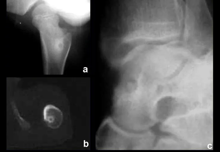
Figura 3: Osteoma osteóide na medular do fêmur (a); tomografia confirmando a localização central (b); osteoma osteóide medular do tálus (c).
Figure 3: Osteoid osteoma in the medullary bone of the femur (a); tomography confirming central location (b); Medullary osteoid osteoma of the talus (c).
  • Medullary : or spongy (endostal), figure 3a, 3b and 3c.
  • Figura 4: Radiografia do antebraço mostrando lesão subperiosteal na face ulnar do rádio, com nicho central do osteoma osteóide e halo de esclerose óssea (a), e tumor ressecado (b).
    Figure 4: Radiograph of the forearm showing a subperiosteal lesion on the ulnar surface of the radius, with a central niche of osteoid osteoma and a halo of bone sclerosis (a), and resected tumor (b).
    Figura 5: Osteoma osteóide subperiosteal na face medial do fêmur.
    Figure 5: Subperiosteal osteoid osteoma on the medial surface of the femur.
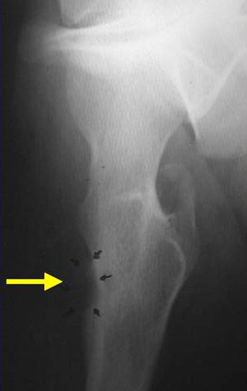
    Figura 6: Nicho de osteoma osteóide na superfície do fêmur, periosteal.
    Figure 6: Osteoid osteoma niche on the surface of the femur, periosteal.
    • Sub-periosteal,  figures 4, 5 and 6.

    It occurs in the first and second decade of life, occurring twice as often in males.

    The clinical picture is nighttime pain (mild pain that the child does not manifest while playing), which improves with the administration of salicylates. Local swelling (fig. 4) and arthralgia may occur.

    Makes differential diagnosis with steoma, osteoblastoma and Brodie’s abscess  (sequestration of chronic osteomyelitis).

    Treatment consists of resection of the central niche (fig. 5). It is not necessary to remove all the surrounding sclerosis. When the bone failure caused by resection poses a risk for fracture (as in the femoral neck (fig. 6), a bone graft must be placed.

    Author: Prof. Dr. Pedro Péricles Ribeiro Baptista

     Orthopedic Oncosurgery at the Dr. Arnaldo Vieira de Carvalho Cancer Institute

    Bdd1b613ee6fcac7694cf648430358ce 3

    Osteoma

    [auto_translate_button]
    Osteoma Benign, slow-growing lesion, with mature bone tissue, with a lamellar structure, well differentiated. It is bone, dense , within the bone, whether in the cortical or medullary region .

    Osteoma

    It can manifest itself in three distinct clinical forms:

    • Exostoses  (dense, homogeneous bone, with an  ivory appearance ): this is the conventional osteoma, restricted to bones of intramembranous origin (facial bones, skullcap), figures 1 to 10.
    Figura 1: Radiografia do crânio com lesão nodular, densa, homogênea, na calota.
    Figure 1: X-ray of the skull with a nodular, dense, homogeneous lesion in the cap.
    Figura 2: Tomografia axial do crânio, com lesão acometendo as duas tábuas, com abaulamento maior da cortical externa.
    Figure 2: Axial tomography of the skull, with the lesion affecting both tables, with greater bulging of the external cortex.
    Figura 3: Reconstrução tomográfica em três dimensões da lesão do crânio.
    Figure 3: Three-dimensional tomographic reconstruction of the skull lesion.
    Figura 4: Nódulo firme, saliente, na base de implantação do cabelo.
    Figure 4: Firm, prominent nodule at the base of hair implantation.
    Figura 6: Ostectomia com formão.
    Figure 5: Asepsis and antisepsis, with plastic field placement. Figure 6: Ostectomy with chisel.
    Figura 8: Fragmentos do osteoma ressecado.
    Figure 7: Regularized surgical bed, without protrusions. Figure 8: Fragments of the resected osteoma.
    Figura 9: Osteoma exofítico da região parietal, aspecto de marfin.
    Figure 9: Exophytic osteoma of the parietal region, ivory appearance.
    Figura 11: Segmento ressecado na base.
    Figure 10: Ivory-looking lesion. Figure 11: Resected segment at the base.
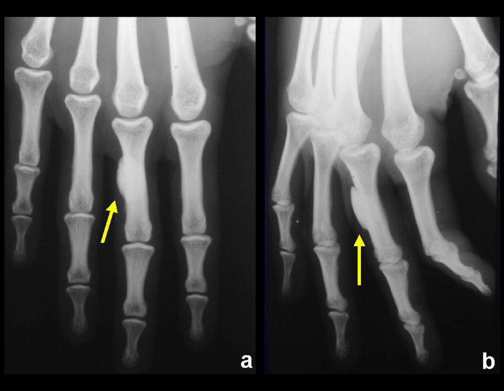
  • Parosteal  (juxtacortical) occurs on the external surface of long or short bones, figure 12.
  • Figura 12: Osteoma da falange proximal, justacortical.
    Figure 12: Osteoma of the proximal phalanx, juxtacortical.
    Figura 13 : Osteoma medular do colo femoral, enostose óssea.
    Figure 13: Medullary osteoma of the femoral neck, bone enostosis.
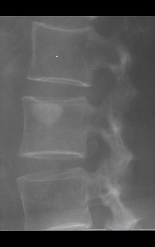
    • Medullary : known as enostosis or bone islet. Lesions histologically similar to mature bone, dense and homogeneous, without significant clinical signs and generally resulting from radiographic findings, figures 13 to 16.
    Figura 14 : Osteoma medular do corpo vertebral, enostose óssea.
    Figure 14: Medullary osteoma of the vertebral body, bone enostosis.
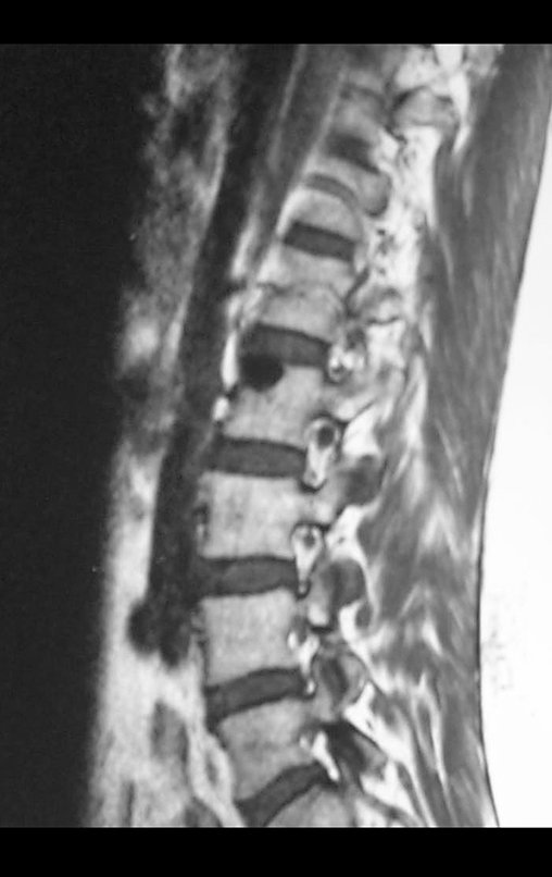
    Figura 15: Rm sagital de osteoma medular do corpo vertebral.
    Figure 15: Sagittal MRI of medullary osteoma of the vertebral body.
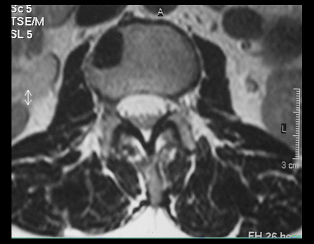
    Figura 16: Rm axial de osteoma medular do corpo vertebral.
    Figure 16: Axial MRI of medullary osteoma of the vertebral body.

    he differential diagnoses, both from a clinical, histological and radiographic point of view, include the following conditions:

    • Bone sclerosis  (inflammatory or post-traumatic cause)
    • Hyperostoses
    • Meningioma
    • Osteoid osteoma.

    Treatment consists of observation when asymptomatic or surgical resection when indicated due to aesthetic or functional changes (compression or obstruction – nasal fossa).

    Author: Prof. Dr. Pedro Péricles Ribeiro Baptista

     Orthopedic Oncosurgery at the Dr. Arnaldo Vieira de Carvalho Cancer Institute

    A4d4ec4aa3c45731396ed6e65fee40b9 3

    Osteochondroma

    [auto_translate_button]
    Osteochondroma is also known as osteo-cartilaginous exostosis . It represents the most common benign bone lesion. Its incidence is even higher than that reported in the literature as many patients have asymptomatic osteochondromas.

    Osteochondroma

    It occurs in the first and second decades, in the metaphyseal region of long bones, radiographically characterized by a tumor that forms cartilage and bone. Characteristically, the central cancellous bone of the exostosis continues with the medullary of the affected bone and the dense, cortical layer of the tumor continues with the normal cortical bone. On the surface of this lesion there is a band of cartilage, through which the lesion grows (hence the name  osteo-chondroma: tumor forming cartilage and bone ).

    It presents with a sessile base (enlarged base) or pedicled shape.

    May be single or multiple (hereditary osteochondromatosis)

    Osteochondromas require surgical treatment ( resection ) when they alter aesthetics, compress vascular-nervous structures or limit function. They generally grow while the patient is in the growth phase.

    When an osteochondroma increases in size after completion of skeletal maturity, it may mean post-traumatic bursitis or malignancy to chondrosarcoma and must be resected with an oncological margin.

    Solitary osteochondroma can have a 1% malignancy rate, whereas multiple osteochondromatosis can reach 10%.

    Among benign cartilaginous lesions, we cannot forget chondroblastoma.

    Author: Prof. Dr. Pedro Péricles Ribeiro Baptista

     Orthopedic Oncosurgery at the Dr. Arnaldo Vieira de Carvalho Cancer Institute

    0ade14765069c05180284d03b3097202 3

    Osteoblastoma

    [auto_translate_button]

    Osteoblastoma

    Benign osteoblastic lesion, locally aggressive, with a histological structure similar to osteoid osteoma but without reactive bone sclerosis and larger in size, generally larger than 1.5 cm

    Osteoblastoma

    Radiographically, it presents large areas of bone rarefaction with few denser foci of ossification.

    It presents two clinical forms:

    • Genuine osteoblastoma : the most common, located in the pedicle of the spine and more rarely in the metaphysis of long bones.
    • Aggressive  (“malignant”) osteoblastoma.

    It affects the first and second decade of life.

    The clinical picture is one of intense pain, which can lead to fractures and functional and neurological deficits, when it occurs in the spine, sometimes with antalgic scoliosis.

    Makes differential diagnosis with:

    • Osteoid osteoma,
    • Aneurysmal bone cyst,
    • Osteosarcoma.

    Treatment consists of en bloc resection of the lesion and bone grafting when necessary, reserving careful curettage for regions such as the spine, where it is generally not possible to resect en bloc. The use of local adjuvants (phenol, electrothermia) has been increasingly used, seeking to avoid recurrence. 

    Author: Prof. Dr. Pedro Péricles Ribeiro Baptista

     Orthopedic Oncosurgery at the Dr. Arnaldo Vieira de Carvalho Cancer Institute

    1 2 3
    Hello! How can we assist you?