This digital library houses the book on Oncology and Orthopedic Oncosurgery.

It includes academic lectures, presentations from national and international congresses, published papers, case discussions, performed surgical procedures, and proprietary techniques developed.

The digital format was chosen because the web allows the inclusion of texts with numerous visual resources, such as images and videos, which would not be possible in a printed book.

The content is intended for students, healthcare professionals, and the general public interested in the field.

Auto Transplantation of the Distal Fibula Growth Plate

Cc75c256acc04ce25a291c4b7a9856c0 2
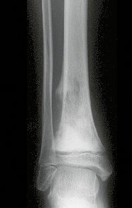
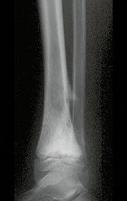
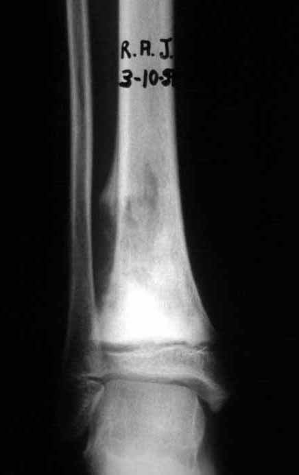
Auto Transplantation of the Distal Fibula Growth Plate. Patient in December 1996, eight years old, with osteosarcoma of the distal meta-epiphyseal region of the tibia, treated with neo-adjuvant chemotherapy (Figures 1 to 4).

Autotransplantation of the distal fibula growth plate. Osteosarcoma of the distal region of the tibia. Tibialization of the distal fibula with preservation of the epiphyseal plate.

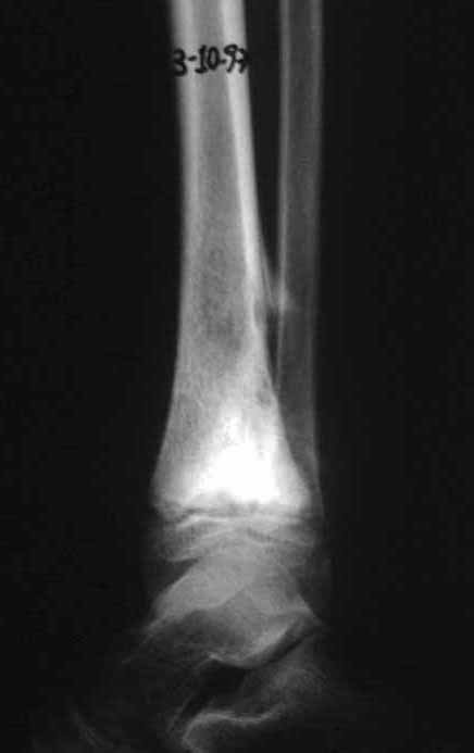
Figura1: Radiografia frente do tornozelo direito, com lesão meta-epifisaria da tíbia, com limites imprecisos e triângulo de Codmann.
Figure 1: Radiograph of the front of the right ankle, with a meta-epiphyseal lesion of the tibia, with imprecise limits and Codmann's triangle.
Figura 2: Radiografia perfil do tornozelo direito, com osteossarcoma da região distal da tíbia.
Figure 2: Profile x-ray of the right ankle, with osteosarcoma of the distal region of the tibia.
Figura 3: Radiografia frente de osteossarcoma do tornozelo direito, em quimioterapia neo-adjuvante.
Figure 3: Frontal radiograph of osteosarcoma of the right ankle, undergoing neo-adjuvant chemotherapy.
Figura 4: Radiografia em perfil de osteossarcoma do tornozelo, em quimioterapia.pré-operatória.
Figure 4: Lateral radiograph of osteosarcoma of the ankle, undergoing preoperative chemotherapy.
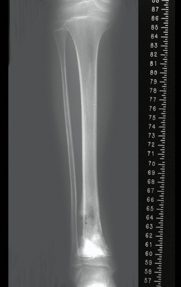
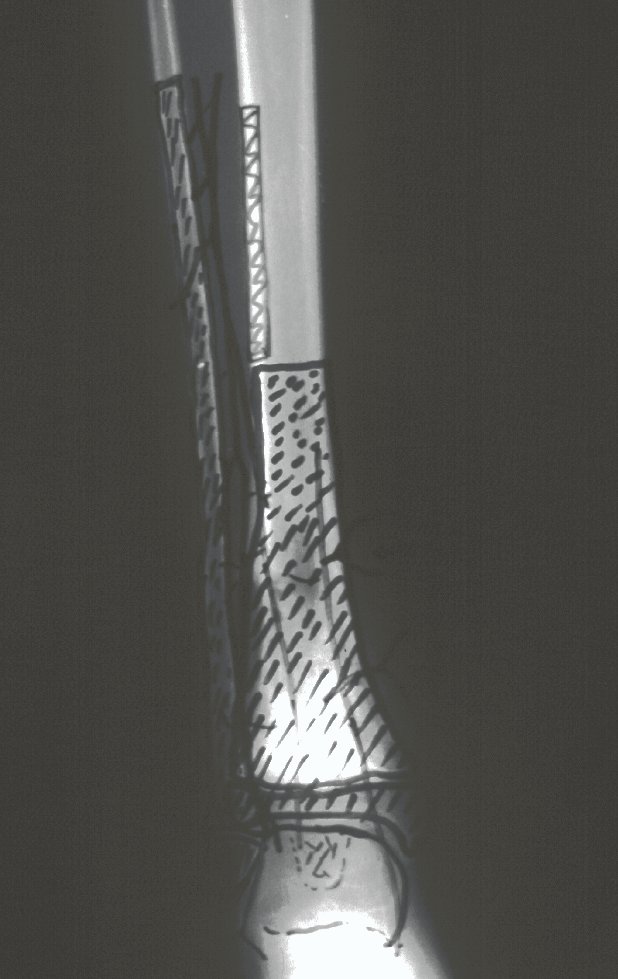
The tomography shows a tumor close to the growth plate, requiring its resection, as an oncological margin (figure 5). How to reconstruct this segment in a seven-year-old child and avoid lower limb discrepancy? Our proposal was to resect the distal 1/3 of the tibia and reconstruct it with the fibula on the same side, transferring the fibula to replace the tibial defect. In this transfer, we would take the vascularized fibula, with the physeal plate, arthrodesing its epiphysis with the talus and nailing the proximal 1/3 of the fibula in the proximal segment of the tibia. We perform x-rays, arteriography and planning for execution (figures 6.7 and 8).
Figura 5: Tomografia do tornozelo mostrando lesão produtora de osso na metáfise da tíbia que ultrapassa a placa fisária e compromete a epífise
Figure 5: Tomography of the ankle showing a bone-producing lesion in the tibial metaphysis that goes beyond the physeal plate and compromises the epiphysis
Figura 6: Escanograma da tibia, para planejamento cirúrgico.
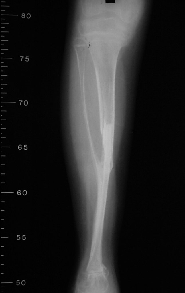
Figure 6: Scanogram of the tibia, for surgical planning.
Figura 7: Angiografia da região distal da tibia, para estudo da vascularização, visando o planejamento cirúrgico.
Figure 7: Angiography of the distal region of the tibia, to study the vascularization, aiming at surgical planning.
Figura 8: Planejamento cirurgico: Ressecção do 1/3 distal da tíbia, tibialização dos 2/3 distais da fíbula com a epífise e a placa de crescimento, abertura de janela na tibia para encaixe do terço médio e preservação da integridade da artéria nutrícia da fíbula.
Figure 8: Surgical planning: Resection of the distal 1/3 of the tibia, tibialization of the distal 2/3 of the fibula with the epiphysis and growth plate, opening of a window in the tibia to fit the middle third and preservation of the integrity of the nutrient artery of the fibula.
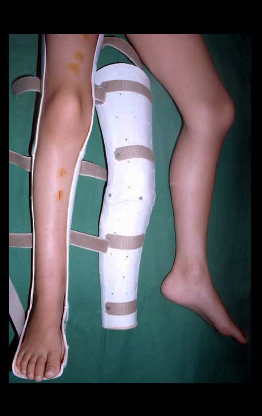
Planning this procedure must include the creation of an orthosis that will serve to immobilize the operated limb. We make a raw foot plaster that will serve as a mold for modeling the orthosis, made of polypropylene (figure 9).
Figura 9: Órtese de polipropileno, confeccionada no pré-operatório, sob molde de gesso, que será utilizada após a cirurgia, para a proteção da reconstrução e início da marcha.
Figure 9: Polypropylene orthosis, made pre-operatively, under a plaster mold, which will be used after surgery, to protect the reconstruction and start of walking.
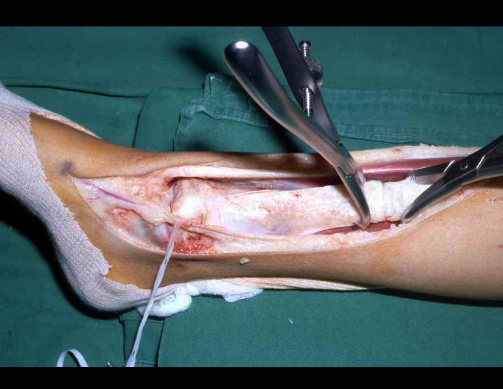
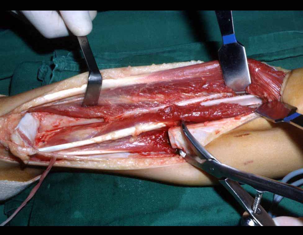
The tumor resection surgery, encompassing the entire distal third of the tibia, and the reconstruction of this segment with autotransplantation of the growth cartilage from the fibula to the tibia are detailed in figures 10 to 20.
Figura 10: Incisão antero-medial do tornozelo e 1/3 distal da tíbia, com preservação dos vasos safenos, e ressecção do tumor.
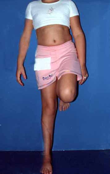
Figure 10: Anteromedial incision of the ankle and distal 1/3 of the tibia, with preservation of the saphenous vessels, and resection of the tumor.
Figura 11: Tumor ressecado, exposição do tálus e da região distal da fíbula para transposição.
Figure 11: Resected tumor, exposure of the talus and the distal region of the fibula for transposition.
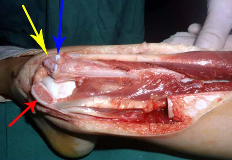
Figura 12: Retirada da cartilagem epifisária da fíbula, seta amarela, com preservação da placa de crescimento da fíbula, seta azul, detalhe do maléolo tibial, seta vermelha.
Figure 12: Removal of the fibula epiphyseal cartilage, yellow arrow, with preservation of the fibula growth plate, blue arrow, detail of the tibial malleolus, red arrow.
Figura 13: Realização de uma cavidade no tálus para colocação da epífise fibular visando obter a artrodese fíbulotalar.
Figure 13: Creating a cavity in the talus to place the fibular epiphysis in order to obtain fibulotalar arthrodesis.
Figura 14: Colocação da epífise fibular na cavidade do tálus, seta amarela e posicionamento proximal da diáfise fibular no 1/3 proximal da tíbia, seta azul.
Figure 14: Placement of the fibular epiphysis in the talus cavity, yellow arrow and proximal positioning of the fibular shaft in the proximal 1/3 of the tibia, blue arrow.
Figura 15: Canaleta aberta lateralmente na tíbia (seta amarela) para permitir o encavilhamento proximal da fíbula (seta azul), sem lesar a artéria nutrícia, mantendo os 2/3 distais da fíbula como um autotransplante vascularizado.
Figure 15: Channel open laterally in the tibia (yellow arrow) to allow proximal nailing of the fibula (blue arrow), without damaging the nutrient artery, maintaining the distal 2/3 of the fibula as a vascularized autotransplant.
Figura 16: Passagem de fio de Kirschner (seta amarela) pelo calcâneo-tálus-epífise fibular-placa de crescimento-diáfise da fíbula-tíbia proximal, estabilizando a reconstrução com uma síntese mínima, geralmente suficiente e de baixa morbidade para as crianças. A seta azul evidencia a placa de crescimento da fíbula, que substituirá a da tíbia.
Figure 16: Passage of a Kirschner wire (yellow arrow) through the calcaneus-talus-fibular epiphysis-growth plate-diaphysis of the fibula-proximal tibia, stabilizing the reconstruction with a minimal synthesis, generally sufficient and with low morbidity for children. The blue arrow highlights the growth plate of the fibula, which will replace that of the tibia.
Figura 17: Peça ressecada, a seta amarela destaca a sindesmose tíbio-fibular.
Figure 17: Resected piece, the yellow arrow highlights the tibiofibular syndesmosis.
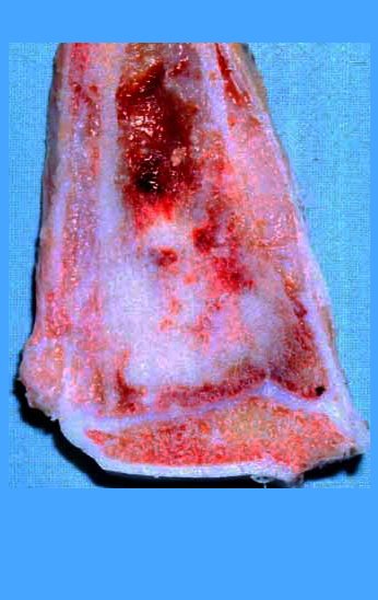
Figura 18: Corte da peça ressecada. A seta azul destaca a reação periosteal e a seta amarela salienta a proximidade do tumor à placa de crescimento.
Figure 18: Section of the dried piece. The blue arrow highlights the periosteal reaction and the yellow arrow highlights the proximity of the tumor to the growth plate.
Figura 19: Região meta-epifisária ampliada.
Figure 19: Enlarged meta-epiphyseal region.
Figura 20: Detalhe da proximidade da placa de crescimento como tumor.
Figure 20: Detail of the proximity of the growth plate to the tumor.
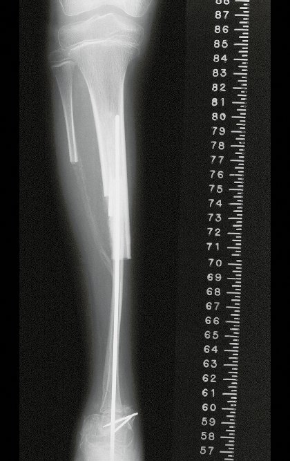
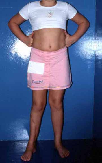
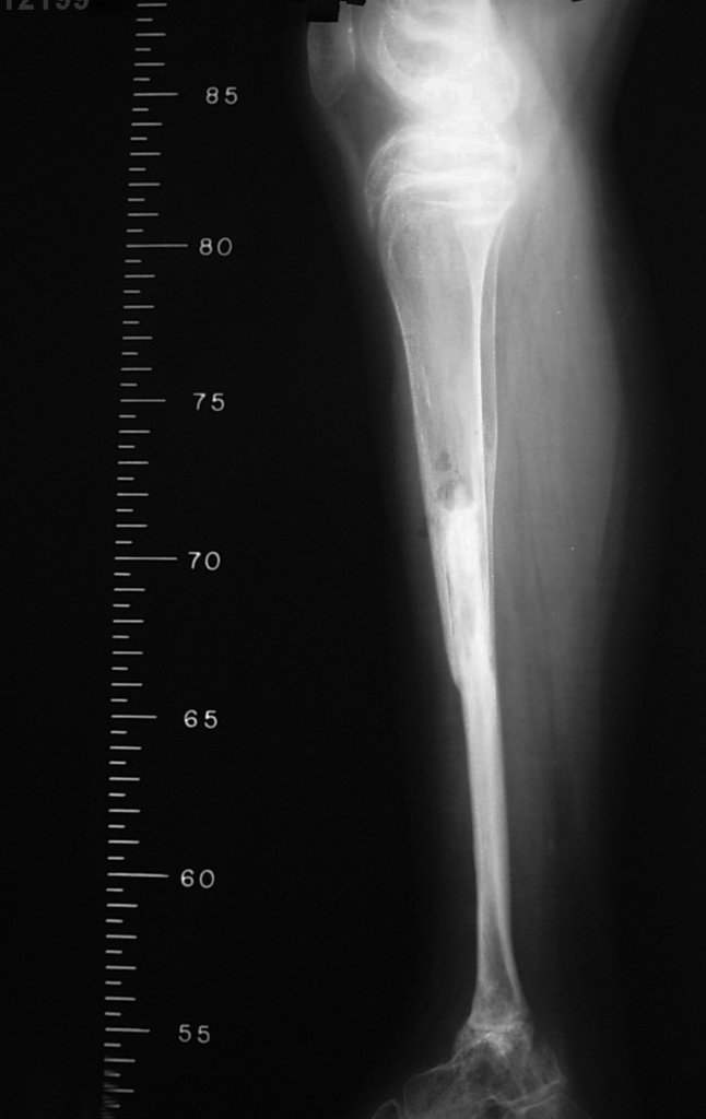
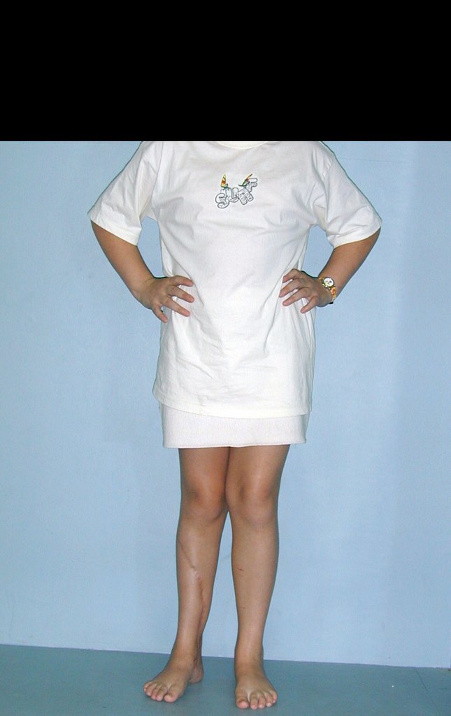
Radiographic documentation and monitoring of the patient’s rehabilitation after reconstruction are illustrated in figures 21 to 46.
Figura 21: Radiografia frente do autotransplante da fíbula vascularizada, transferindo a cartilagem de crescimento para substituir a que foi ressecada da tíbia.
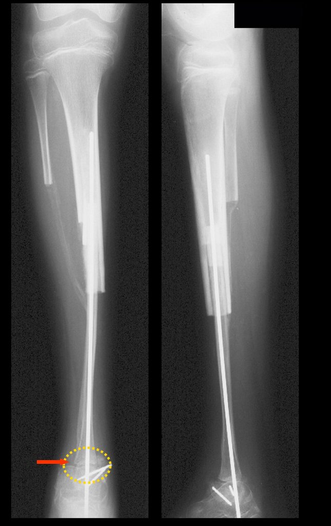
Figure 21: X-ray of the autotransplantation of the vascularized fibula, transferring the growth cartilage to replace that which was resected from the tibia.
Figura 22: Radiografia perfil da reconstrução com a fíbula vascularizada, fixada com fio intramedular.
Figure 22: Profile radiograph of the reconstruction with the vascularized fibula, fixed with intramedullary wire.
Figura 23: Aspecto clínico após seis meses.
Figure 23: Clinical appearance after six months.
Figura 24: Paciente com carga parcial, após seis meses da reconstrução, em quimioterapia adjuvante.
Figure 24: Patient with partial load, six months after reconstruction, undergoing adjuvant chemotherapy.
Figura 25: Pós-operatório de onze meses, alinhamento dos MMII, carga com apoio.
Figure 25: Eleven months post-operative, lower limb alignment, weight bearing with support.
Figura 26: Carga com apoio, após onze meses.
Figure 26: Load with support, after eleven months.
Figura 27: Alinhamento do membro operado, perfil, carga com apoio após onze meses.
Figure 27: Alignment of the operated limb, profile, load with support after eleven months.
Figura 28: Flexão com carga total após um ano da cirurgia.
Figure 28: Flexion with full load one year after surgery.
Figura 29: Alinhamento e função após um ano da cirurgia.
Figure 29: Alignment and function one year after surgery.
Figura 30: Carga total monopodal após um ano da cirurgia, paciente já fora de quimioterapia.
Figure 30: Total single-leg weightlifting one year after surgery, patient no longer undergoing chemotherapy.
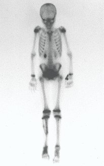
Figura 31: Cintilografia óssea, fase tardia, mostrando captação no 1/3 distal da perna, confirmando a presença de vascularização da fíbula transplantada.
Figure 31: Bone scintigraphy, late phase, showing uptake in the distal 1/3 of the leg, confirming the presence of vascularization of the transplanted fibula.
Figura 32: Cintilografia óssea da perna destacando a captação da fíbula e a ossificação do trajeto da transferência proximal, promovendo uma fusão proximal.
Figure 32: Bone scintigraphy of the leg highlighting capture of the fibula and ossification of the proximal transfer path, promoting proximal fusion.
Figura 33: Radiografia frente, após três meses da reconstrução.
Figure 33: Front X-ray, three months after reconstruction.
Figura 34: Radiografia perfil, após três meses da reconstrução.
Figure 34: Profile x-ray, three months after reconstruction.
Figura 35: Paciente após um ano e seis meses, fora de quimioterapia, bom alinhamento.
Figure 35: Patient after one year and six months, out of chemotherapy, good alignment.
Figura 36: Carga total, monopodal, após um ano e seis meses.
Figure 36: Full load, single leg, after one year and six months.
Figura 37: Radiografia frente, em 1998.
Figure 37: Front X-ray, in 1998.
Figura 38: Radiografia perfil, em 1998.
Figure 38: Profile x-ray, in 1998.
Figura 39: Radiografia frente, após um ano da cirurgia. Fíbula integrada e já tibializada com significativo espessamento cortical.
Figure 39: Front X-ray, one year after surgery. Fibula integrated and already tibialized with significant cortical thickening.
Figura 40: Radiografia perfil, após um ano da cirurgia. Fíbula já tibializada
Figure 40: Profile x-ray, one year after surgery. Fibula already tibialized
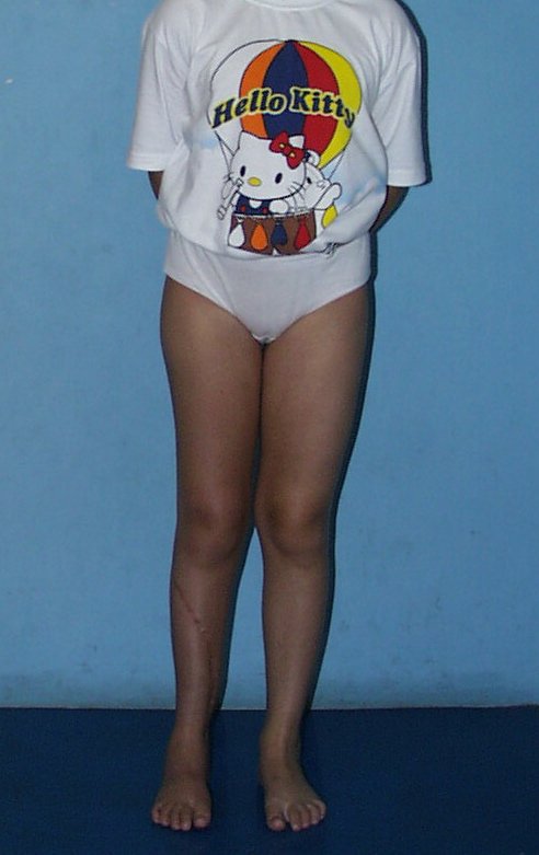
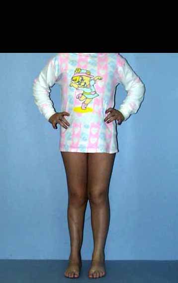
Figura 41: Paciente após um ano e sete meses da reconstrução. Bom alinhamento dos MMII.
Figure 41: Patient one year and seven months after reconstruction. Good alignment of the lower limbs.
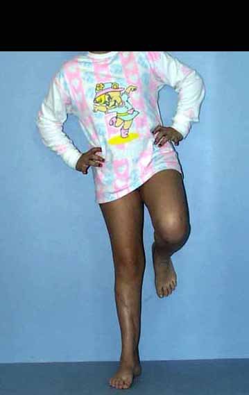
Figura 42: Carga total, monopodal, após um ano e sete meses da cirurgia.
Figure 42: Full load, single leg, one year and seven months after surgery.
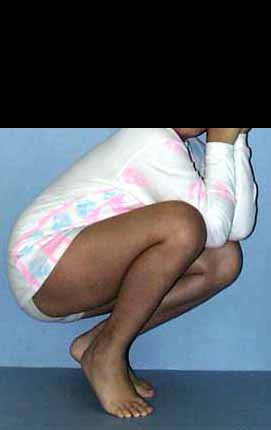
Figura 43: Flexão com carga total, após um ano e sete meses.
Figure 43: Flexion with full load, after one year and seven months.
Figura 44: Radiografia após um ano e sete meses, fíbula tibializada e fise viável, solução biológica que permite a equalização dos membros com o crescimento.
Figure 44: Radiograph after one year and seven months, tibialized fibula and viable physis, biological solution that allows limbs to equalize with growth.
Figura 45: Pós-operatório da reconstrução evidenciando a placa de crescimento da fíbula, transferida para substituir a da tíbia, que foi ressecada.
Figure 45: Post-operative reconstruction showing the fibula growth plate, transferred to replace the tibia, which was resected.
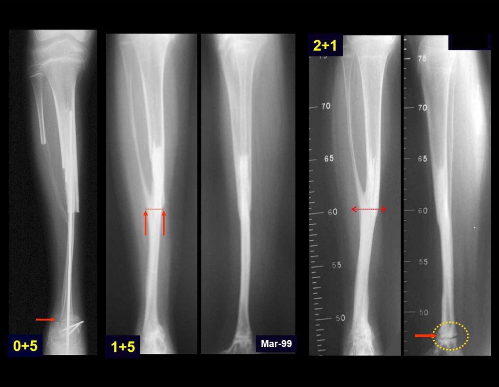
Figura 46: Radiografias ilustrando a evolução do autotransplante de cartilagem de crescimento, desde o pós-operatório precoce até após dois anos e um mês da reconstrução.
Figure 46: Radiographs illustrating the evolution of growth cartilage autotransplantation, from the early postoperative period to two years and one month after reconstruction.
This technique of autotransplantation of the fibula, with its growth plate, to replace the distal segment of the tibia, in young children, is an excellent alternative for autologous biological reconstruction that preserves the growth of the limb, avoiding discrepancies. It was published in the Brazilian Journal of Orthopedics and Traumatology in November 1998, vol. 33 – number 11.

Author: Prof. Dr. Pedro Péricles Ribeiro Baptista

 Orthopedic Oncosurgery at the Dr. Arnaldo Vieira de Carvalho Cancer Institute

Leave a Reply

Hello! How can we assist you?