• Rua General Jardim, 846 cj 41 Higienópolis, São Paulo - SP

Author Archives: drpprb@gmail.com

This digital library houses the book on Oncology and Orthopedic Oncosurgery.

It includes academic lectures, presentations from national and international congresses, published papers, case discussions, performed surgical procedures, and proprietary techniques developed.

The digital format was chosen because the web allows the inclusion of texts with numerous visual resources, such as images and videos, which would not be possible in a printed book.

The content is intended for students, healthcare professionals, and the general public interested in the field.

drpprb@gmail.com
0d9d154fb5b905d3f6d606f8b6cbb750 3

Chondrosarcoma or Chondroma

[auto_translate_button]

Chondrosarcoma or Chondroma. For a better understanding of the differential diagnosis of chondroma and grade I chondrosarcoma, let’s discuss the case:

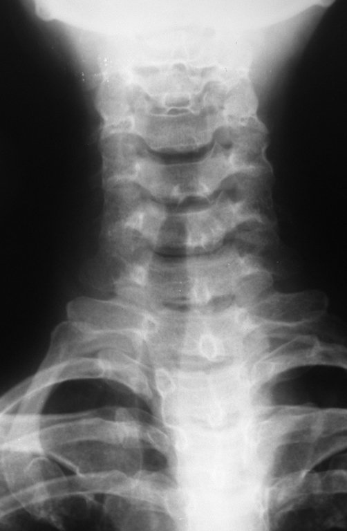
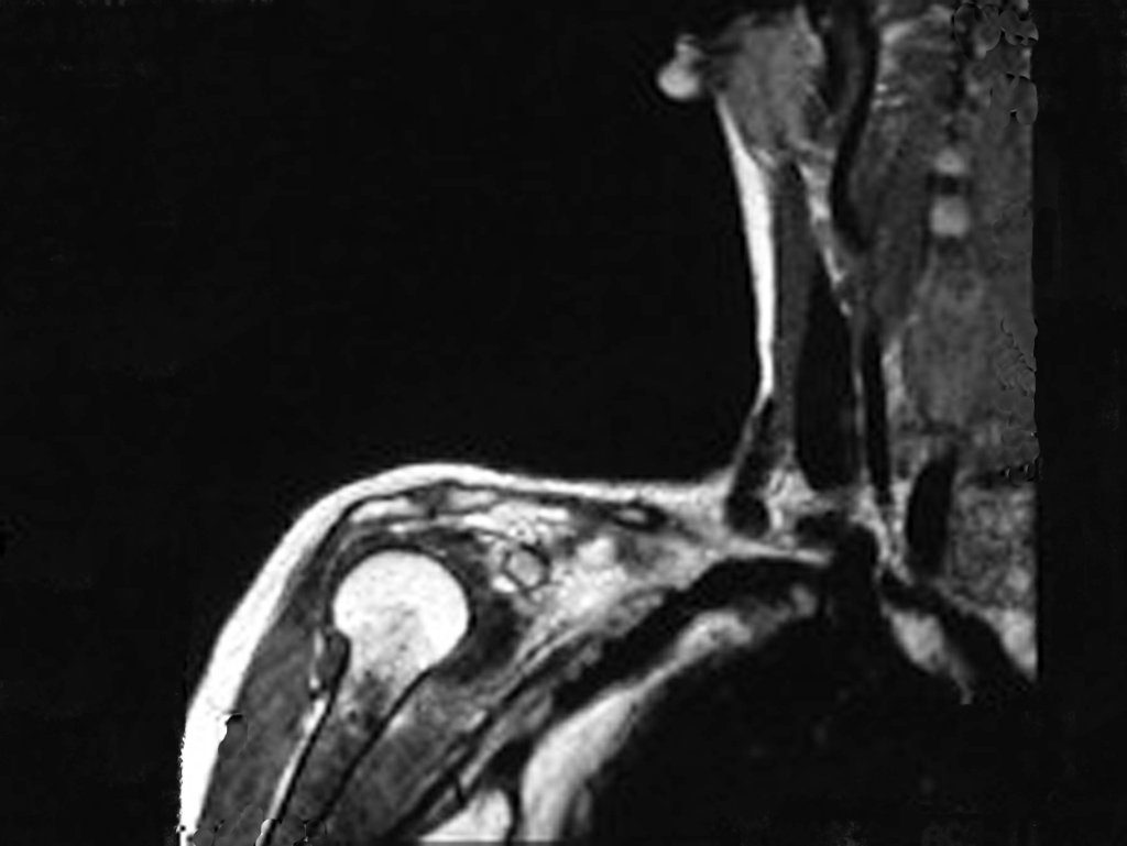
Female patient, 39 years old , odontologist , right handed. The patient refers pain in his right shoulder for about eight months. The first doctor performs radiographs of the cervical spine and indicated physical therapy for cervical spine ( Figure 1). Not getting better, performs magnetic resonance imaging of the ervical spine, which showed no cervical lesions(Figure 2).

CHONDROSARCOMA and CHONDROMA: Differential Diagnosis, Management and Treatment.

Figura 1: Radiografia (RX) da coluna cervical, sem alterações.
Figure 1: Radiography of the cervical spine without lesions.
Figura 2: Ressonância (RM) da coluna cervical sagital (sag) T1, normal.
Figure 2: Magnetic Resonance (MR)) of the sagittal cervical spine (sag) T1, normal.

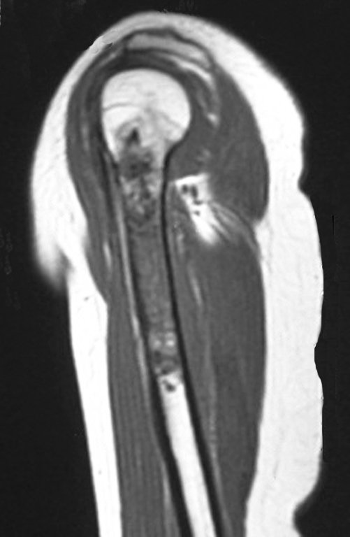
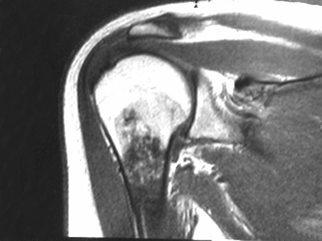
However, analysis of this examination showed lesions in the proximal humeral metaphysis, characterized by low signal and intermediate signal in T1 and high signal on T2 ( Figures 3 and 4 ).

Figura 3: RM com lesão na metáfise proximal do úmero direito, apresentando sinal intermediário e áreas de baixo sinal em T1, que devem corresponder a focos de calcificação.
Figure 3: MR with lesions in the proximal metaphysis of the right humerus, with intermediate signal and low signal areas in T1, which must correspond to foci of calcification.
Figura 4: RM coronal (cor) T2, lesão heterogênea com áreas de alto sinal entremeadas com áreas de baixo sinal (focos de calcificação).
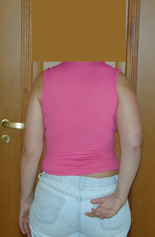
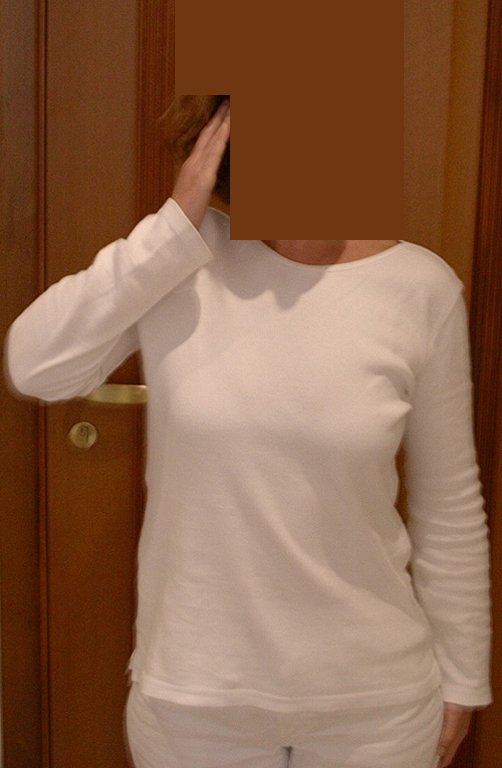
Figure 4: coronal MR (cor)T2, heterogeneous lesion with areas of high signal intensity interspersed with low signal areas (calcified foci).
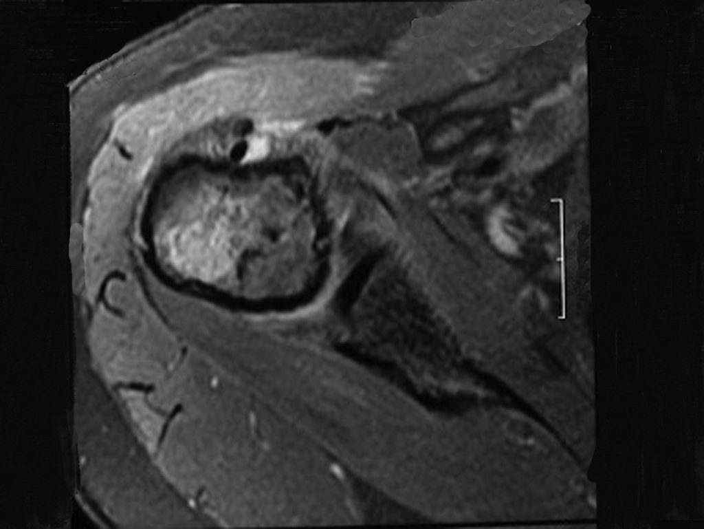
After a week , new resonance examination was conducted to evaluate this finding. This humeral resonance imaging showed a solid lesion , heterogeneous, with low signal and intermediate signal on T1, replacing the bone marrow fat (Figure 4a ). In the sagittal T1 spir, we see escaloped erosion of the endostal cortex.(Figure 4.b ). Careful analysis of magnetic resonance images, showed the aggressive characteristics of the lesion: with erosion of endosteal cortex; areas with hyposignal and areas of hyperintensity , heterogeneous pattern, with contrast uptake and calcification foci , which are most evident in the resonance PD ( proton density ) (Figures 5.a – 5.d ).
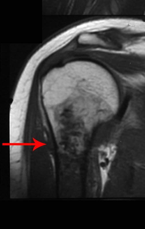
Figura 4.A: Ressonância coronal T1, evidenciando lesão meta-diafisária, sólida, heterogênea, com baixo sinal e sinal intermediário em T1, substituindo a gordura da medular óssea.
Figure 4.A: Coronal T1 MR , showing meta-diaphyseal injury, solid, heterogeneous, with low signal and intermediate signal on T1, substituting fat from bone marrow .
Figura 4.B: Ressonância em sagital T1 Spir, verifica-se erosão da cortical interna, lesões em saca bocado.
Figure 4.B : MR in sagittal T1 Spir , there is erosion of the endostal cortex, scalloped erosion.
Figura 5.B: RM ax DP: Lesão heterogênea com áreas de alto sinal e baixo sinal.
Figure 5: The: Color MR T1, irregular metaphyseal lesion with low signal areas. Figure 5B: MR ax DP: heterogeneous lesion with areas of high signal and low signal.
Figura 5.D: RM cor spir, com forte captação de contraste.
Figure 5.C: MRT1 Spir, scalloped erosion of the endostal cortex is observed. Figure 5.D: MR cor, spir color, with strong contrast enhancement.

The radiograph of this region, held on 24 July 2003 , two weeks later , highlighted the presence of this metadiafisiary injury, occupying two thirds of the proximal humerus.

This image shows scalloped areas due to the erosion of the endostal cortex, making the secondary enlargement of the bone marrow and areas of condensation dotted with cotton wool spots aspect , suggesting calcified foci (Figure 6.a ).

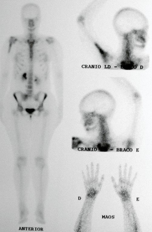
After this finding , it was referred to oncologist , requesting tests to investigate and stage the lesion . Scintigraphy performed: uptake was observed only in the right humerus (Figure 6.b).

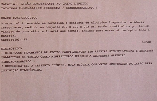
Then a biopsy which revealed  cartilage tissue without atypia , suggesting new biopsy ( Figure 7).

Figura 6.A: Rx do úmero com lesão meta-diafisária, com focos de calcificação, alargamento da medular e erosão da cortical interna.
Figure 6.A: Rx humerus with meta-diaphyseal lesion with calcified foci, bone marrow enlargement and erosion of the endostal cortex
Figura 6.B: Cintilografia evidenciando hiper-captação na região proximal do úmero direito.
Figure 6.B : Scintigraphy showing hyper-uptake in the proximal region of the right humerus.
Figura 7: Relatório da anatomia patológica identificando tecido cartilaginoso, sugerindo nova biópsia.
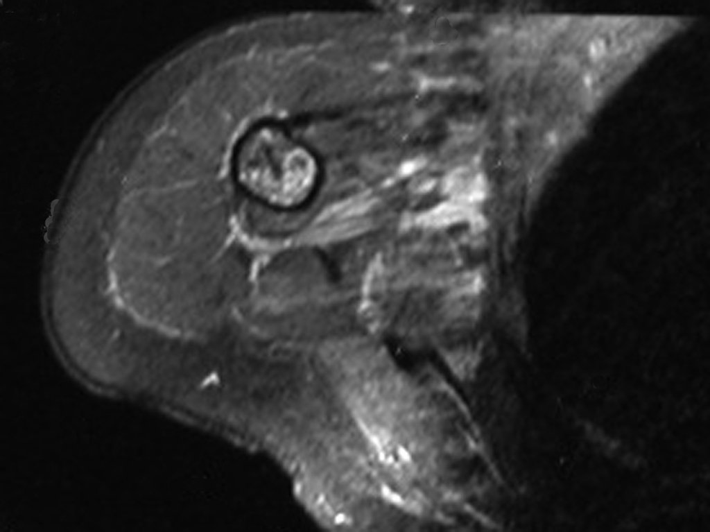
The shoulder pain and imaging findings of aggressive lesion with calcified foci, indicate the possibility of Chondrosarcoma. In this case the differential diagnoses between Chondroma and more remotely from Bone Infarction should be discarded due to the following considerations: 1- The patient went to the doctor because of progressive pain, was not a casual examination found. 2- The rays show metaphyseal enlargement, endostal cortex erosion and foci of calcification, which associated with the clinical symptoms indicates that it is active lesion, with local aggressiveness and points to the diagnosis of Chondrosarcoma. 3- The MR complement the image data and awaken the reasoning, concluding the same diagnosis. 4- The biopsy revealed that it was a “cartilaginous tissue,” there is no logic to suggest new biopsy in this situation. A second biopsy, in this case, became in an academic discussion because of the histopathological diagnosis of Chondroma and Chondrosarcoma grade I is often difficult, and the conduct of the treatment here is surgical. As we know that surgery is the only treatment that can cure Chondrosarcoma, this case must be approached and treated surgically as a Chondrosarcoma, regardless of whether the eventual biopsy comes with the previous diagnosis of Chondroma. The biopsy may have its indication just to confirm that it is a cartilage injury. The first doctor chose to perform a biopsy, with Jamshidi needle. The result of the pathological examination was cartilaginous lesions without atypia. Concerned about the aggressive image of the lesion did not feel peaceful in treating the case as a chondroma and not of assume a policy of treating as a Chondrosarcoma. Proposed to observe patient for two months. After this period the patient returns, still with the same clinical feature. The doctor indictes new MR, observing the same previous aspects, the biopsy scar and a new findin:the presence of extra-cortical tumor, infected all subcutaneous tissue (Figures 8a 8.b, 8.c and 9 ).
Figura 8.A: Rm cor T1, observa-se o levantamento periosteal e lesão extra cortical.
Figure 8A: Color MR T1, we observe the survey periosteal and cortical extra injury.
Figura 8.C: Rm cor T2, com supressão de gordura evidencia tumor extra cortical, contaminando o espaço sub-periósteo.
Figure 8.B: MR ax T1, path contamination of the biopsy.
Figure 8.C: MR color T2 with fat suppression shows extra cortical tumor, contaminating the sub-periosteal space.
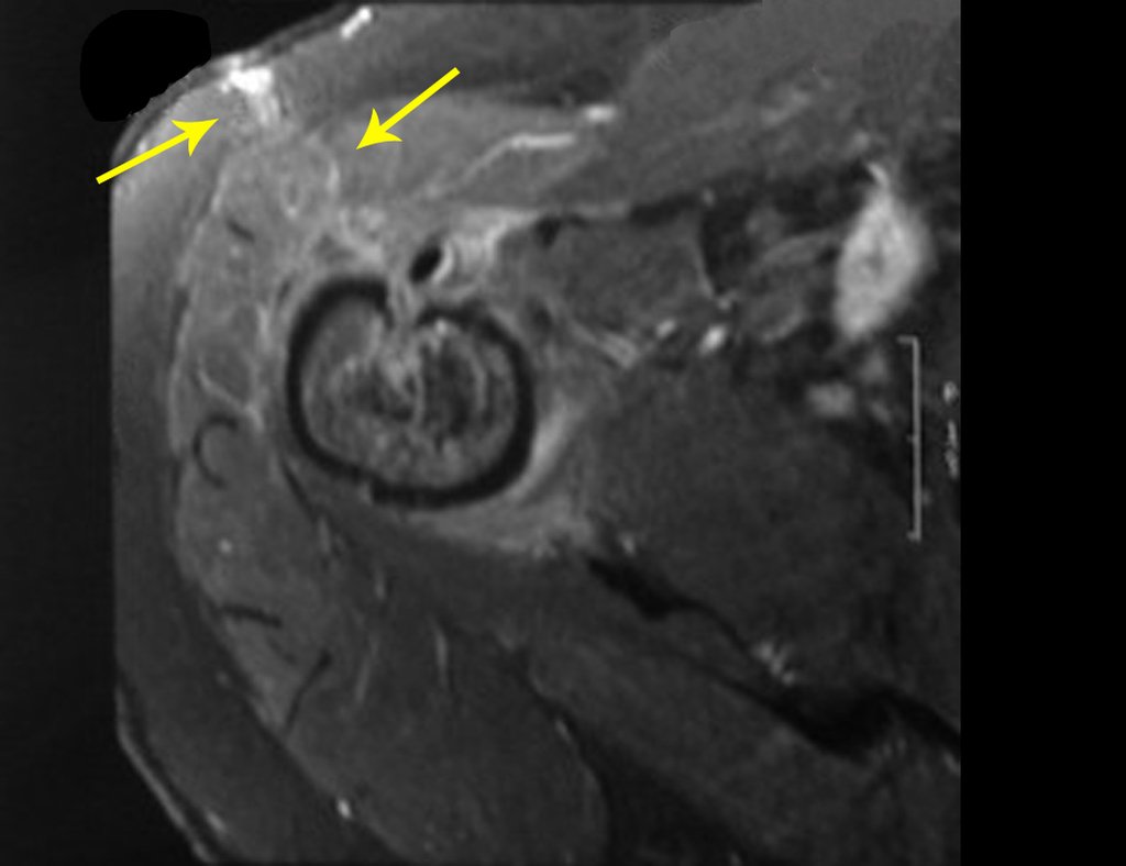
Figura 9: Rm ax T1, com saturação de gordura, evidencia o edema da cicatriz da biópsia e a implantação de tumor nos tecidos moles, no trajeto da biópsia.
Figure 9: Rm ax T1 with fat saturation: evidence of edema in scar biopsy and tumor implantation in soft tissue at the biopsy path.
After this last exam , the doctor offers to perform another biopsy. The patient decides to consult another professional , seeking a second opinion and joint us. The specialist in orthopedic oncology, must complete the diagnosis and define the conduct at this time . Must not launch a new biopsy because what action it will take if this biopsy is not conclusive for Chondrosarcoma ? What to do if the result is Chondroma ? With clinical data that revealed progressive pain, imaging with locally aggressive lesion and even the pathology of cartilage injury, the expert has all the parameters to indicate the treatment of this injury as Chondrosarcoma , for the histopathological diagnosis of Chondroma and Chondrosarcoma Grade I it is very difficult and sometimes controversial (Figures 10 and 11).
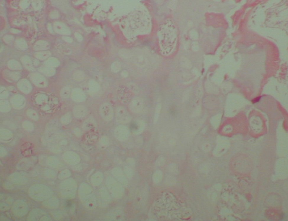
Figura 10: lâmina de condroma
Figure 10: Chondroma blade
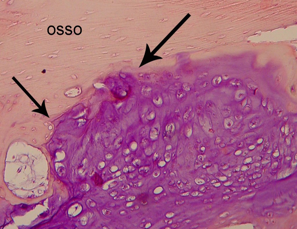
Figura 11: Lâmina de condrossarcoma grau I. Tecido cartilaginoso, com vacúolos contendo mais de um núcleo. Imagem de erosão do tecido ósseo pelo avanço do tumor
Figure 11: Chondrosarcoma Grade I: cartilaginous tissue, with vacuoles containing more than one core. Arrows show the erosion of the bone tissue due to tumour invasion

The same slide, presented to the same pathologist, after some time, may have changed the report of Chondroma to Chondrosarcoma to Grade I or Grade I Chondrosarcoma to Chondroma. Still, if this same slide is displayed to other pathologists, we can get the two different diagnoses. We know that the final diagnosis of bone tumors must have CLINICAL-RADIOLOGICAL and pathologic correlation. The pathologist usually is only analyzing the blade. Who is leading the case is who has all the data. We must therefore enhance the imaging and all the clinical picture in this situation.

After these considerations, surgical treatment is necessary. Chondrosarcoma is unresponsive to chemotherapy or radiotherapy. It can be cured with surgical resection with oncologic margin, because unfortunately relies on locally when this margin is not achieved. In relapse can occur dedifferentiation, invasion of adjacent tissues that impede the limb salvage surgery, as well as providing the occurrence of metastases.

You can not miss the opportunity to heal this injury with the appropriate surgery.

Careful analysis of the images of this case indicates the need for wide resection with margin and replacement with nonconventional prosthesis.

It is contraindicated intralesional curettage, even with adjuvant site and fill with cement, because the recurrence and dedifferentiation are frequent with this conduct.

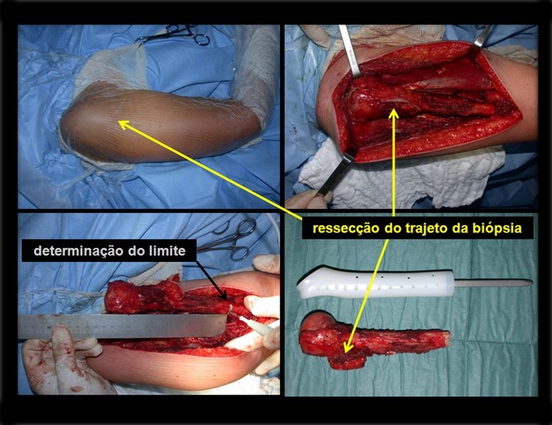
After this clarification to the patient, we perform a resection of two proximal humerus thirds, including the skin and the path of the biopsy, because in addition of Chondrosarcoma can deploy soft tissue, this was already happening in this case in the biopsy path.

For the reconstruction of the humerus, we employ a nonconventional endoprosthesis built in polyethylene. These are lighter than metal, have elasticity similar to that of bone and allow drilling holes, where necessary, to reattach the remaining ligaments and muscles. Around the polyethylene is a fibrotic reaction involving the prosthesis and fixed definitely the soft tissues reinserted.

In detail, we present the steps of the surgery, reconstruction with endoprosthesis and pathological anatomy of the piece (figures 12 to 23).

Figura 12: Paciente posicionada em decúbito dorsal horizontal. Assepsia e antissepsia, com exposição de todo o braço.
Figure 12: Patient positioned in the supine position. Area cleaning and disinfection with exposure around the arm.
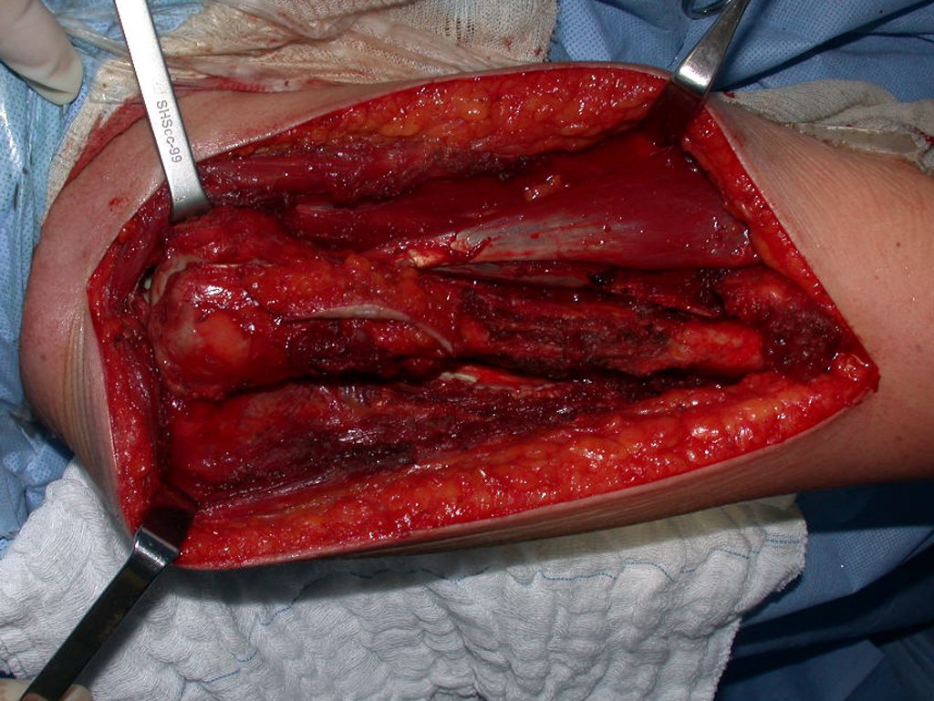
Figura 13: Incisão antero-lateral do braço direito, abertura de pele, tecido celular sub-cutâneo e hemostasia. Dissecção do segmento afetado, incluindo todo o trajeto da biópsia.
Figure 13: anterolateral aprouch on right arm, opening skin, subcutaneous tissue and hemostasis. Dissection of the affected segment, including all the way biopsy.
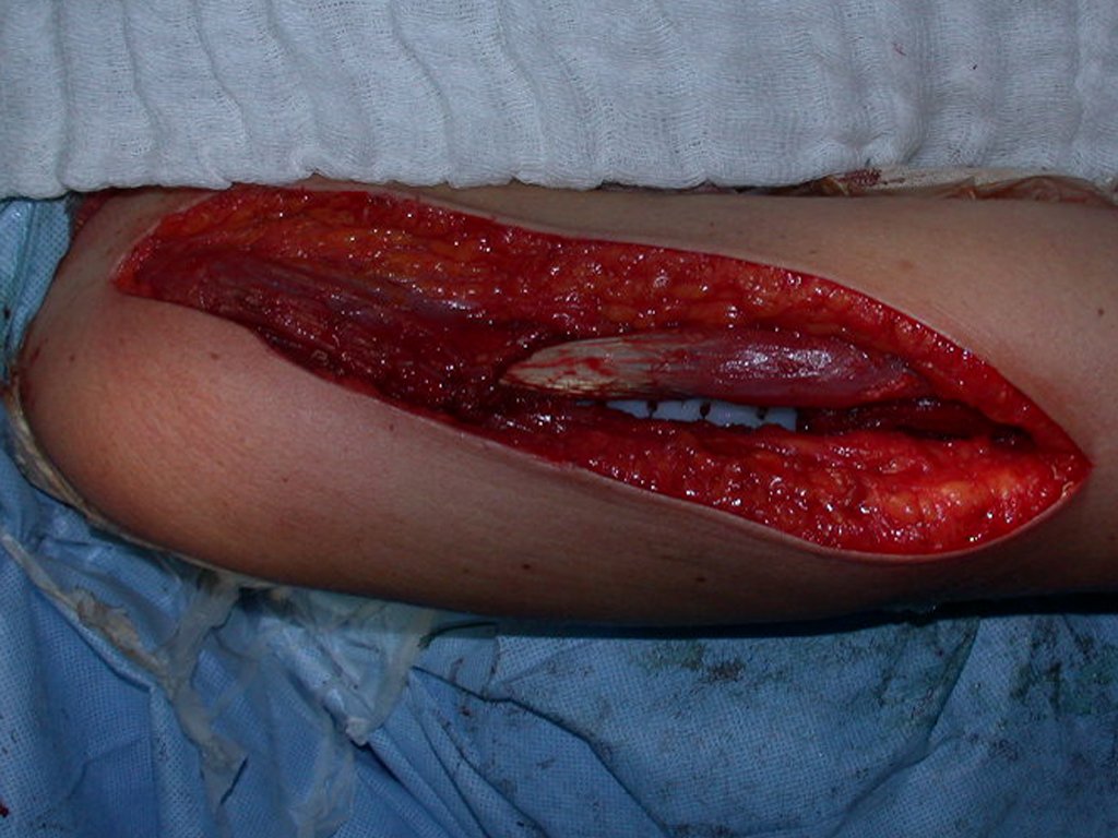
Figura 14: Liberado os dois terços da extremidade proximal do úmero. Dissecção e hemostasia. Em detalhe o tendão longo do músculo bíceps, inserido na glenóide.
Figure 14: Released two thirds of the proximal humerus. Dissection and hemostasis. In detail the long tendon of the biceps muscle, inserted into the glenoid.
Figura 15: Mensuração do segmento a ser ressecado.
Figure 15: Measurement segment to be resected.
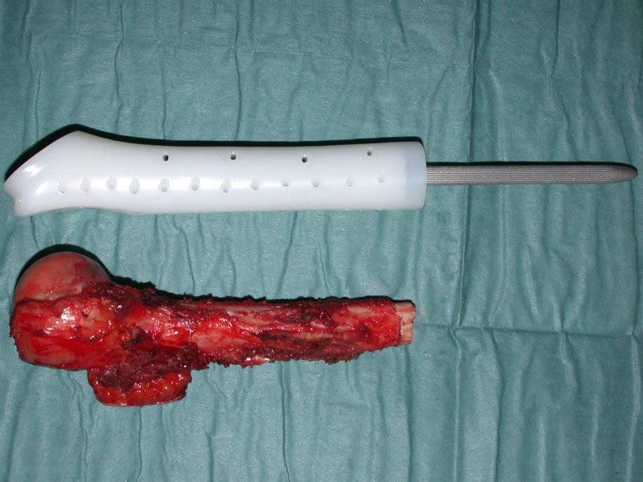
Figura 16: Segmento ressecado em conferência com a endoprótese confeccionada em polietileno e titânio, desenhada por Roberto Fabroni e produzida pela IMPOL, utilizada na reconstrução.
Figure 16: Segment resected and nonconventional endoprosthese made of polyethylene and titanium stem, designed by Roberto Fabroni and produced by IMPOL used in the reconstruction.
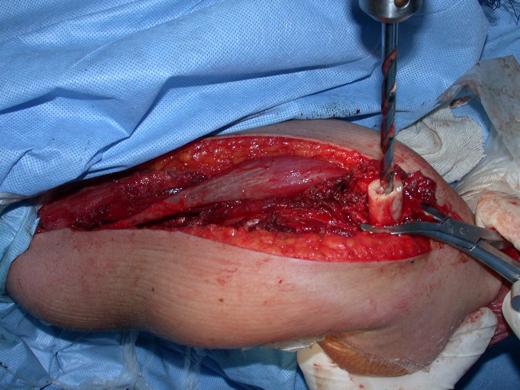
Figura 17: Após a osteotomia, realizamos o alargamento do canal medular, onde será cimentada a prótese.
Figure 17: After osteotomy, performed the enlargement of the spinal canal, where it will be cemented prosthesis.
Figura 18: Após a cimentação da haste, realizamos a reinserção da cápsula articular e dos músculos remanescentes, nos orifícios do polietileno da prótese, para obtermos a melhor funcão.
Figure 18: After cementing the stem, held the reintegration of the joint capsule and the remaining muscles in polyethylene holes of the prosthesis, to obtain the best user function.
Figura 19: Reinserção do músculo deltóide e aproximação do subcutâneo.
Figure 19: Reintegration of the deltoid muscle and subcutaneous approach.
Figura 20: Em destaque o local da biópsia, a pele e todo o trajeto sendo ressecado, a mensuração do segmento a ser ressecado e a peça ressecada em bloco, com margem oncológica.
Figure 20: Highlights the biopsy site, skin and all the way being resected, the segment measurement to be resected and the resected specimen block with oncologic margin.
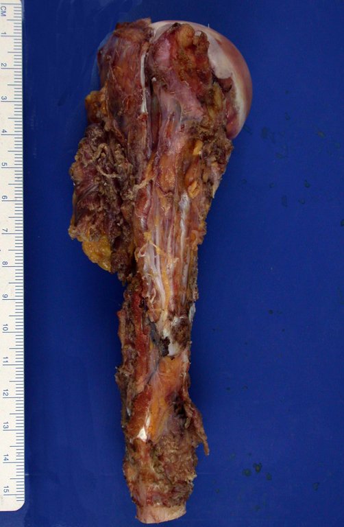
Figura 21.A: Segmento ressecado, incluindo o tecido celular subcutâneo e pele, do trajeto da biópsia.
Figure 21.A: resected segment, including the subcutaneous tissue and skin, the biopsy path.
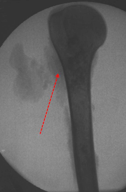
Figura 21.B: Radiografia da peça documentando os tecidos moles ressecados.
Figure 21b: X-ray of the resected piece documenting the dry soft tissue.
Figura 22.A: Corte da peça. Observamos o tumor ocupando o segmento proximal do úmero.
Figure 22.A: Cut of the piece. We observe the tumor occupying the proximal segment of the humerus.
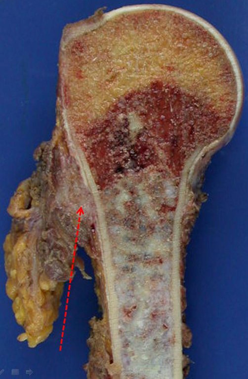
Figura 22.B: Em maior aumento, a seta aponta a disseminação do tumor nos tecidos moles.
Figure 22.B: In greater increase, the arrow indicates the spread of the tumor in soft tissue.
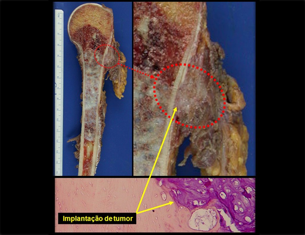
Figura 23: Corte da peça ressecada com o trajeto da biópsia contaminado, a seta destaca a lesão extra cortical e a ossificação do periósteo e a lâmina da Histologia destaca a erosão da cortical pelo tumor.
Figure 23: Cut of the resected piece with the path of the contaminated biopsy, the arrow highlights the extra cortical injury and ossification of the periosteum and the blade of Histology highlights the erosion of the cortical tumor.

The oncologic surgery should first seek resection in order to obtain margins decreasing the possibility of local recurrence . Reached this goal , the best reconstruction should be performed to restore function , the closer to normal point. In nonconventional endoprostheses, made ​​to reconstruct tumor resection, we can not be expected the same function as conventional prostheses used in arthritis or in other indications, since in each case there will be a loss of muscles and healthy soft tissues, larger or smaller, resected due to need to obtain oncologic margin.

Physiotherapy driven professional who knows the surgery is critical to achieve a good functional results (Figures 24 to 27).

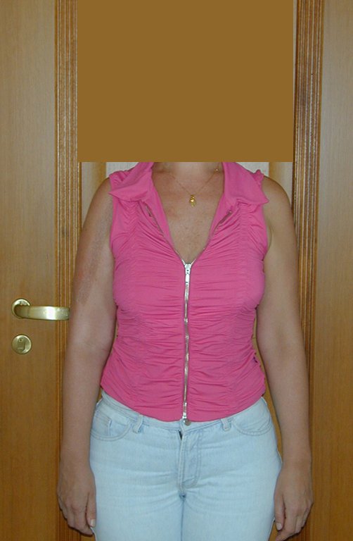
Figura 24: Pós operatório de dois meses. observa-se boa cicatrização, diminuição do volume do ombro devido à perda de tecidos ressecados como margem e hipotrofia muscular
Figure 24: Post-operative two months. observed good healing, decreased shoulder volume due to loss of tissue resected margin and as muscle hypotrophy.
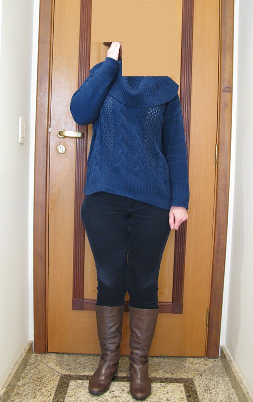
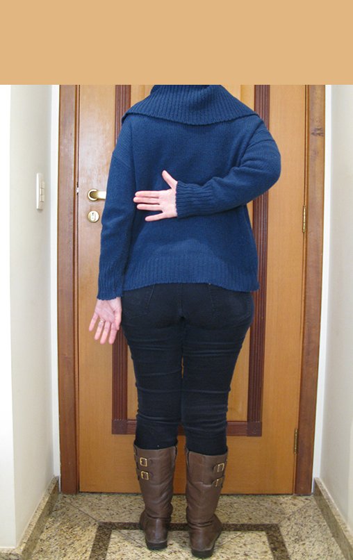
Figura 25.A: A paciente consegue alcançar o queixo com a mão.
Figure 25.A: The patient can achieve his mouth with his hand.
Figura 25.B: Limitação da rotação interna. A paciente alcança a nádega.
Figure 25.B: Limitation of internal rotation. The patient attains buttock
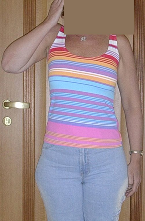
Figura 26.A: Pós operatório de sete meses, a paciente alcança a orelha.
Figure 26.A: Post operatively seven months, the patient reaches the ear.
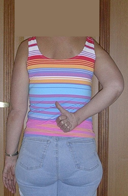
Figura 26.B: Melhora da rotação interna, alcançando L2.
Figure 26.B: Improved internal rotation, reaching L2.
Figura 27.A: Após um ano, alcançando a região da orelha.
Figure 27.A: After a year, reaching the region of the ear.
Figura 27.B: Melhora da rotação interna, após um ano consegue chegar a T11.
Figure 27.B: Improved internal rotation after a year can reach T11.
We can observe the funtional outcome three years after surgery ( Video 1 ).
Video 1: Patient after three years of surgery and performing his professional duties.

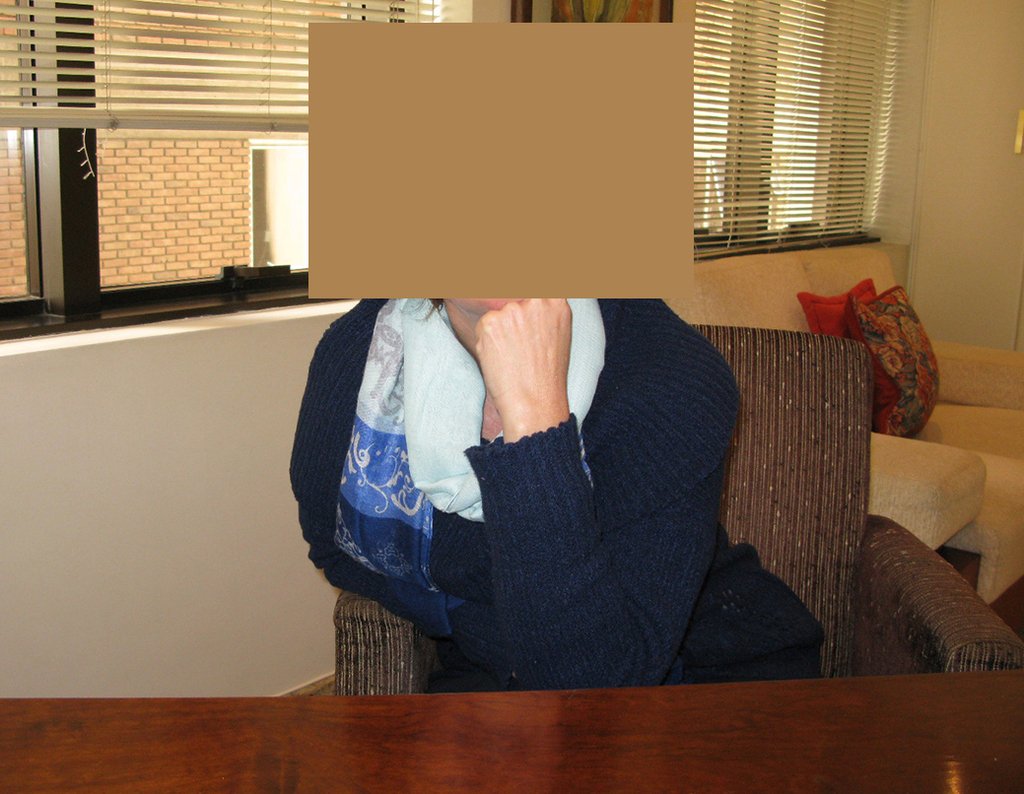
After ten years and seven months the patient did not present any complaint. Leans casually in his chair on the right elbow operated arm (Figure 28) , can raise his hand to his mouth (Figure 29Aa), good internal rotation (Figure 29b).

Figura 28: Paciente após dez anos de cirurgia, apoiando descontraidamente sobre o braço operado.
Figure 28: Patient after ten years of surgery, resting casually on the operated arm.
Figura 29.A: Elevação da mão até a orelha, dez anos após.
Figure 29.B: Hand lift to the ear, ten years after.
Figura 29.B: Após dez anos da cirurgia, com excelente rotação interna, alcançando a escápula. Sem dificuldade para o exercício profissional de odontóloga
Figure 29.A: After ten years of operation, with excellent internal rotation, reaching the scapula. No problem for the professional practice of odontologist

The patient has good function and works very well , without any difficulty , in their professional activities as Odontologist ( Video 2 ).

Video 2: Patient with ten years after surgery, without complaint and exercising his profession of odontologist all these years.

The patient has good function and plays very well, without any difficulty, their professional activities (Video 2).

The patient has good function and plays very well, without any difficulty, their professional activities (Video 2).

REVIEW:     

Chondrosarcoma is the primary malignant bone tumor more frequent  after osteosarcoma 23,24). The central subtype is the most common and affects more than five times the periferic (3), there are also rare subtypes of clear and mesenchymal cells (2).

Normally arises in the bones of endochondral origin and mainly at the root of the limbs (shoulder, pelvis, rib and spine (1)) are rare in the membranous origin (24,11,15,14). It is slow growing and often the patient seeks treatment when the lesion presents major. This tumor can affect any age, with prevalence between 30 and 40 years (7, 11, 22), with reference in the literature since three years (15) to 73 years old (1).

It is a malignant tumor of mesenchymal nature, producing interstitial substance and cells that assume aspect of hyaline cartilage, with varying degrees of immaturity and frequent calcification foci and can occur in different locations.

Can be classified according to location: A- Central , B- cortical fair (Paraosteal or Periosteal) (23,2,24,6,3), C-Peripheral (or exophytic, which occurs in the cartilaginous cap of an osteochondroma) (28)  and D- Soft Tissue Chondrosarcoma (13); on the histology in: A- degree of anaplasia: are classified into grades I, II and III, dedifferentiated; B-, C- and D-:mesenchymal clear cell; as to the origin may still be: 1 primary and 2 secondary (which originates at the site of a preexisting benign cartilaginous lesion as in Oilier disease (Enchondromatosis) or Maffucci syndrome processing for Chondrosarcoma is common (20 to 30 %) (2.28), can also occur as Solitary Osteochondroma (in less than 1%, or multiple 10%) (2), and more rarely secondary to Paget’s disease.

Pain can be insidious symptoms for several years, progressing to slow growth, volume increase, mobility restriction getting skin sometimes reddish and warm (23). The first symptom is often a fracture in pathological bone (2.24).

The radiograph shows transparent radio metaphyseal lesions replacing the bone marrow that extend into the epiphysis or diaphysis , eroding the internal cortical ( lesions in piecemeal ) , inflating or expanding the medullary portion of the bone , but remaining bounded by cortical that thick.

The appearance of calcifications ( dotted such as cotton balls ( 5 ) or rings ) is frequently (23, 2 , 24, 13 , 6, 28) . These are due to the degeneration of the cartilage that receives new vascularization and calcified . This process is accelerated in Chondrosarcoma and slow in benign cartilage lesions and low grade.

The bone mapping assists in  lesion staging. MR  and CT are important for the evaluation of the intramedullary extension and extra osseous lesion ( 2).

The diagnosis of well-differentiated Chondrosarcoma presents difficulties and histological data from clinical history, location, imaging findings should be valued for diagnostic conclusion and definition of proper behavior (23, 14, 12 ) . The irregularity of histological minutiae in the array and the number of cells within the chondroid matrix, the core of hyperchromasia changes , polymorphism and atypical mitotic when located in members of roots must be considered grade I Chondrosarcomas , although these same histological aspects can be found in benign Chondromas of hand and feet.  In microscopic descriptions are similar to Central Chondrosarcomas ( 23).

For the diagnosis we must also differentiate the pathological , clinical and radiological similarities with other injuries.

The diferential diagnosed with aneurysmal bone cyst is on the multiloculated character; with Chondroma , the Osteochondroma , Chondroblastoma , the Paraosteal and Periosteal Osteosarcoma (with cortical just Chondrosarcoma ) ( 16); Myositis Ossificans ; Fibroma Chondromyxoid ; T.G.C. and Non-Hodgkin Lymphoma ( 23 , 6, 28) . The Clear Cell Chondrosarcoma has intra lesional formation of reactive bone may cause confusion with Osteosarcoma . The Mesenchymal Chondrosarcoma is formed by small round cells that resemble Hemangiopericytoma and Ewing’s sarcoma ( 14). The central Chondroma of long bones, Chondrosarcoma and Bone Infarction are often difficult to diagnose , requiring periodically clinical and radiographic evaluation to monitor the evolution of injury and definition of conduct.

A biopsy can often not definitive for diagnosis ( 23, 28 , 12).

The treatment of Chondrosarcoma is exclusively surgery (25 ) , should be chosen a wide resection , including the path of the biopsy (21, 13). Radiation therapy is ineffective ( 6) in controlling this cancer. For cases of grade III can be argued chemotherapy indication to the protocol used for  large cell high-grade sarcomas . Mesenchymal Chondrosarcoma in which present predominance of small cell undifferentiated , discussed the chemotherapy lies with the treatment protocol of Ewing ‘s sarcoma. In both cases the response to chemotherapy is usually poor ( 6). The treatment of this cancer should be individualized for each clinical subtype.

Complications occur hematogenous metastases to the lungs ( 28) , may also present lymphatic dissemination and local recurrence. Many chondrosarcomas feature local invasion trend (14 ), reaching enormous sizes becoming inoperable and causing death by this local complications propagation.

The local recurrence increases the incidence of lung metastases ( 21).

EXERCISES:

1- What are the radiographic features of Central Chondrosarcoma ?

a) Intra and extra medullary ossification.

b) Diaphyseal lesion with bone thinning and triangle of Codman with coarse lamellar reaction.

c) Areas of bone thinning , internal cortical erosion and calcification foci.

d) Bone condensing areas with periosteal reaction onion skin.

Answer: c) the cartilaginous tissue is more radiopaque than bone and thus presents itself as causing bone thinning and enlargement of medullary lesions in the bone marrow cousing cortical erosion . This growing cartilaginous tissue gets vascular sprouts and cartilage comes into regression due to calcification.

2- What are the characteristics of the MR of Chondrosarcoma ?

a) hyper- signal in T1, T2 and low signal with contrast enhancement.

b) hypo- signal in T1, hypo- signal in T2 and capture the contrast.

c)hypo- signal in T1, hyper- signal in T2 and without contrast uptake.

d)low signal on T1, high signal on T2 and capture the contrast.

Answer: d ) the cartilage tissue has low to intermediate signal on T1 . Intermediate down the cartilage and the calcified foci . Displays contrast uptake by increasing the local metabolism due to cancer.

3- What are the main differential diagnosis of Central Chondrosarcoma ?

a) Bone infarction and chondroma.

b) Osteochondroma and Ewing’s sarcoma

c) Osteomyelitis and T.G.C.

d) Osteosarcoma and condroblastoma.

Answer: a ) Bone  Infarction causes damage to the bone marrow , but does not cause erosion of the inner cortex and has no evolutionary character of pain. It is usually a diagnosis finding on occasional ray.The same applies to the Chondroma that does not evolve and is only cartilaginous remains of development.

4- What is the treatment for the central chondrosarcoma ?

a) Intralesional curettage and autologous bone graft.

b) Wide resection and replacement with nonconventional endoprosthesis.

c)Intralesional curettage , location adjuvant with liquid nitrogen and homologous bone graft.

d)Intralesional curettage , local adjuvant electrotherm and bone cement.

Answer : b ) The wide resection provides oncological treatment and reconstruction with nonconventonal endoprosthesis  provides the best restoration of function.

5- Histologically it is difficult differential diagnosis between:

a)Osteosarcoma and Eosinophilic granuloma.

b)Grade I Chondrosarcoma and Chondroma.

c)T.G.C and Ewing’s sarcoma.

d) Osteoblastomas and Enchondroma.

Answer: b ) The central long bones Chondroma and chondrosarcoma grade I are often difficult to histological diagnosis , it requires the radiographic evaluation for the definition and conduct.

BIBLIOGRAPHY

ACKERMAN, L.V.; SPJUT, H.J. Tumors of bone and cartilage. Atlas of tumor pathology. Washington, Air Force Inst. Pathology, 1962, fasc, 4.
CANALE, S.T. Cirurgia ortopédica de Campbell. Barueri: Manole; 2006
DAHLIN, D.C. Tumores óseos . Barcelona: Ediciones Toray S/A; 1982
DORFMAN, H.D.; CZERNIAK, B. Bone tumors. St Louis, C.V. Mosby Co., 1997, cap. 7, p.410.
EDEIKEN, J.; HODES, P.J. Diagnóstico radiológico de las enfermedades de los huesos. Buenos Aires, Panamericana, 1977, cap. 15.
ETCHEBEHERE, M. Tumores cartilaginosos malignos: Condrossarcomas. In: Camargo O.P. Clínica Ortopédica. Rio de Janeiro: Med si; 2002. p. 753-759
FELDMAN, F. Cartilaginous tumors and cartilage-forming tumor like conditions of the bonés and soft tissues. In:Diseases of the Skeleton System (Roentgen Diagnosis). Part. 6 – Bone Tumors, New York, Springer-Verlag, 1977,p.177.
FLETCHER, C.D.M., Unni K.K., OMS – Merters F. (Eds.): World Health Organization. Classification of Tumors. Pathology and Genetics of Tumors of Soft Tissue and Bone. IARC Press: Lyon 2002.
GREENSPAN, A. Radiologia ortopédica. Rio de Janeiro: Guanabara; 2001.
HENDERSON, E.D.; Le PAGE, G. A. Apud FELDAMAN, F. Cartilaginius tumors and cartilage forming tumor like conditions of the bone and soft tissues. In: Disease of the Skeletal System (Roentgen Diagnosis). Part. 6 – Bone tumors, New York, Springer Verlag, 1977, p.182.
HUVOS, A.G. Bone tumors Diagnosis, Treatment and Prognosis. Philadelphia, W. B. Saunders Co., 1979, p. 13.
JAFFE, H.L. Tumores y estados tumorales oseos y articulares. México: La Prensa Medica Mexicana; 1966.
JESUS-GARCIA, R. – Reynaldo Jesus-Garcia
LICHTENSTEIN, L. Barcelona: Talleres Gráficos Ibero-Americanos; 1975.
LICHTESTEIN, L. Bone Tumor. 4 Ed St. Louis, C.V. Mosby Co., 1972, cap. 15.
LICHTESTEIN, L.; BERNSTEIN, D. Unusual benign and malignant chondroid tumors of bone. Cancer, 12:1142, 1959.
MARCOVE, R.C. Condrosarcoma: Diagnóstico y tratamiento. In: Clínicas Ortopécias de Norteamérica. Tumores del aparato musculosquelético. Buenos Aires, Panamericana, 1977, cap. 7.
MARCOVE, R.C. et al. Chondrosarcoma of the pélvis and upper end of the femur. Na analisys of factors influencing survival time in113 cases.  J. Bone Joint Surg., 54A:61, 1972.
MARCOVE, R.C.; SHOJI, H,; HARLEN, M. Altered carbohidrate metabolism in cartilaginous tumors. Contemp. Surg. 5:53, 1974.
McFARLAND, G.B.Jr.; McKINLEY, L.M.; REED, R.J. Dedifferentiation of low grade chondrosarcomas. Clin. Orthop., 122:157, 1971.
MENENDEZ, L.R. Orthopaedic knowledge update: Actualizaciones en cirugía ortopédica y traumatología. Barcelona: Ars Medica; 2003.
O’NEAL, L.W.; ACKERMAN, L. V. Chondrossarcoma of boné. Cancer, 5:551, 1952.
PRÓSPERO, J.D. Tumores Ósseos. São Paulo, Roca, 2001, cap. II.
ROBBINS. Patologia estrutural e funcional. Rio de Janeiro: Guanabara; 1996.
ROMSDAHL, M.; EVANS, H.L.; AYALA, A.G. Surgical treatment of  chondrosarcoma. In: Managment of primary bone and soft tissue tumors. Chicago, Year book med. Publisher Inc., 1977, p. 125.
SCHAJOWICZ, F. Tumores y Lesiones Seudotumorales de Huesos y Articulaciones. Buenos Aires: Editora Médica Panamericana; 1982.
TORNBERG, D.N.; RICE, R.W.; JOHNSTON, A.D. The ultrastructure of chondromyxoid fibroma. Clin. Orthop. Rel. Research, 95:295, 1973.

Autor : Prof. Dr. Pedro Péricles Ribeiro Baptista

 Oncocirurgia Ortopédica do Instituto do Câncer Dr. Arnaldo Vieira de Carvalho

373f32fe149adc95e1dae88d8993a678 2

History of Orthopedic Surgeons’ Training.

History of Orthopedic Surgeons' Training. Treatment of Bone Tumors

History of Orthopedic Surgeons’ Training. In the mid-20th century, Doctors Walter Edgard Maffei and Bartolomeu Bartolomei, both short-statured physicians who turned out to be giants of Medicine, could be seen walking through the hospital corridors.

In January 1952, Maffei sought to structure the Department of Pathology, welcoming José Donato de Próspero, who had just graduated from USP in 1951. At the time, Professor Donato noticed an orthopedic surgeon frequently visiting the pathology department, which puzzled him initially: How could a “bone plumber” dare to examine specimens and look through a microscope?

The persistence of this character piqued Donato’s curiosity. He observed, with intrigue, that only two conditions seemed to be diagnosed in the field of orthopedics, as the specimens received were often diagnosed either as osteomyelitis or osteosarcoma. No one dissected or studied the amputation specimens in a rigorous manner.

At that time, the Department of Pathology was located on the top floor of the Central Laboratory, and the phenomenon that was Bartolomeu insisted on studying those specimens, invading Maffei’s sanctuary daily.

In 1958, Huppert Sissons, a pathologist from the Royal Institute of London with a special dedication to bone pathology, visited the hospital.

This visitor spent four weeks frequenting the Departments of Pathology and Orthopedics, closely followed by Donato de Próspero. They became close friends and corresponded for years. From then on, Donato de Próspero began to invade Bartolomeu Bartolomei’s space, becoming a regular attendee at orthopedic meetings and requesting permission from the department to archive the X-rays that interested him. The study of this rich material was shared with radiologist Alfonso Vitule.

The collection of X-rays gathered by Donato de Próspero became a valuable archive that is still under the custody of the current Orthopedic Oncology Group. This concern with documentation was crucial for the knowledge and growth of the specialty, which is now compiled in the book “Bone Tumors” by Editora Rocca.

At that time, X-rays, when stored at all, were kept in the trunks of Bartolomeu Bartolomei’s three Ford Galaxy cars; he was a true magician.

In 1963, Elio Consentino completed his training in orthopedics and began his professional career at Santa Casa de São Paulo, where he worked alongside Bartolomeu and Donato. Later, they would create the Bone Tumor Group, initiating the training of numerous orthopedic surgeons in the specialty of orthopedic oncological surgery.

In 1970, this group of bone pathology enthusiasts received a visit from Fritz Schajowicz, who invited José Donato de Próspero to join the select group of nine pathologists from different nations who would develop the International Classification of Bone Tumors at the request of the World Health Organization.

In the following years, many letters, exchanges of slides, and meetings took place to conclude the work, and in 1978, the WHO Classification was published.

Throughout all these years, orthopedic meetings continued to be held, now divided into specialty subgroups. In 1994, Pedro Péricles Ribeiro Baptista took over the leadership of the Orthopedic Oncology Group.

Doctors Claudia Kazue Yamaguchi, Hea Yung Yo, Guinel Hernandez Filho, and Abdalla Youssef Skaf from radiology, José Donato de Próspero and Maria Fernanda Carriel Amary from pathology, Cássio Perleizzon from radiotherapy, Schlomon Lewin and Akemi Ogawa from nuclear medicine, Andréa Basta from physiotherapy, Pedro Péricles Ribeiro Baptista, Alex Guedes, and Eduardo Ribeiro Adriano, among others, actively participated in the Multidisciplinary Team, attending the anatomical-clinical meetings held every Wednesday from 1:00 PM to 3:00 PM.

Several doctors contributed to the strengthening of this Group, and many completed their training through six-month to one-year internships.

The following doctors trained at the institution and contributed to orthopedic oncology: Bartolomeu Bartolomei (1954-1973 SP), Elio Consentino (1975–2006 SP), Teruo Yoneda (1979–1981 SP), Eduardo Salim Haddad Filho (1981–1982 SP), Javier Perez Torres (1982–1985 Colombia), Moisés Cohen (1983-1984 SP), Pedro Péricles Ribeiro Baptista (1983–2015 SP), Valter Pena (1983-1984 SP), Reynaldo Jesus-Garcia Filho (1985-1985 SP), Paulo Alencar (1985-1985 PR), Eduardo Bertacchi Uvo (1986-1986 ES), Paulo Taniguchi (1986-1986 Ba), José Custódio de Moura (1987-1987 SP), Gilbert Sotomayor Alvear (1988-1989 Ecuador), Maria Guadalupe Repper (1988-1988 Mexico), Ernesto Luiz Michanie (1989-1990 Argentina), Carlos Henrique Ribeiro do Prado (1991-1991 GO), Marcos Hajime Tanaka (1991-1993 SP), Alejandro Enzo Cassone (1992-1992 SP), Cláudio Luiz Wanderley Saab (1993-1993 MS), Julio Cesar Goyano (1993-1993 GO), Lucíola Assunção Alves (1993-1993 MA), Florindo Volpe Neto (1994-1996 RS), Giancarlo Polesello (1995-1995 PR), Marcos Sanmartin Fernandez (1996-1996 Spain), Rogério de Andrade Amaral (1997-1997 GO), Alex Guedes (1998-1999 BA), Ricardo Fontes Lavieri (1998-1998 SP), Roberto Reggiane (1998-1998 MG), José Augusto Sá Lopes (1998-1998 PI), Ricardo Marzola (2001-2001 SP), Fabrício Lenzi Chiesa (2001-2002), Israel Vargas (2001-2001 Honduras), André Rensi de Mello (2001-2001 SP), Pedro Calabrese (2002-2003 Paraguay), Carlos Arturo Izquierdo (2002-2002 Colombia), Fábio Romualdo (2002-2002 RN), Esdras Fernando Furtado (2002-2003 PB), Adriano Ughini (2003-2003 RS), Aureliano Duarte Bezerra (2003-2003 PE), Érico Raimundo Guimarães Fantini (2003-2003 MG), Rodrigo Pólo da Costa (2003-2003 AM), Eduardo Ribeiro Adriano (2003-2006 SP), Pablo de Andrade Lima Filho (2004-2004 PE), Rafael Monteiro Macedo (2004-2004 TO), Flávio Leão Rabello (2005-2006 GO), Gustavo Henrique Ferreira (2005-2006 MS), Marcelo Rodrigues Alves (2005-2006 RJ), Alexandre Ferracioli Fusão (2006-2007 SC), Daniela Cruvinel Petto (2006-2007 SP), and Leonardo Silva Galloni (2006-2007 RS).

Author: Prof. Dr. Pedro Péricles Ribeiro Baptista

 Orthopedic Oncosurgery at the Dr. Arnaldo Vieira de Carvalho Cancer Institute

Hello! How can we assist you?