• Rua General Jardim, 846 cj 41 Higienópolis, São Paulo - SP

Author Archives: drpprb@gmail.com

This digital library houses the book on Oncology and Orthopedic Oncosurgery.

It includes academic lectures, presentations from national and international congresses, published papers, case discussions, performed surgical procedures, and proprietary techniques developed.

The digital format was chosen because the web allows the inclusion of texts with numerous visual resources, such as images and videos, which would not be possible in a printed book.

The content is intended for students, healthcare professionals, and the general public interested in the field.

drpprb@gmail.com

Fd4eeaf6abdc74d8c3a37fbfbb055cd9 3

Osteosarcoma

[auto_translate_button]
Osteosarcoma is an immature neoplasm that forms osteoid and neoplastic bone produced by malignant osteoblasts, with a strongly sarcomatous stroma. Histologically, it may appear as osteoblastic, chondroblastic, fibroblastic or teleangectatic, depending on the similarity of its cells. After myeloma, it is the most common primitive bone neoplasm.

Osteosarcoma

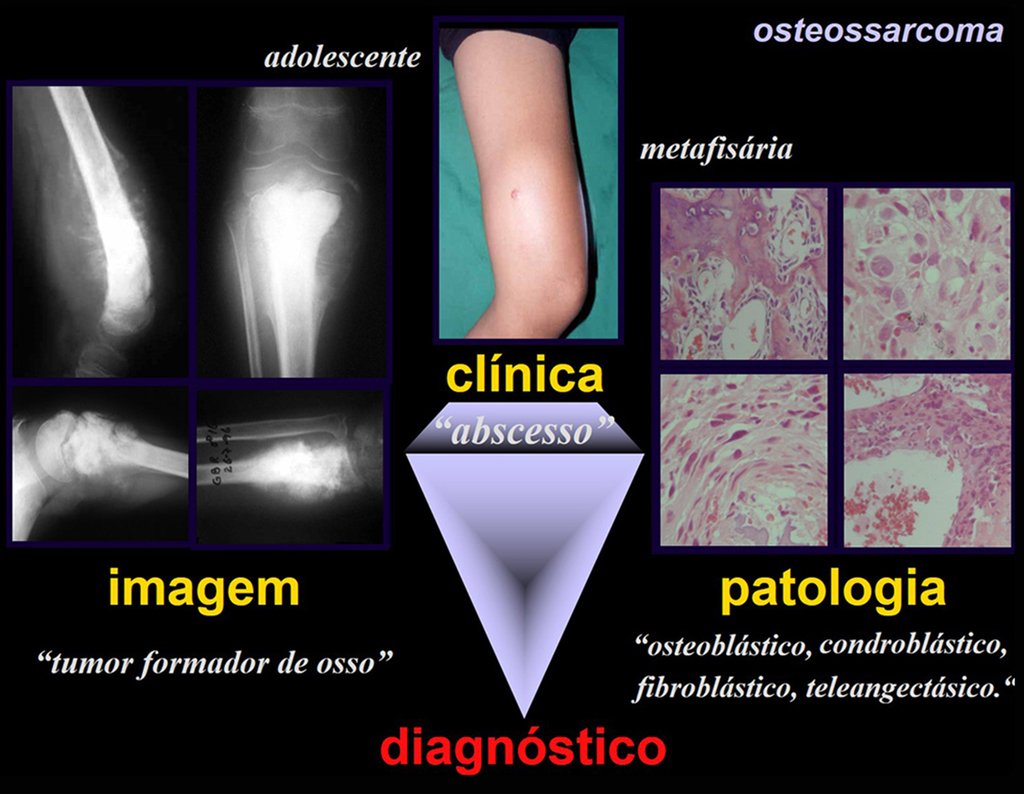
It affects young people in the first and second decade, affecting the metaphyseal region of long bones, close to the growth plate. Clinically, it presents  pain  in the affected region,  heat  due to hyperemia,  tumor  due to increased local volume plus some  functional limitation .

Radiographically, it presents a lesion in the metaphyseal region characterized by areas of new bone formation interspersed with areas of bone rarefaction, with imprecise limits, with cortical erosion and a thin lamellar periosteal reaction, forming Codman’s triangle. The presence of Codman’s triangle denotes the existence of an extra-cortical tumor.

Para o diagnóstico dos tumores ósseos é preciso fazer a correlação entre a clinica, a radiologia e a anatomia patológica.
To diagnose bone tumors, it is necessary to make a correlation between the clinic, radiology and pathological anatomy.

Codman’s triangle (figure 2) is a radiographic sign that occurs in other conditions such as osteomyelitis, eosinophilic granuloma, Ewing’s sarcoma and other tumors. It can appear whenever a lesion occurs inside a bone and grows outside of it. When the lesion  (or pus in osteomyelitis)  surpasses the cortical bone, it causes detachment of the periosteum that was attached to the cortical bone. The lesion therefore grows and this detachment stimulates the periosteum to produce bone. In this way, a layer of mineralization occurs as if a neocortical was formed. This process can occur successively, forming true layers (lamellae) (figures 3a and 3b). These layers can be thin  (thin lamellar periosteal reaction – characterizing the growth speed of the lesion)  or thick   (thick lamellar periosteal reaction – characterizing slow growth) . As the lesion progresses, this attempted “barrier” ruptures and a triangle appears radiographically delimited  externally by the periosteum  (which was successively detached and removed from the bone cortex),  internally by the  bone cortex and at the  base by the  expanding tumor.

         The imaging study of the lesion can be more detailed using tomography (figure 3c), which allows the evaluation of erosion of the bone cortex and extra-cortical involvement. Magnetic resonance imaging is important to define the extent of the tumor, both in the medullary and extracortical areas (figures 4d, 4e and 4f). This exam also allows you to evaluate the relationship between the tumor and the soft parts (muscles, vessels and nerves) and is essential for surgical planning.

Figura 2: Triangulo de Codman (presente em osteossarcoma, osteomielite, sarcoma de Ewing e granuloma eosinófilo).
Figure 2: Codman's triangle (present in osteosarcoma, osteomyelitis, Ewing's sarcoma and eosinophilic granuloma).
Figura 3: a) osteossarcoma com triângulo de Codman; b) reação periosteal lamelar fina; c) tumor produtor de osso.
Figure 3: a) osteosarcoma with Codman’s triangle; b) thin lamellar periosteal reaction; c) bone-producing tumor.
Figura 4: d) lesão heterogênea, limites imprecisos; e) epífise sem lesão; f) lesão extra cortical.
Figure 4: d) heterogeneous lesion, imprecise limits; e) epiphysis without injury; f) extra-cortical lesion.
The current treatment of osteosarcoma consists of preoperative polychemotherapy, for approximately 3 to 4 cycles, followed by complete resection of the tumor and complemented with postoperative chemotherapy. It is currently possible to resect the tumor (figures 5, 6 and 7) and reconstruct the affected segment with prostheses or biological solutions (figure 6) (autologous or homologous bone graft). Ablative surgeries (amputations) are reserved for cases of advanced tumor, of large volume, compromising the skin and/or vascular-nervous bundle or in those cases that did not obtain a good response to pre-operative chemotherapy and whose conservative resection, with preservation of the limb , implies a risk of local recurrence of the lesion.
Figura 5: a) osteossarcoma com fratura consolidada, pós quimioterapia.
Figure 5: a) osteosarcoma with healed fracture, post chemotherapy.
Figura 6: b) ressecção transepifisária; c) corte axial da placa de crescimento, removida com a lesão.
Figure 6: b) transepiphyseal resection; c) axial cut of the growth plate, removed with the lesion.
Figura 7: d) reconstrução com autotransplante de fíbula vascularizada, preservando sua placa de crescimento, encaixada em enxerto homólogo e estabilizada com placa angulada e dispositivo de fixação interna extensível.
Figure 7: d) reconstruction with autotransplantation of a vascularized fibula, preserving its growth plate, embedded in a homologous graft and stabilized with an angled plate and an extensible internal fixation device.

Author: Prof. Dr. Pedro Péricles Ribeiro Baptista

 Orthopedic Oncosurgery at the Dr. Arnaldo Vieira de Carvalho Cancer Institute

Condroma

Juxtacortical Chondroma

[auto_translate_button]

Chondroma: Benign Tumor of Cartilage in Bones

Chondroma is a benign cartilage tumor that often affects the short bones of the hands and feet (fig. 18). Essentially, it is a mass that forms from cartilaginous tissue. It can appear as a single lesion or, in more severe cases, affect several bones, forming what is known as enchondromatosis.

Unilateral enchondromatosis is a specific form of bone dysplasia, called Ollier’s disease (fig. 19a; 19b; 19c), characterized by the presence of multiple chondromas on one side of the body. On the other hand, Maffucci Syndrome is an even rarer condition, characterized by the presence of multiple enchondromas associated with hemangiomas.

Although less common, chondroma can also develop in long bones, such as the distal femur (fig. 20), proximal humerus and tibia. In these locations, the differential diagnosis between chondroma and other conditions, such as bone infarction and central chondrosarcoma, can be challenging. Bone infarction, generally painless, can be detected by radiographic examinations, while central chondrosarcoma is symptomatic, with erosion of the internal cortical bone. In these cases, clinical and radiographic observation is crucial before any therapeutic intervention.

Radiographically, the chondroma usually appears as an area of ​​rarefaction, which may present points of calcification and eventually cause bone deformities.

Typical treatment for chondroma involves curettage of the lesion and, if necessary, bone grafting. When the chondroma affects the external surface of the bones, it is known as juxtacortical chondroma and is generally treated with surgical parietal resection (fig. 21).

In short, chondroma is a benign condition, but its presence and behavior must be carefully monitored and treated to avoid complications.

Author: Prof. Dr. Pedro Péricles Ribeiro Baptista

 Orthopedic Oncosurgery at the Dr. Arnaldo Vieira de Carvalho Cancer Institute

7651301cabf91a1be8e3cf0b72e8734f 3

Osteoid Osteoma

[auto_translate_button]
Osteoid Osteoma Osteoid osteoma is a benign osteoblastic lesion, smaller than one centimeter, with precise limits and with reactive bone sclerosis around osteoid tissue, with highly vascularized stroma and histologically mature bone.

Osteoid osteoma

It is a lesion that is preferably located in the cortex of long bones or in the pedicle of the spinal column (compact bones). It can occur in three different locations in the bone:

  • Cortical  : the vast majority, figures 1 and 2a, 2b and 2c.
Figura 1: Quadro clínico de tumefação devido a osteoma osteóide da cortical medial da tíbia .
Figure 1: Clinical picture of swelling due to osteoid osteoma of the medial cortical bone of the tibia.
Figura 2: Aumento de densidade da cortical medial, devido à esclerose óssea (a); corte tomográfico mostrando a lesão de rarefação na cortical, circundada por halo de esclerose (b) e nicho de osteoma osteóide com a esclerose, já ressecado (c).
Figure 2: Increased density of the medial cortex, due to bone sclerosis (a); tomographic section showing the rarefaction lesion in the cortex, surrounded by a halo of sclerosis (b) and an osteoid osteoma niche with sclerosis, already resected (c).
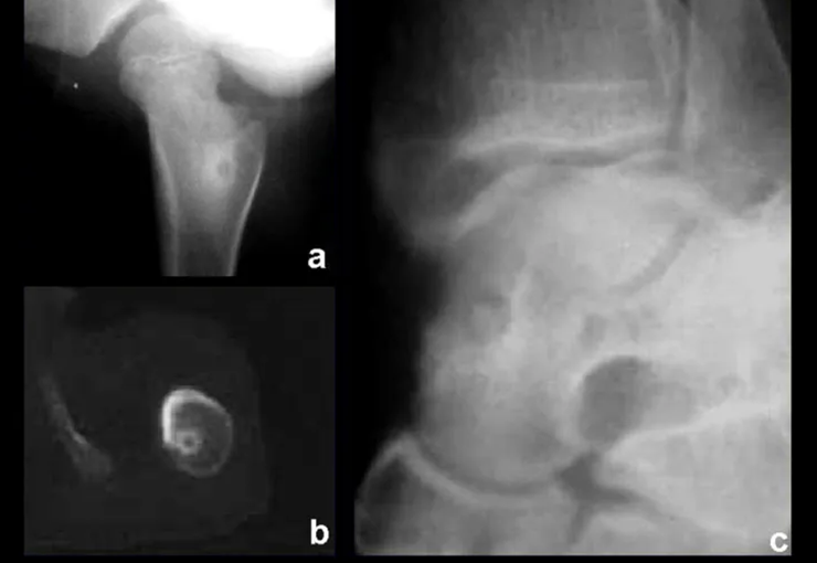
Figura 3: Osteoma osteóide na medular do fêmur (a); tomografia confirmando a localização central (b); osteoma osteóide medular do tálus (c).
Figure 3: Osteoid osteoma in the medullary bone of the femur (a); tomography confirming central location (b); Medullary osteoid osteoma of the talus (c).
  • Medullary : or spongy (endostal), figure 3a, 3b and 3c.
  • Figura 4: Radiografia do antebraço mostrando lesão subperiosteal na face ulnar do rádio, com nicho central do osteoma osteóide e halo de esclerose óssea (a), e tumor ressecado (b).
    Figure 4: Radiograph of the forearm showing a subperiosteal lesion on the ulnar surface of the radius, with a central niche of osteoid osteoma and a halo of bone sclerosis (a), and resected tumor (b).
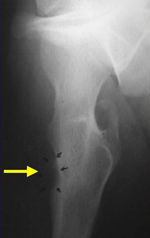
    Figura 5: Osteoma osteóide subperiosteal na face medial do fêmur.
    Figure 5: Subperiosteal osteoid osteoma on the medial surface of the femur.
    Figura 6: Nicho de osteoma osteóide na superfície do fêmur, periosteal.
    Figure 6: Osteoid osteoma niche on the surface of the femur, periosteal.
    • Sub-periosteal,  figures 4, 5 and 6.

    It occurs in the first and second decade of life, occurring twice as often in males.

    The clinical picture is nighttime pain (mild pain that the child does not manifest while playing), which improves with the administration of salicylates. Local swelling (fig. 4) and arthralgia may occur.

    Makes differential diagnosis with steoma, osteoblastoma and Brodie’s abscess  (sequestration of chronic osteomyelitis).

    Treatment consists of resection of the central niche (fig. 5). It is not necessary to remove all the surrounding sclerosis. When the bone failure caused by resection poses a risk for fracture (as in the femoral neck (fig. 6), a bone graft must be placed.

    Author: Prof. Dr. Pedro Péricles Ribeiro Baptista

     Orthopedic Oncosurgery at the Dr. Arnaldo Vieira de Carvalho Cancer Institute

    Bdd1b613ee6fcac7694cf648430358ce 3

    Osteoma

    [auto_translate_button]
    Osteoma Benign, slow-growing lesion, with mature bone tissue, with a lamellar structure, well differentiated. It is bone, dense , within the bone, whether in the cortical or medullary region .

    Osteoma

    It can manifest itself in three distinct clinical forms:

    • Exostoses  (dense, homogeneous bone, with an  ivory appearance ): this is the conventional osteoma, restricted to bones of intramembranous origin (facial bones, skullcap), figures 1 to 10.
    Figura 1: Radiografia do crânio com lesão nodular, densa, homogênea, na calota.
    Figure 1: X-ray of the skull with a nodular, dense, homogeneous lesion in the cap.
    Figura 2: Tomografia axial do crânio, com lesão acometendo as duas tábuas, com abaulamento maior da cortical externa.
    Figure 2: Axial tomography of the skull, with the lesion affecting both tables, with greater bulging of the external cortex.
    Figura 3: Reconstrução tomográfica em três dimensões da lesão do crânio.
    Figure 3: Three-dimensional tomographic reconstruction of the skull lesion.
    Figura 4: Nódulo firme, saliente, na base de implantação do cabelo.
    Figure 4: Firm, prominent nodule at the base of hair implantation.
    Figura 6: Ostectomia com formão.
    Figure 5: Asepsis and antisepsis, with plastic field placement. Figure 6: Ostectomy with chisel.
    Figura 8: Fragmentos do osteoma ressecado.
    Figure 7: Regularized surgical bed, without protrusions. Figure 8: Fragments of the resected osteoma.
    Figura 9: Osteoma exofítico da região parietal, aspecto de marfin.
    Figure 9: Exophytic osteoma of the parietal region, ivory appearance.
    Figura 11: Segmento ressecado na base.
    Figure 10: Ivory-looking lesion. Figure 11: Resected segment at the base.
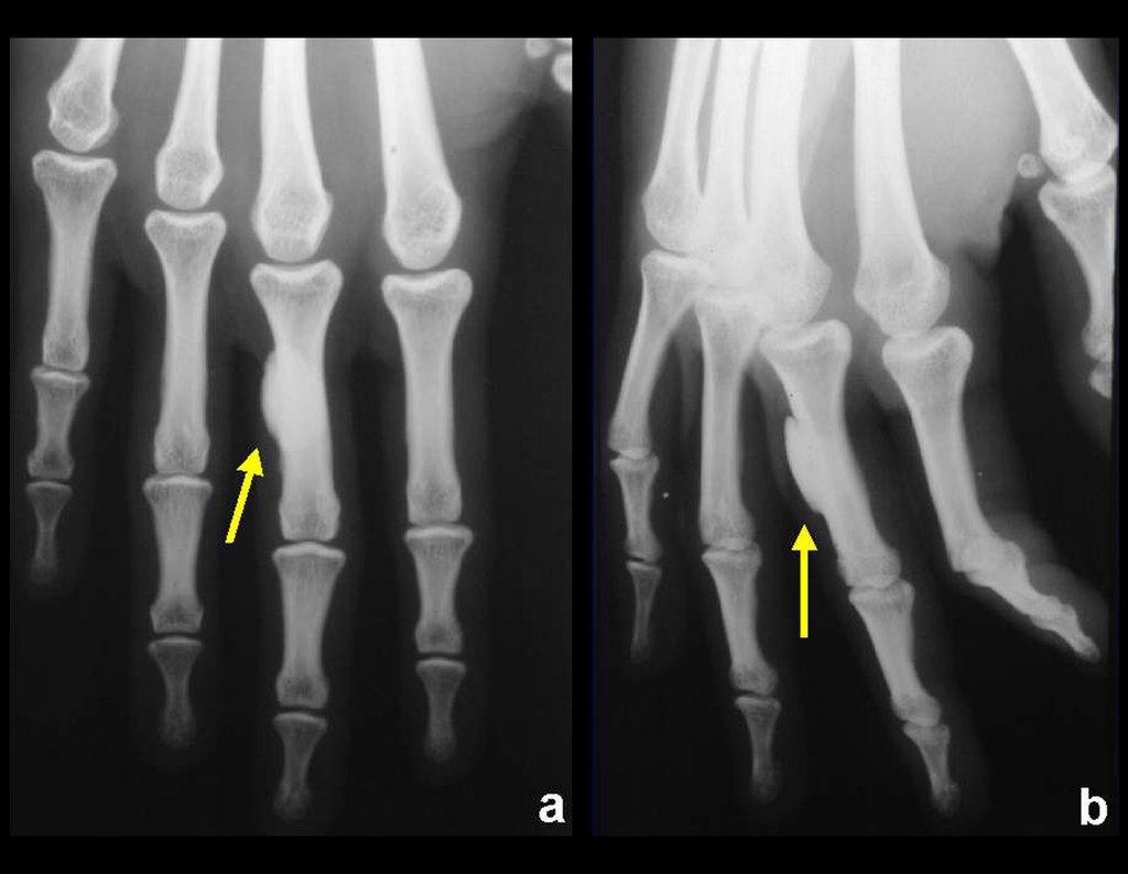
  • Parosteal  (juxtacortical) occurs on the external surface of long or short bones, figure 12.
  • Figura 12: Osteoma da falange proximal, justacortical.
    Figure 12: Osteoma of the proximal phalanx, juxtacortical.
    Figura 13 : Osteoma medular do colo femoral, enostose óssea.
    Figure 13: Medullary osteoma of the femoral neck, bone enostosis.
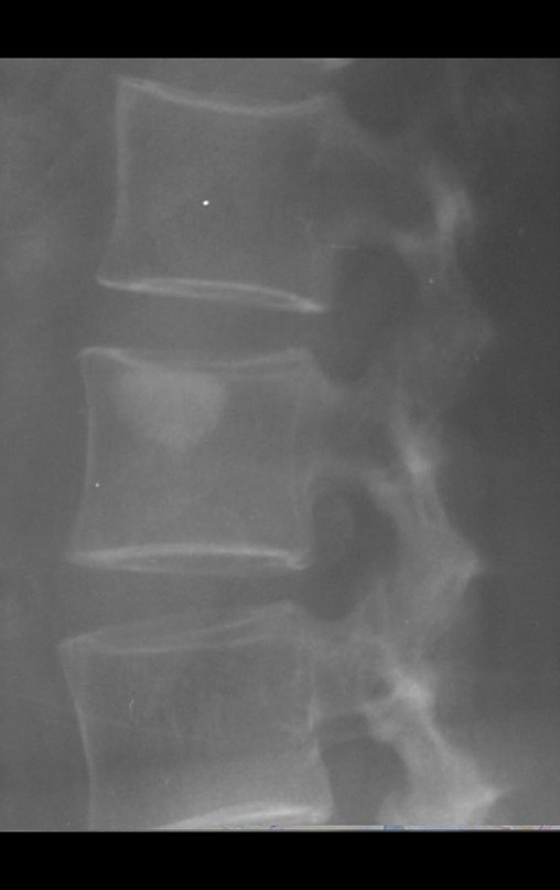
    • Medullary : known as enostosis or bone islet. Lesions histologically similar to mature bone, dense and homogeneous, without significant clinical signs and generally resulting from radiographic findings, figures 13 to 16.
    Figura 14 : Osteoma medular do corpo vertebral, enostose óssea.
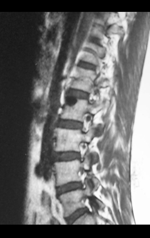
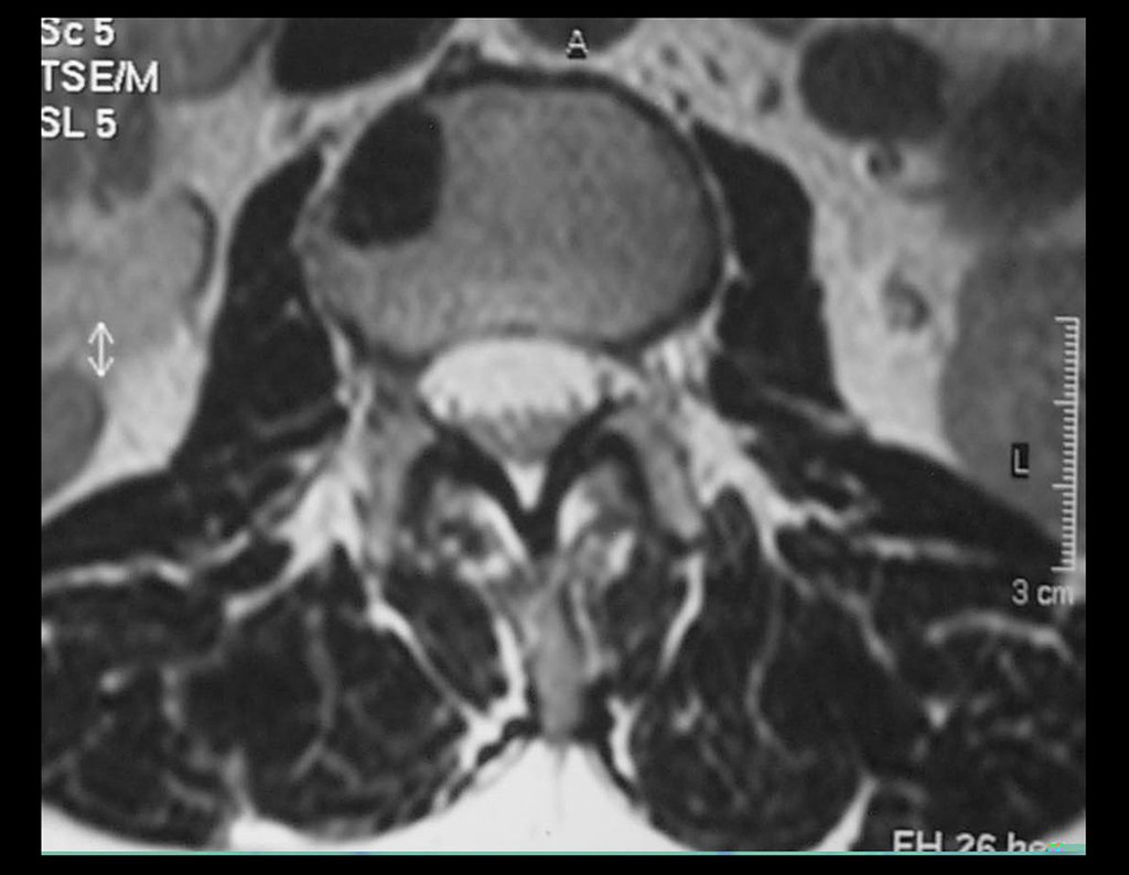
    Figure 14: Medullary osteoma of the vertebral body, bone enostosis.
    Figura 15: Rm sagital de osteoma medular do corpo vertebral.
    Figure 15: Sagittal MRI of medullary osteoma of the vertebral body.
    Figura 16: Rm axial de osteoma medular do corpo vertebral.
    Figure 16: Axial MRI of medullary osteoma of the vertebral body.

    he differential diagnoses, both from a clinical, histological and radiographic point of view, include the following conditions:

    • Bone sclerosis  (inflammatory or post-traumatic cause)
    • Hyperostoses
    • Meningioma
    • Osteoid osteoma.

    Treatment consists of observation when asymptomatic or surgical resection when indicated due to aesthetic or functional changes (compression or obstruction – nasal fossa).

    Author: Prof. Dr. Pedro Péricles Ribeiro Baptista

     Orthopedic Oncosurgery at the Dr. Arnaldo Vieira de Carvalho Cancer Institute

    A4d4ec4aa3c45731396ed6e65fee40b9 3

    Osteochondroma

    [auto_translate_button]
    Osteochondroma is also known as osteo-cartilaginous exostosis . It represents the most common benign bone lesion. Its incidence is even higher than that reported in the literature as many patients have asymptomatic osteochondromas.

    Osteochondroma

    It occurs in the first and second decades, in the metaphyseal region of long bones, radiographically characterized by a tumor that forms cartilage and bone. Characteristically, the central cancellous bone of the exostosis continues with the medullary of the affected bone and the dense, cortical layer of the tumor continues with the normal cortical bone. On the surface of this lesion there is a band of cartilage, through which the lesion grows (hence the name  osteo-chondroma: tumor forming cartilage and bone ).

    It presents with a sessile base (enlarged base) or pedicled shape.

    May be single or multiple (hereditary osteochondromatosis)

    Osteochondromas require surgical treatment ( resection ) when they alter aesthetics, compress vascular-nervous structures or limit function. They generally grow while the patient is in the growth phase.

    When an osteochondroma increases in size after completion of skeletal maturity, it may mean post-traumatic bursitis or malignancy to chondrosarcoma and must be resected with an oncological margin.

    Solitary osteochondroma can have a 1% malignancy rate, whereas multiple osteochondromatosis can reach 10%.

    Among benign cartilaginous lesions, we cannot forget chondroblastoma.

    Author: Prof. Dr. Pedro Péricles Ribeiro Baptista

     Orthopedic Oncosurgery at the Dr. Arnaldo Vieira de Carvalho Cancer Institute

    0ade14765069c05180284d03b3097202 3

    Osteoblastoma

    [auto_translate_button]

    Osteoblastoma

    Benign osteoblastic lesion, locally aggressive, with a histological structure similar to osteoid osteoma but without reactive bone sclerosis and larger in size, generally larger than 1.5 cm

    Osteoblastoma

    Radiographically, it presents large areas of bone rarefaction with few denser foci of ossification.

    It presents two clinical forms:

    • Genuine osteoblastoma : the most common, located in the pedicle of the spine and more rarely in the metaphysis of long bones.
    • Aggressive  (“malignant”) osteoblastoma.

    It affects the first and second decade of life.

    The clinical picture is one of intense pain, which can lead to fractures and functional and neurological deficits, when it occurs in the spine, sometimes with antalgic scoliosis.

    Makes differential diagnosis with:

    • Osteoid osteoma,
    • Aneurysmal bone cyst,
    • Osteosarcoma.

    Treatment consists of en bloc resection of the lesion and bone grafting when necessary, reserving careful curettage for regions such as the spine, where it is generally not possible to resect en bloc. The use of local adjuvants (phenol, electrothermia) has been increasingly used, seeking to avoid recurrence. 

    Author: Prof. Dr. Pedro Péricles Ribeiro Baptista

     Orthopedic Oncosurgery at the Dr. Arnaldo Vieira de Carvalho Cancer Institute

    D0812edf466bc20c650b452513b583e7 3

    Bone Metastasis

    [auto_translate_button]
    Bone Metastasis The lung is the largest filter in our body, through which all circulation passes and therefore any metastatic embolus that reaches the venous circulation has a high probability of suffering stasis in the lung and developing metastatic lesions. This is why the lung is the biggest site for metastases in our body. The liver represents a filter for the digestive system, which through the portal circulation can receive metastatic emboli from that system.

    Bone Metastasis

        Skeletal tissue represents the third major filter, which, due to its slow sinusoidal circulation, favors the shelter of metastatic emboli that may reach the bone.

             Currently, with the increase in the survival of patients with different neoplasms, resulting from increasingly earlier diagnoses, advances in chemotherapy with a variety of increasingly effective drugs and the control of side effects, the number of patients who have their primitive disease controlled and that metastasize to the skeleton is increasing.

             The tumors that most frequently produce bone metastases are breast carcinoma in women, prostate carcinoma in men, and lung carcinoma. kidney and thyroid in both.

             The bone injuries that most frequently give clinical manifestations, requiring orthopedic surgery, occur in the femur, humerus, vertebrae, pelvis, scapula and tibia, in that order.

             The natural history of this condition is painful and the diagnosis can generally occur due to a pathological bone fracture that causes functional impotence, limitations in activities of daily living, dependence on others, bedsores and multiple organ failure due to the patient being bedridden.

             The role of the oncology orthopedist aims to operate on the metastatic lesion as early as possible in order to alleviate pain, restore function and improve the patient’s quality of life.

             For this purpose, resection and reconstruction surgeries are performed using osteosyntheses with cement or endoprostheses.

             Radiotherapy can eventually be used as a palliative measure, aiming to relieve pain for patients who are not clinically fit for surgery. 

             In relation to multiple myeloma, which is the most common primitive tumor of the bone, a tumor of the SRE, we must add that it is treated with chemotherapy and is also highly sensitive to radiotherapy. However, in cases that develop significant bone injuries or risk of fracture, orthopedic surgical management is similar to the treatment of bone metastases.

    Click here to download PDF article on male breast tumor metastasis.

    Click here to see hypernephroma metastasis treatment.

    Author: Prof. Dr. Pedro Péricles Ribeiro Baptista

     Orthopedic Oncosurgery at the Dr. Arnaldo Vieira de Carvalho Cancer Institute

    Lesões Ósseas Pseudotumorais

    Pseudotumor Bone Lesions

    [auto_translate_button]

    Pseudotumor Bone Lesions

    The group of diseases known as pseudotumor bone lesions corresponds to the set of bone changes that mimic, from a radiographic point of view, tumor lesions.

    Pseudotumor Bone Lesions

    The injuries that are part of this group are:

    1. Simple bone cyst
    2. Aneurysmal bone cyst
    3. Juxtacortical bone cyst (Intraosseous ganglion)
    4. Metaphyseal fibrous defect  ( non-ossifying fibroma )
    5. Eosinophilic granuloma
    6. Fibrous dysplasia  /  osteofibrodysplasia
    7. Myositis ossificans
    8. Brown tumor of hyperparathyroidism
    9. Intraosseous epidermoid cyst
    10. Giant cell reparative granuloma
    Lesões Ósseas Pseudotumorais
    What is this child's diagnosis?
    • Simple Bone Cyst

    The  Simple Bone Cyst  is a cavity, initially unicameral, filled with clear liquid surrounded by a membrane, with vascularized connective tissue showing osteoclastic giant cells, and there may be some areas of hemorrhage or fissures with content rich in cholesterol.

    Diagrama do C.O.S.
    COS Diagram
    • It occurs between 5 and 15 years of age, with a slight predominance in males. It most frequently affects the proximal metaphyseal region of the humerus and femur.

      Although its etiology is still unknown, we have seen contrast inside vessels when we infiltrate the cavity, which makes us assume that it is a vascular phenomenon.

      The fracture is often the first manifestation of the cyst, which has often evolved asymptomatically.

      It appears as a radio-transparent lesion in the metaphyseal region of long bones, centrally located, not exceeding the width of the epiphyseal line. With growth, the simple bone cyst moves away from the growth plate, occupying a meta-diaphyseal position, and can erode and fracture the cortex.

                  Its treatment is generally non-operative, classically performed with a series of three corticosteroid injections, at intervals of four weeks. In load-bearing bones, particularly in the femoral neck region, we must consider the possibility of surgical treatment, with curettage and bone grafting.

      Definition:

        Unicameral cavity filled with clear or bloody fluid and limited by a membrane of variable thickness, with vascularized connective tissue showing osteoclastic giant cells and some areas with recent or old hemorrhage or fissures with cholesterol-rich content (OMS)

      Incidence:

      In the treatment of musculoskeletal tumors, we observed a predominance of cases in the age group between 5 and 15 years, with a slight predominance of cases in males, and the majority involving the proximal metaphyseal region of the humerus and femur. The vast majority are referred due to an episode of fracture caused by trauma at the site of the injury or as an x-ray finding during an eventual x-ray taken due to some trauma suffered by the patient.

      Etiology:

      Although its recognition from a radiographic point of view is simple, its etiology is still unknown.

      Clinical Assessment:

      Most patients present asymptomatically, and fractures are often the reason for their first consultation with an orthopedist. Some patients report sporadic episodes of pain or functional limitation before the presence of a bone cyst is diagnosed.

      Radiographic Characteristics:

      The Simple Bone Cyst presents as a radiolucent lesion in the metaphyseal region of long bones, centrally located, mainly in the proximal region of the humerus and femur and close to the epiphyseal line. They are well-defined lesions, with sclerotic edges, rarely crossing the limits of the cortex or the limits of the bone, expanding, thinning the cortex, but almost never breaking it. In some cases, the “fallen fragment” sign can be observed, which represents fragments of the cortical wall loose within the cyst.

      Treatment:

      COS treatment depends on its location and size, in the vast majority of cases it can be conservative and non-operative. In general, treatment for the upper limb is less surgical and more conservative, whereas treatment for the lower limb tends to be more surgical, in an attempt to avoid a fracture. The classic treatment consists of infiltrations with corticosteroids (depomedrol), observing whether or not bone content is formed inside. If there is an imminent fracture in a load-bearing bone, we should seriously consider the possibility of intra-lesional treatment filling the cavity with either an autologous or homologous graft.

      1-  Click to see more :   http://bit.ly/cisto_ósseo_simples

      2-  Reconstruction of the femoral neck with fracture due to bone cyst

      • Aneurysmal Bone Cyst

      Definition:

      Expansive, blood-filled osteolytic lesion between variable-sized spaces separated by connective tissue septa containing trabeculae of bone or osteoid tissue and osteoclastic giant cells.

      Incidence:

      Aneurysmal Bone Cysts are more frequent in the first three decades of life, with their peak incidence occurring around 10 to 15 years of age, with a slight predominance in females than in males.

      Etiology:

      The origin and etiology are still unknown

      Clinical Assessment:

      Patients normally present with mild pain at the site of the injury and inflammatory signs such as increased volume and heat at the site are often observed. When there is a compromise in the spine, there may be neurological symptoms of spinal cord or nerve root compression. The evolution is very variable with a slow and progressive increase in volume or in some cases rapidly expansive. It frequently affects the lower limbs and vertebrae, including the sacrum and the pelvis, mainly in the iliopubic branch.

      Radiographic Characteristics:

      It presents an insufflative and radiotransparent lesion mainly in the metaphyseal or diaphyseal region of long bones, with the presence of septa scattered throughout its content, with thinning and expansion of the cortex, which may be eccentric or central.

    Diagrama do cisto ósseo aneurismático
    Aneurysmal bone cyst diagram

    Treatment:

    The treatment of choice has been intralesional curettage and filling with autologous graft. Oftentimes, it may or may not be possible to associate intra-lesional adjuvant treatment with the application of phenol, or electro cauterization or the use of cryotherapy. Rarely and in some specific cases, it is possible to resect the compromised bone without compromising function, as in cases of involvement of the rib, fibula and metacarpal and metatarsal bones. Recurrence is very rare.

    Cisto aneurismático da fíbula; lesão sendo ressecada e leito operatório após a retirada da lesão.
    Aneurysmal cyst of the fibula; lesion being resected and operating bed after removal of the lesion.
    Peça ressecada; radiografia pós operatória e paciente com carga monopodal, boa função sem instabilidade.
    Dry piece; Post-operative x-ray and patient with single-leg weight bearing, good function without instability.

    1-  Click here to see more :    http://bit.ly/cisto_aneurismático

    • Eosinophilic granuloma

    (Histiocytosis X, Langerhans Cell Granuloma, Reticuloendotheliosis)

    Eosinophilic Granuloma is   a pseudo-tumor lesion, also of unknown etiology, characterized by intense proliferation of reticulohistiocytic elements with a variable number of eosinophils, neutrophils, lymphocytes, plasma cells and multinucleated giant cells. It presents frequent areas of necrosis, as well as the presence of numerous fatty cells.

    Among reticuloendotheliosis, eosinophilic granuloma presents as a single lesion, preferentially affecting the diaphyseal and metaphyseal regions of long bones. These single lesions may resolve spontaneously over time, ranging from months to years. They are rarely disabling or cause pathological fractures.

    Reticuloendotheliosis in the multiple form characterizes Hand-Schuller-Christian disease, which presents multiple lesions in the skullcap and frequently affects other tissues, with Diabetes insipidus (due to involvement of the parapituitary gland), exophthalmos and lesions in the liver and spleen occurring.

    In the most severe form, Letterer-Siwe, the frequent clinical findings are fever, otitis media, frequent history of bacterial infections and, in some cases, we observe anemia, hepatosplenomegaly, hemorrhage with no apparent cause, lymphadenopathy and disseminated bone lesions. Evolution is often fatal due to severe systemic involvement.

    The radiographic appearance of eosinophilic granuloma itself is a radiolucent lesion, with a rounded or ovoid shape, with delimited and well-defined edges, in the diaphyseal region of long bones and, sometimes, in the metaphyseal region, causing cortical erosion and a periosteal reaction. onion skin”, mimicking the periosteal reaction of Ewing Sarcoma, but in eosinophilic granuloma it is of the thick lamellar type.

    When it affects the spine, it causes collapse of the  “Calvé vertebrae” vertebra , but rarely leads to neurological impairment.

    Vértebra plana de Calvé – Granuloma eosinófilo do úmero – Aspecto clínico do braço
    Flat vertebra of Calvé – Eosinophilic granuloma of the humerus – Clinical appearance of the arm
    Aspecto intra-operatório, com erosões na cortical – material de curetagem
    Intra-operative appearance, with cortical erosions – curettage material

    In single lesions, treatment is curettage and bone grafting when necessary. 

    Definition:  Non-neoplastic lesion of unknown etiology, characterized by an intense proliferation of reticulohistiocytic elements with a variable number of eosinophils, neutrophils, lymphocytes, plasma cells and multinucleated giant cells. Frequent areas of necrosis, as well as the presence of fat cells, especially in old and multiple lesions.

    Incidence:  Reticuloendotheliosis presents several forms of involvement, but is mainly divided into three basic forms: Eosinophilic Granuloma (75%), Hand-Schuller-Christian (15%) and Letterer-Siwe (10%).

    Eosinophilic granuloma: 5 to 20 years

    Hand-Schuller-Christian: 3 to 5 years

    Letterer-Siwe: 1 to 3 years

    Etiology:  Reticuloendotheliosis does not have a known etiology, however some authors relate it to a probable viral or immunological cause, due to the presence of an inflammatory phenomenon with the formation of a hyperplastic granulomatous process, often similar to neoplastic processes.

    Clinical Manifestations:  The natural history of the evolution of this disease will depend on one of the three forms in which it presents itself.

    – Eosinophilic granuloma: most of the time it presents as a single lesion, preferentially affecting the diaphyseal and metaphyseal region of the long bones, and more rarely we also see cases with multiple involvement, which can be simultaneous or consecutive, starting in adolescence and dragging itself into young adulthood. Single injuries often end up resolving spontaneously over time, ranging from months to years, and are rarely disabling or lead to a pathological fracture.

    – Hand-Schuller-Christian: normally presents with multiple lesions, which are more difficult to treat and evolve in a more disabling way than Eosinophilic Granuloma. They frequently present secondary involvement of other tissues, frequently progressing to Diabetes insipidus (involvement of the parapituitary gland), exophthalmos due to orbital involvement and involvement of the liver and spleen.

    – Letterer-Siwe: the most frequent clinical findings are fever, otitis media and a frequent history of bacterial infections, and in some cases there is anemia, hepatosplenomegaly, bleeding with no apparent cause, lymphadenopathy and disseminated bone lesions. Evolution is often fatal due to systemic involvement.

    Radiographic Aspects:  The lesions have a radio-transparent appearance, with a rounded and ovoid shape, with well-defined and well-defined edges, and trabeculae within them can often be visible. They frequently affect the diaphyseal region of long bones and less commonly in the metaphyseal region, causing cortical erosion and slight cortical expansion. It is possible to visualize a small periosteal lift with an “onion skin” reaction similar to that of Ewing Sarcoma and osteomyelitis.

    When the involvement is in the spine, it rarely leads to neurological impairment, although there is a collapse of the vertebra, presenting a flattening and known as “Calvé’s flat vertebra”.

    In more serious cases, such as Hand-Schüller-Christian Syndrome and Letterer-Siwe Syndrome, disseminated radio-transparent lesions are observed in the cranial vault.

    Treatment and Prognosis : The treatment and prognosis of the disease depend directly on the degree of involvement and clinical manifestations. In single lesions, the treatment of choice is curettage and in large defects, filling with cancellous bone. In some cases where there is no impairment of function or aesthetic impairment, resection of the compromised bone can be performed, such as the ribs, clavicle, and upper part of the fibula. In cases of multiple and systemic involvement, part of the treatment is carried out with the use of chemotherapy drugs and corticosteroid therapy. 

    1-  Click to see more:   http://bit.ly/granuloma_eosinoófilo-por

    2-  Case of polyostotic eosinophilic granuloma :   http://bit.ly/Granuloma_Eosinófilo_do-Rádio


    • Cortical fibrous defect / Non-ossifying fibroma

                          The cortical fibrous defect is a benign non-neoplastic bone lesion, of unknown cause, which is characterized by fibrous proliferation in a small area of ​​cortical bone. Non-ossifying fibroma is the same process, with a larger size.

                            The cortical fibrous defect generally does not present any clinical symptoms or signs. In the vast majority of cases it is diagnosed in an x-ray examination carried out for some reason. When it takes on the characteristics of a non-ossifying fibroma, it may manifest as mild pain, a protrusion noticeable by the patient or, less frequently, a fracture. 

    Fibroma não ossificante, acompanhado clinicamente, sem intervenção.
    Non-ossifying fibroma, clinically monitored, without intervention.

     They are usually found in the metaphysis of long bones, mainly in the distal femur and proximal tibia.

                          On radiographs, the cortical fibrous defect appears as a small radio-transparent lesion, measuring approximately 1 to 2 cm, eccentric, as it is located in the cortex of the metaphysis of long bones. At first they are rounded and over time they become oval, with the largest diameter along the longitudinal axis in relation to the involved bone. The process is superficial and restricted to the cortex, easily determined by computed tomography.


    • Fibrous Dysplasia and Osteofibrodysplasia

                      Fibrous dysplasia is a pseudotumor lesion, characterized by failure in the development of one or more bones, which remain with partial replacement of the bone by fibrous tissue amid irregularly arranged osteoid beams. Radiographically, the lesion is radio transparent, with the appearance of “ground” glass.

                     It can manifest itself in two clinical forms: solitary (monostotic) and multiple (polyostotic), and skin pigmentation may occur.

    Displasia fibrosa no colo femoral – Mancha café com leite e puberdade precoce (Albright)
    Fibrous dysplasia in the femoral neck – Café au lait spot and precocious puberty (Albright)
    Evolução com deformidade e arqueamento do fêmur – Paciente adolescente com encurtamento pela deformidade angular.
    Evolution with deformity and bowing of the femur – Adolescent patient with shortening due to angular deformity.

    Albright described the syndrome with “fibrous osteitis” in multiple bones, accompanied by skin patches and precocious puberty in females. Endocrine disorders such as hyperthyroidism, diabetes, Cushing’s syndrome, in addition to hypertension and mental retardation, may be associated. The association of polyostotic fibrous dysplasia and soft tissue myxomas is known as Mazabraud syndrome.

                      Monostotic fibrous dysplasia most of the time presents no symptoms. It is a congenital bone modeling defect, most frequently manifesting itself in the first and second decade of life. Deformity, fracture or casual finding on x-rays are the main occurrences that lead the patient to an appointment. 

                     Treatment, when necessary, must be surgical, as there is no clinical treatment for any of the forms of fibrous dysplasia. The lesion or lesions generally stop progressing with growth and usually cease with puberty. Surgical intervention will be indicated for orthopedic corrections when there are deformities or risks of imminent fracture. 

                        The malignancy of fibrous dysplasia is rare, and cases of transformation to osteosarcoma and chondrosarcoma may occur.

                        Single lesions of fibrous dysplasia may resemble desmoplastic fibroma, central chondrosarcoma, or adamantinoma of long bones. When the patient presents with cysts, the differential diagnosis with adamantinoma of long bones may be difficult on radiographic examination.

                        Histologically, the main differential diagnoses are desmoplastic fibroma, adamantinoma of long bones and osteofibrodysplasia.

    Lesão cortical pequena; aumento da lesão, evolução agressiva e deformidade; aspecto clínico da deformidade.
    Small cortical lesion; increase in injury, aggressive evolution and deformity; clinical aspect of the deformity.
    Acesso cirúrgico amplo, com margem; ressecção da lesão; tibialização da fíbula para a reconstrução.
    Wide surgical access, with margin; resection of the lesion; tibialization of the fibula for reconstruction.
    Pós operatório imediato; consolidação e espessamento da fíbula rx frente; Rx perfil; fíbula completamente tibializada.
    Immediate post-operative period; consolidation and thickening of the fibula front rx; Rx profile; completely tibialized fibula.
    When osteofibrodysplasia manifests itself in the first years of life, it is necessary to intervene early to stop the progression of the lesion and prevent deformities.
    Osteofibrodisplasia com um ano – Planejamento cirúrgico com 3 anos
    Osteofibrodysplasia at one year – Surgical planning at 3 years
    Cirurgia= tibialização da fíbula – Integração do enxerto e função.
    Surgery = tibialization of the fibula – Integration of the graft and function.

    See the complete technique.

                        In osteofibrodysplasia, the histological appearance is very similar to that of fibrous dysplasia. Only intracortical location in the tibia or fibula will enable the differential diagnosis.


    • Brown Tumor of Hyperparathyroidism


              It is a metabolic disease that, when not diagnosed early, can lead to a process that simulates a tumoral appearance, with numerous lesions due to foci of bone resorption due to primary hyperparathyroidism. It is a non-neoplastic lesion that presents numerous multinucleated giant cells.

              It is caused by a parathyroid adenoma and has a slow and insidious onset, predominating in female adults, presenting diverse clinical manifestations such as nausea, indigestion, loss of appetite, recurrent renal calculosis and fractures with no apparent cause or due to mild trauma and , eventually, psychic changes.

               Hypercalcemia accompanied by hypercalciuria, hypophosphatemia, hyperphosphaturia and increased parathyroid hormone in the blood close the laboratory diagnosis. When left untreated, increasingly intense bone changes can occur, up to generalized fibrocystic osteitis or Von Recklinghausen’s disease of the bones.

               The initial lesions of hyperparathyroidism occur as foci of subperiosteal bone resorption in the phalanges of the hands and resorption of the “lamina dura” of tooth implantation in the alveoli.

    Múltiplas lesões no esqueleto, paciente há 6 anos sem diagnóstico (“metástase de câncer à esclarecer)
    Multiple skeletal lesions, patient undiagnosed for 6 years (“cancer metastasis to be clarified)
    Reabsorção sub-periosteal das falanges; fratura do fêmur, adenoma da paratireoide (seta preta) .
    Sub-periosteal reabsorption of the phalanges; femur fracture, parathyroid adenoma (black arrow).
    Osteossíntese e consolidação da fratura, carga total após 3 meses; paciente diagnosticada corretamente e restabelecida.
    Osteosynthesis and fracture consolidation, full weight-bearing after 3 months; patient correctly diagnosed and restored.
     Treatment consists of excision of the parathyroid adenoma, taking care to replace calcium, as the skeleton is avid and cramps may occur due to hypocalcemia. After parathyroid surgery, fractures heal quickly and bone injuries regress quickly.

    Author: Prof. Dr. Pedro Péricles Ribeiro Baptista

     Orthopedic Oncosurgery at the Dr. Arnaldo Vieira de Carvalho Cancer Institute

    Introdução Ao Estudo Dos Tumores Ósseos

    Introduction To The Study

    [auto_translate_button]

    Introduction to the Study of Bone Tumors. The philosophy of this chapter is to present our experience and a form of objective reasoning. To treat it, you must first make the correct diagnosis.

    We begin the approach to bone tumors seeking to convey “how I think” about musculoskeletal injuries.

    Introduction to the Study of Bone Tumors

    Introdução Ao Estudo Dos Tumores Ósseos
    . Diagnosis of Musculoskeletal Neoplasms

    Firstly, we need to frame the condition we are evaluating within one of the five major chapters of Pathology:

                 1- CONGENITAL MALFORMATIONS

                 2- CIRCULATORY DISORDERS

                 3- DEGENERATIVE PROCESSES

                 4- INFLAMMATORY

                 5-  NEOPLASTICS

              If the case was classified within the neoplasms chapter, our objective is to establish the diagnosis so that we can institute treatment. It is essential to establish an accurate diagnosis.

              To be diagnosed, it is necessary to know and learn the universe of tumors already described.

              Are we, as medical students, not already aware of all musculoskeletal neoplasms?

              We usually convey, in our classes, that our brain can store information randomly. However, if when we assimilate knowledge we try to do it in an orderly way, it will be “stored” in “folders”, these in “drawers” ​​and we will have a “ file ” . This way we can retrieve the information more easily.

              We will therefore help you build this  file,  organizing the  “HD” .

              Firstly, we need to remember that the neoplasm originates from a cell that already exists in our body. This cell, when reproducing, undergoes changes in its genetic code, due to different factors (radiation, viruses, mutations, translocations, etc.) and this then becomes the “ mother cell ” of the neoplasm.

              As we already learned histology at the Faculty, we are knowledgeable about all possible neoplasms. We just need to add some concepts to safely name and classify all the tumors already described.

    The term carcinoma          was reserved  for malignant neoplasms whose primitive cells originate from the ectodermal layer and  sarcoma  for those from the mesoderm. 

              If we take our thigh as an example and do an exercise, remembering all the cells that make it up, starting with the skin and going deeper into the subcutaneous tissue, muscles, etc., up to the bone marrow of the femur, we will have reviewed all the cells of the locomotor system. and therefore we will be able to name all musculoskeletal neoplasms.

              Let’s do this exercise. Starting with the skin, we remember  squamous cell carcinoma ,  basal cell carcinoma  and  melanoma . These neoplasms are most frequently treated by dermatologists and plastic surgeons and only rarely require the help of an orthopedist.

              Below the skin, all structures are derived from the mesoderm and therefore we will add the suffix  oma for benign  lesions   and  sarcoma  for  malignant ones .

              Therefore, below the skin we have the subcutaneous cellular tissue (fat) whose most representative cell is the lipocyte. If the lesion is made up of cells similar to the typical lipocyte, we will have a  lipoma , consisting of disordered cells, with atypical mitoses, a  liposarcoma . In this same subcutaneous tissue we have fibroblasts, fibrohistiocytes and consequently  fibroma ,  fibrosarcoma ,  fibrohistiocytoma  of low and high degree of malignancy.

              Another structure that makes up our thigh are the striated muscles, (voluntary muscles), thus giving rise to  rhabdomyosarcoma . Smooth muscles, found in the locomotor system, are located around the vessels and, although they are rare, we also find  leiomyosarcoma .

              Nervous tissue is represented here by the axons of peripheral nerves. These axons have a sheath, whose cells were described by Schwann, from which  Schwannoma can originate .

               In soft tissues, remembering, as derived from lymphatic tissue,  lymphangioma  and  lymphangiosarcoma ; vascular tissue,  hemangioma  and  angiosarcoma .

              The bone is covered by the periosteum, whose function is to form bone tissue, in addition to protecting, innervating and nourishing. Trauma can lead to the formation of a sub-periosteal hematoma which, if mature, homogeneous ossification occurs, can be translated as a  periosteoma  (“osteoma”). Low-grade surface osteosarcoma  known as  paraosteal osteosarcoma  (grade I) as well as high-grade osteosarcoma can be derived from this same bone surface  .

              In our exercise we now reach the medullary region of the bone. This region is made up of fat, which can then lead to  intraosseous liposarcoma  and red bone marrow, from which we can have all neoplasms of the ERS ( Reticulum Endothelial System ) such as  plasma cell myeloma ,  lymphocytic lymphoma ,  Ewing’s sarcoma .

              If we remember, deep in our memory, the histology of endochondral ossification, we will find several precursor cells. One of them is large  (giant)  made up of cells with several nuclei, responsible for bone resorption, the osteoclast and consequently we have  osteoclastoma , better known as  giant cell tumor  ( GCT ). From the chondroblast the  chondroblastoma ; osteoblast  , osteoblastoma ; from the chondrocyte the  chondroma , the  chondrosarcoma ; and so on, we will be able to deduce all the neoplasms described. It will be enough to name them based on the knowledge of the normal cell, adding  oma  to the benign lesion and  sarcoma  to the malignant one.

              We consider this form of introduction to be important, as this way we will be better helped to remember what we already know and arrive at the diagnosis.

              The World Health Organization groups these injuries according to the tissue they try to reproduce, classifying them into:

    I –  Tumors that form bone tissue

           Benign:  Osteoma – Osteoid Osteoma – Osteoblastoma

           Intermediate :  Aggressive Osteoblastoma

           Malignant :  Central Osteosarcoma – Parosteal – Periosteal – High Grade

    II –  Cartilage-forming tumors

           Benign :  Chondroma (enchondroma) – Osteochondroma – Chondroblastoma – Chondromyxoid Fibroma

           Malignant :  Primary – Secondary – Juxtacortical – Mesenchymal – Dedifferentiated – Clear Cell Chondrosarcoma

    III –  Giant Cell Tumors   (GCT) (Osteoclastoma)

    IV –  Bone Marrow Tumors

             Malignant :  Ewing Sarcoma – Lymphocytic Lymphoma – Plasmocyte Myeloma – PNET

    V –  Vascular Tumors

            Benign :  Hemangioma – Lymphangioma – Glomus tumor

            Intermediate :  Hemangioendothelioma – Hemangiopericytoma

           Malignant :  Angiosarcoma

    VI –  Connective Tissue Tumors

             Benign :  Fibroma – Lipoma – Fibrohistiocytoma

             Malignant :  Fibrosarcoma – Liposarcoma – Malignant fibrohistiocytoma – Leiomyosarcoma – Undifferentiated sarcoma

    VII –  Other tumors

               Benign :  Schwannoma – Neurofibroma

               Malignant :  Chordoma – Adamantinoma of the long bones

    VIII –  Metastatic Tumors in the Bone

                Carcinomas: breast, prostate, lung, thyroid, kidney, neuroblastoma, melanoma, etc.

    IX –  Pseudotumor Lesions

             Simple bone cyst (COS)

             Aneurysmal bone cyst (AOC)

             Juxta-articular bone cyst (intraosseous ganglion)

             Metaphyseal fibrous defect (Non-ossifying fibroma)

             Fibrous dysplasia

             Eosinophilic granuloma

            “Myositis ossificans”

            Brown tumor of hyperparathyroidism

            Intraosseous epidermoid cyst

            Giant cell reparative granuloma

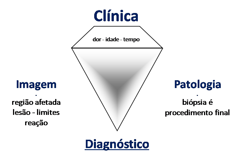
             All of these lesions mentioned have  clinical particularities ,  imaging characteristics , and  histological aspects  that need to be analyzed together to correlate each one of them.

             This is fundamental, as we can have radiologically and/or histologically similar lesions but with different diagnoses.

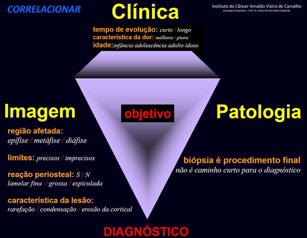
             Therefore, imaging studies and histology must always be correlated with the clinical picture, for the correct diagnosis.

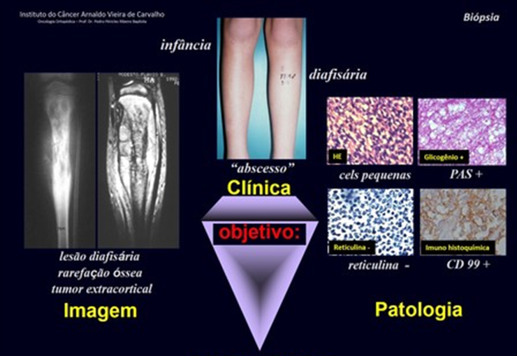
    EXEMPLO 1: Clínica = adolescente, dor, calor, tumor , circulação colateral ("abscesso"), na região metafisária dos ossos longos. Imagem= lesão de limites imprecisos, agressiva, heterogênea, produtora de tecido ósseo na metáfise. Patologia: células malignas semelhantes à osteoblastos, condroblastos, fibroblastos ou teleangiectásico = OSTEOSSARCOMA.
    EXAMPLE 1: Clinic = adolescent, pain, heat, tumor, collateral circulation ("abscess"), in the metaphyseal region of long bones. Image= lesion with imprecise limits, aggressive, heterogeneous, producing bone tissue in the metaphysis. Pathology: malignant cells similar to osteoblasts, chondroblasts, fibroblasts or teleangiectatic = OSTEOSARCOMA.
    EXEMPLO 1: Lesão de limites imprecisos, agressiva, heterogênea, produtora de tecido ósseo, na região metafisária dos ossos longos em adolescente é a identidade do OSTEOSSARCOMA.
    EXAMPLE 1: A lesion with imprecise limits, aggressive, heterogeneous, producing bone tissue, in the metaphyseal region of long bones in adolescents is the identity of OSTEOSSARCOMA.

    In this example, if the biopsy diagnosis is chondrosarcoma, fibrosarcoma, or aneurysmal bone cyst, the physician managing the case should review with the pathology/multidisciplinary team.

    The biopsy may not show newly formed bone tissue and, therefore, will not diagnose chondroblastic osteosarcoma or fibroblastic osteosarcoma, nor teleangiectatic osteosarcoma.

    When the pathologist does not have data on the patient’s history, physical examination and images, he is restricted to the material he received, which is a sample of the tumor. If you have access to this data, you will be able to make the correct diagnosis, without the need to repeat the biopsy.

    Repeating the biopsy delays treatment, increases local aggression and will not guarantee obtaining a sample with newly formed bone tissue.

    The pathologist will not be wrong if he makes the report only with the diagnosis of what is on the slide, when he is not aware of the patient’s data and exams.

    But the doctor, who manages the case, will make a big mistake if he does not clarify the case, as he is the one who has all the patient’s data, clinical picture, history, physical examination, laboratory and imaging tests.

    Para o diagnóstico dos tumores ósseos é preciso fazer a correlação entre a clinica, a radiologia e a anatomia patológica.
    To diagnose bone tumors, it is necessary to make a correlation between the clinic, radiology and pathological anatomy.

    Author: Prof. Dr. Pedro Péricles Ribeiro Baptista

     Orthopedic Oncosurgery at the Dr. Arnaldo Vieira de Carvalho Cancer Institute

    Granuloma Eosinófilo: Histórico

    Eosinophilic Granuloma

    [auto_translate_button]

    Eosinophilic granuloma

    Eosinophilic Granuloma: History: In 1938, Schairer diagnosed a lesion in the skull of a child as eosionphilic myeloma or eosinophilic osteomyelitis 1 . This condition was later described as a new clinical entity by Otani and Ehrlich in 1940 under the name Solitary Granuloma of Bone 2 .

    Farber and Green, in 1942, demonstrated that the lesion could occur in a localized or multiple manner in the skeleton and possibly be related to Hand-Schuller-Christian disease and Letter-Siwe disease 3 .

    In 1944, Jaffe and Lichtenstein introduced the term eosinophilic granuloma of bone 4 . The relationship between this lesion and the systemic forms of the disease was confirmed by Lichtenstein in his 1953 publication, encompassing them under the name Histiocytosis X 5 .

    Currently, this entity is called Langerhans Cell Histiocytosis, which has four distinct clinical forms: Eosinophilic Granuloma, a form restricted to the skeleton, which can be localized or multiple; Hand-Schuller-Christian, chronic and disseminated form; Letter-Siwe, acute or subacute disseminated form and Hashimoto-Pritzker, postnatal form with spontaneous resolution 6 .

    Introduction:  Solitary eosinophilic granuloma of the bone is the most common of the four forms of presentation of Langerhans Cell Histiocytosis, representing between 60% and 80% of cases 7 .

    Among benign bone lesions, it is a rare entity, accounting for less than 1% 8 . It preferentially affects children and adolescents with a male predominance 2:1 9 . Around 80% of patients are under 21 years of age and the majority of these are between five and 15 years of age 6,7,9 .

    Some patients may begin with an isolated bone lesion and later develop multiple bone lesions. These cases can eventually evolve into systemic forms of the disease. When this occurs, it generally happens within the first six months of diagnosis and practically never after a year of evolution, which is a criterion for good prognosis, when no new lesions appear after this period of clinical follow-up 10 .

    Hand-Schuller-Christian Syndrome is   the chronic form of Langerhans cell histiocytosis, characterized by systemic involvement with multiple bone lesions, mainly in the skull, exophthalmos and diabetes insipidus, affecting children over 3 years of age 10 .

    Letter-Siwe Syndrome affects   children under three years of age, it is the acute or subacute form, also with systemic involvement. It presents with fever, otitis media, recurrent bacterial infections, anemia, hemorrhages, viceromegaly, diffuse and painful adenopathy with skin involvement similar to seborrheic eczema and generalized ostelitic lesions, with frequent progression to death 10 .

    Hashimoto-Pritzker Syndrome is   a form of Langerhans cell histiocytosis that affects the skin exclusively. It affects children in the first month of life, manifesting with eczematous eruptions that resolve spontaneously 6 .

    Etiology:  Unknown.

    Genetics:  No significant reports regarding this.

    Definition:  Eosinophilic Granuloma is a pseudotumor lesion, of unknown etiology, characterized by bone rarefaction that can be solitary or multiple. Microscopically, it presents a profile of mononuclear histiocytic cells, presenting antigens of dendritic origin, known as Langerhans cells, amidst the variable quantity of leukocytes, eosinophils, lymphocytes and giant cells.

    Epidemiology:  Eosinophilic Granuloma mainly affects the axial skeleton, in this order: skull, pelvis, vertebrae, ribs, mandible, clavicle and scapula.

    In the appendicular skeleton the femur, proximal region and diaphysis, humerus and tibia 9 . It most often affects the diaphysis or metadiaphyseal region, being rare in the epiphysis 7 .

    The spine represents 10% of cases in the pediatric population, the majority in the lumbar region.

    In adults, it occurs more frequently in the ribs and less frequently in the spine, respectively 25% and 3% 6 .

    Clinical picture:  The most frequent symptom of Eosinophilic Granuloma is localized, throbbing, short-lasting pain, worsening at night associated with local heat and edema. When it affects the skull, this pain can be confused with other causes of headache.

    Compromise of vertebral bodies can produce painful scoliosis. Any angular deviations are small, less than 10 0 , as the vertebral flattening is usually uniform and rarely produces neurological symptoms. 

    In other forms of Langerhans Cell Histiocytosis, systemic symptoms may be present such as fever, skin rush and diabetes insipidus. Hepatosplenomegaly can occur in Letter-Siwe syndrome, which is the most severe form of the disease 6

    Classification:  Eosinophilic granuloma can   manifest itself in two clinical forms:  Solitary  or  Multiple .

    Laboratory tests:  Laboratory changes that can be found are an increase in ESR and CRP, and mild eosinophilia may occasionally occur on the blood count.

    Imaging tests:  The radiographic image is of bone rarefaction, rounded or oval, which begins in the medullary bone and progresses with erosion of the cortical bone.

    In the initial phase, the edges are irregular and poorly defined.

    In the late phase, slight sclerosis may occur around the lesion. In long bones, there is an evident periosteal reaction that appears as multiple thick lamellar layers, which characterizes slow-evolving benign lesions or the reaction of osteomyelitis.

    This type of solid periosteal reaction differentiates Eosinophilic Granuloma from Ewing’s Sarcoma, where the periosteal reaction is thin lamellar, due to the rapid evolution of the malignant tumor.

    Another radiographic difference between these two lesions is that Ewing’s tumor early presents extra-cortical tumor tissue, with a large volume, which does not occur in Eosinophilic Granuloma.

    In flat bones, such as the skull or pelvis, erosion affects both cortices in an irregular and asymmetrical manner, producing the visual impression of a hole within another hole, called a double contour lesion.

    In the mandible, destruction of the alveolar bone produces the radiographic impression of floating teeth.

    In the spine, the disease affects the vertebral body, with flattening occurring in 15% of cases, producing the so-called flat vertebra of Calvè 6 . The posterior elements and intervertebral discs are preserved, even when the injury occurs in more than one vertebra.

    Pathologic anatomy:

    Macroscopic appearance:  it has a soft, gelatinous consistency, yellowish in color, necrotic liquefaction is common.

    Microscopic appearance:  They appear as clusters of large histiocytic cells, with a slightly basophilic cytoplasm, globose, lobulated or indented nucleoli, in these cases similar to a bean seed, which correspond to Langerhans cells.

    These clusters are interspersed with giant cells, lymphocytes, numerous eosinophils and areas of necrosis, simulating an abscess. Electron microscopy shows typical cytoplasmic granules called Birbeck bodies 11,12 .

    Immunohistochemistry shows positivity for S-100 protein, vimentin and CD1a 11,13 .

    Diagnosis:

    Differential diagnosis:  The main radiological differential diagnoses of Eosinophilic Granuloma are Osteomyelitis and Ewing Tumor.

    When the lesion occurs in the skull, it must be differentiated from an epidermoid cyst or metastasis. The main histological differential diagnoses are Osteomyelitis and Lymphoma.

    Staging:

    Treatment:  The literature presents reports that expectant treatment or biopsy alone can be indicated as an effective therapeutic strategy for isolated skeletal injuries 7,14 .

    Eosionophilic granuloma can resolve spontaneously, especially in children. The capacity for the affected bone to rebuild itself exists, as most patients are affected before skeletal maturity, therefore with great potential for remodeling by the growth physes, which are normally not affected 15,16 .

    In our experience, there was resolution in five cases, which regressed only with percutaneous biopsy. The same happens after vertebral collapse in spinal injuries, probably due to the leakage of the contents of the lesion, resembling the drainage of an abscess, with surgical indication in the spine being extremely rare. Cases have been reported where there was complete restitution of the vertebral body height 15 . In our series we had two cases that presented this evolution.

    Although there may be spontaneous resolution, the time required is unpredictable, and there may be significant morbidity secondary to intense pain and functional limitation.

    Currently, the best therapeutic approach for Eosionophilic Granuloma is to perform a percutaneous biopsy, if possible with immediate diagnosis by frozen section, followed by intralesional corticosteroid infusion (methylpredinisolone – 40mg to 120mg depending on the size of the lesion) 7 . The anatomopathological result must be subsequently confirmed by histology in paraffin blocks.

    Eosionophilic Granuloma can take up to three months to regress, and it may be necessary to repeat the infusion 6 . In our experience, we had only one case, of an isolated injury to the humerus, which required complementation of the initial treatment in which we performed oral corticosteroid therapy, prednisolone 5mg/24h, for four months. In polyostotic Eosinophilic Granuloma, systemic corticosteroid therapy is used.

    When an incisional biopsy is necessary, corticosteroids can be applied locally after curettage of the lesion, which facilitates the resolution of the process. This curettage must be careful, carried out by opening in the form of a narrow slit, longitudinal to the bone, trying not to add greater local fragility. Eventually, the cavity can be filled with a bone graft, but this is generally unnecessary due to the great potential for regeneration that exists.

    Radiofrequency was proposed as a percutaneous treatment for Eosionophilic Granuloma, being applied in a second stage, two to four weeks after the biopsy 17 . The author restricts the technique to small injuries that are at least one centimeter away from the neural or visceral structures, warning of the risk of fractures in the load-bearing limbs. This approach, in addition to increasing costs and causing local morbidity, does not add any advantage to the treatment. The biopsy itself may have been curative, and the infusion of corticosteroids has greater justification, as this is indicated both in isolated cases and in multiple lesions. To date, there are no studies comparing percutaneous techniques with corticosteroid infusion in relation to the use of radiofrequency that justify their use. 

    Historically, radiotherapy was used in low and fractionated doses for the treatment of Langerhans Cell Histiocytosis. Currently, the indication of radiotherapy for benign lesions is controversial.

    In cases of eosinophilic granuloma with more than one skeletal lesion, without visceral involvement, systemic treatment may be indicated for a period of approximately six weeks with corticosteroid therapy (2 mg/kg) and Vinblastine (6 mg/kg).

    Prognosis:  Solitary lesions of eosinophilic granuloma evolve well in 97% of cases, with biopsy alone or in addition to corticosteroid infusion or surgical treatment 6 .

    Bibliography:

    1 SCHAIRER, E. Ueber eine eigenartige Erkrankung des kindlichen Schädels.  Zentralbl Allg Patho Pathol. Anat.,  71:113, 1938.

    2 Otani S, Ehrlich JC; Solitary granuloma of bone simulating primary neoplasm. Am J Pathol 16:479–90. 1940

    3 Green WT, Faber S; “eosinophilic or solitary granuloma” of bone. J Bone Joint Surg (Am) 24:499-526. 1942

    4 Jaffe HL, Lichtenstein L; Eosinophilic granuloma of bone. Arch Pathol 37: 99-118. 1944.

    5 Lichtenstein L.: Histiocytosis Pathol. 56:84, 1953

    6 Schwartz HS.  Orthopedic Knowledge Update: Musculoskeletal Tumors 2. American Academy of orthopedic Surgeons, Rosemont, Illinois. Chapter 12 (128-32), 2007

    7 Mavrogenis AF, Abati CN, Bosco G, Ruggieri P. Intralesional Methylprednisolone for Painful Solitary Eosinophilic Granuloma of the Appendicular Skeleton in Children. J PediatrOrthop 2012;32:416–422

    8 Chadha M, Agarwal A, Agarwal N, et al. Solitary eosinophilic granuloma of the radius. An unusual differential diagnosis. Acta Orthop Belg. 2007; 73:413–417.

    9 Campanacci, M. Bone and Soft Tissue Tumors; Springer-Verlag Wien New York. Second Edition, (54); 857-75. 1999.

    10 SCHAJOWICZ, F. Buenos Aires: Osseous Tumors; Talleres de editorial Médica Panamericana SA (9); 464-80. 1981.                  

    11 CHRISTIAN, HA Defects in membranous bones, exosphthalmos and diabetes insipidus: in a usual syndrome of dyspituitarism: a clinical study.  Med.Clin. North. Am.,  3:849, 1920.

    12 ARCECI, RJ; BRENNER, M.K.; PRITCHARD, J. Controversies and new approaches to the treatment of Langerhans cell histiocytosis.  Hemtol. Oncol. Clinic. North. Am., 12:339, 1998

    13 ALBRIGHT, F.; REIFNSTEIN, EC  The parathyroid glands and metabolic disease.  Baltmore, Williams & Wilkins, 1948.

    14 Plasschaert F, Craig C, Bell R, et al. Eosinophilic granuloma. A different behavior in children than in adults. J Bone Joint Surg Br 2002;84:870–872.

    15 Greenlee JD, Fenoy AJ, Donovan KA, et al. Eosinophilic granuloma in the pediatric spine. Pediatr Neurosurg. 2007; 43:285–292.

    16 Sessa S, Sommelet D, Lascombes P, et al. Treatment of Langerhans cell histiocytosis in children. Experience at the Children’s Hospital of Nancy. J Bone Joint Surg Am. 1994; 76:1513–1525.

    17 Corby RR, Stacy GS, Peabody TD, et al. Radiofrequency ablation of solitary eosinophilic granuloma of bone. Am J Roentgenol.2008;190:1492–1494.

    Diagnosis of tumors

    2. Parameters:

    We must analyze the following aspects of the injury:

    1) Identify the compromised bone or bones;

    2) Regarding the number of injuries:  

              2.1) Located in a bone: monotopic;

              2.2) A lesion in several bones: monotopic and polyostotic;

              2.3) Multiple lesions in one bone: polytopic and monostotic;

              2.4) Multiple lesions in different bones: polytopic and polyostotic.

    3) Regarding location in the bone:

              3.1) Epiphysis, metaphysis or diaphysis;

              3.2) Cortical, spongy, subperiosteal, paraosteal or juxta-cortical region;

              3.3) Central or eccentric.

    4) Limits of bone injury:

              4.1) Precise, imprecise, infiltrative or permeative, surrounded or not by reactional sclerosis;

              4.2) It goes beyond the cortex with an extra-osseous lesion;

              4.3) It reaches the soft tissues (yes/no) (displaces/infiltrates);

              4.4) Exceeds the growth line.

    5) Regarding other aspects of the injury:

              5.1) Destructive (osteolytic)

              5.2) Condensing or osteogenic

              5.3) Multiloculated, “in soap bubbles”

              5.4) Calcifications: focal, diffuse, striated

    6) Type of periosteal reaction:

              6.1) In thin slices – “in onion skins”

              6.2) In thick sheets

              6.3) Spiculates – “in sunbeams” or “in a comb”

              6.4) Periosteal survey interrupted by the tumor – Codman’s Triangle


    3. Diagnosis:

     Study methods for pathological anatomical examination:

     Cytology:

    It is the study of desquamated cells obtained from secretions, excretions or obtained with needles and making “imprints” (printing tissue fragments on slides). It should rarely be used to diagnose bone neoplasia. Its importance lies mainly in the cytohistological correlation.

    Punch-biopsy:

    Collection of material with trephines for inclusion in paraffin and microscopic examination. Although the material obtained by this method is small, when it is collected from a significant area of ​​the neoplasia and by an orthopedist with experience in handling these lesions, it makes a definitive diagnosis possible. The location for obtaining this material must be planned by the surgeon, in order to prevent disruption of the tumor’s balance in neighboring tissues, preventing its spread.

    Incisional biopsy:

    It is the most used method for diagnosing bone tumors. The biopsy site must be planned, not only in terms of the area that will enable a better histological diagnosis but also to predict future resection of the tumor, which should include the skin of the biopsied region. The biopsy should not be performed in inappropriate locations of the tumor, such as areas of necrosis, hemorrhage, Codman’s triangle or in areas that only present peritumoral reactional bone sclerosis.

    Frozen biopsy

    It is performed during the surgical procedure. This method is not recommended when there is bone tissue. The possibility of a diagnostic error is high in this situation. Diagnostic errors in numerous bone lesions with multinucleated giant cells, in the various tumors of undifferentiated cells, small cells and round cells, the impossibility of a histological differential diagnosis when there is neoformed bone tissue in the fracture callus, osteosarcoma and myositis ossificans, are some examples that contraindicate the method. Frozen examination may be useful in cases of metastatic lesions and even so, the speed of the method will not alter the operative approach.

    Microscopic study:

    Fragments obtained by puncture or incisional biopsy must be embedded in paraffin and subsequently stained with hematoxylin-eosin. Special methods such as PAS (Periodic Acid Schiff) and silver impregnation to study reticulin are usually used for differential diagnosis, for example, between Ewing Sarcoma, Lymphomas and PNET (Primitive Neuroectodermal Tumor). PAS, demonstrating glycogen and a scarce amount of reticulin, are common for diagnosing Ewing’s sarcoma. In Lymphomas, reticulin is abundant and PAS is negative. Immunohistochemistry techniques with immunoperoxidase are entering the routine of anatomopathological examinations. They are mainly indicated in the search for the diagnosis of the organ of origin of metastatic neoplasms in the bones. The use of markers that allow identifying the origin of the neoplastic cell is increasingly used in daily practice. Examples are PSA, to identify neoplasia originating from the prostate, CK7 for primitive lung neoplasia, CK20 for primitive digestive tract neoplasia and estrogen and progesterone receptors for breast neoplasia.

    Surgical parts:

    Routinely a surgical specimen must be examined externally and at the cuts. Externally for analysis of surgical margins in order to verify whether the neoplasm was completely extirpated. In the sections, we verified the involvement of the bone, extension and dimensions of the neoplasm and its main macroscopic characters for adequate microscopic study. (Figure 1) 

    When the study of a surgical resection is of a patient undergoing preoperative chemotherapy, particularly in osteosarcoma and Ewing’s sarcoma, the study of the specimen must follow a systematized examination, as the purpose is to analyze the response of the neoplasm to therapy. The study stages will be as follows:

    A) Slices will be made of the surgical piece along its entire length with a maximum thickness of 0.5 cm,

    B) One or more slices must be reproduced on a computer “scanner” or photographed and x-rayed,

    C) This reproduction must be gridded from the proximal to the distal end,

    D) The fragments from each checkered area must be thoroughly examined under a microscope in order to quantify the necrosis of the neoplasm and the persistence of histologically viable tumor cells,

    E) The final report of the study of the entire specimen must be graded according to the response to preoperative chemotherapy according to the Huvos criteria.

    Huvos Criteria:

    Grade I: Up to 50% tumor necrosis;

    Grade II: 50 to 90% tumor necrosis;

    Grade III: Above 90% necrosis;

    Grade IV: 100% tumor necrosis – Absence of histologically viable neoplastic cells.

    With this degree, the oncologist will be able to guide post-operative treatment taking into account the worst statistical prognosis in cases of cranes I and II and better in those of III and IV.

    Diagnóstico dos tumores

    2. Parâmetros:

    Devemos analisar os seguintes aspectos da lesão:

    1) Identificar o osso ou ossos comprometidos;

    2) Quanto ao número de lesões:  

              2.1) Localizada em um osso: monotópica;

              2.2) Uma lesão em diversos ossos: monotópica e poliostótica;

              2.3) Múltiplas lesões em um osso: politópica e monostótica;

              2.4) Múltiplas lesões em diversos ossos: politópica e poliostótica.

    3) Quanto à localização no osso:

              3.1) Epífise, metáfise ou diáfisa;

              3.2) Região cortical, esponjosa, subperiostal, paraosteal ou justa-cortical;

              3.3) Central ou excêntrica.

    4) Limites da lesão no osso:

              4.1) Precisos, imprecisos, infiltrativo ou permeativo, circundado ou não por esclerose reacional;

              4.2) Ultrapassa a cortical com lesão extra-óssea;

              4.3) Atinge as partes moles (sim/não) (desloca/infiltra);

              4,4) Ultrapassa a linha de crescimento.

    5) Quanto ais aspectos da lesão:

              5.1) Destrutiva (osteolítica)

              5.2) Condensante ou osteogênica

              5.3) Multiloculada, “em bolhas de sabão”

              5.4) Calcificações: focais, difusas, estriadas

    6) Tipo de reação periostal:

              6.1) Em lâminas finas – “em casca de cebola”

              6.2) Em lâminas grossas

              6.3) Espiculadas – “em raios de sol” ou “em pente”

              6.4) Levantamento periostal interrompido pelo tumor – Triângulo de Codman


    3. Diagnóstico:

     Métodos de estudo para exame anátomo patológico:

     Citologia:

    É o estudo de células descamadas obtidas em secreções, excreções ou obtidas com agulhas e realizando-se “imprints” (impressão de fragmentos de tecidos em lâminas). Raramente devera ser utilizado para diagnóstico de uma neoplasia óssea. Sua importância reside principalmente, na correlação cito-histológica.

    Punção-biópsia:

    Coleta de material com trefinas para inclusão em parafina e exame microscópico. Embora o material obtido por este método seja pequeno, quando é colhido em área significativa da neoplasia e por ortopedista com experiências no manuseio destas lesões, possibilita o diagnóstico definitivo. O local de obtenção deste material deve ser planejado pelo cirurgião, a fim de impedir ruptura do equilíbrio do tumor nos tecidos vizinhos, evitando sua disseminação.

    Biópsia incisional:

    É o método mais utilizado para diagnóstico de tumores ósseos. O local da biópsia deve ser planejado, não só quanto à área que possibilitará melhor diagnóstico histológico como para prever a futura ressecção do tumor, na qual deverá ser incluída a pele da região biopsiada. A biópsia não deverá ser realizada em locais inadequados do tumor como áreas de necrose,  hemorragia, no triângulo de Codman ou em áreas que apresentam apenas esclerose óssea reacional peritumoral.

    Biópsia de congelação

    É realizada durante o ato cirúrgico.  Este método não é indicado quando existir tecido ósseo. A possibilidade de erro diagnóstico é grande, nesta situação. Os erros de diagnóstico nas numerosas lesões ósseas com células gigantes multinucleadas, nos diversos tumores de células indiferenciadas, de células pequenas e redondas, a impossibilidade de diagnóstico diferencial histológico quando há tecido ósseo neoformado no calo de fratura, osteossarcoma e miosite ossificante, são alguns exemplos que contra-indicam o método. O exame em congelação poderá ser útil em casos de lesões metastáticas e mesmo assim, a rapidez do método não alterará a conduta operatória.

    Estudo microscópico:

    Os fragmentos obtidos por punção ou biópsia incisional devem ser incluídos em parafina e posteriormente faz-se a coloração com hematoxilina-eosina. Métodos especiais como PAS (Acido periódico de Schiff) e impregnação pela prata para estudo da reticulina são usualmente utilizados para diagnóstico diferencial, por exemplo, entre Sarcoma de Ewing, Linfomas e PNET (Tumor neuroectodérmico primitivo). O PAS, demostrando glicogênio e a escassa quantidade de reticulina, são usuais para diagnóstico de  sarcoma de Ewing. Nos Linfomas a reticulina é abundante e o PAS é negativo. Técnicas de imuno-histoquímica com imunoperoxidase estão entrando na rotina dos exames anatomopatológicos. São principalmente indicadas na procura do diagnóstico do órgão de origem de neoplasias metastáticas nos ossos. O uso de marcadores que permitem identificar a origem da célula neoplásica é cada vez mais usado na prática diária. Exemplos são o PSA, para identificar neoplasia originária da próstata, CK7 para neoplasia primitiva do pulmão, CK20 para neoplasia primitiva do tudo digestivo e receptores de estrógeno e progesterona para neoplasia da mama.

    Peças cirúrgicas:

    Rotineiramente uma peça cirúrgica deve ser examinada externamente e aos cortes. Externamente para análise das margens cirúrgicas com a finalidade de se verificar se a neoplasia foi completamente extirpada. Aos cortes verificamos o comprometimento do osso, extensão e  dimensões da neoplasia e seus caracteres macroscópicos principais para adequado estudo microscópico. (Figura 1) 

    Quando o estudo de uma ressecção cirúrgica for de paciente submetido a quimioterapia pré-operatória, particularmente no osteossarcoma e no sarcoma de Ewing,  o estudo da  peça deverá obedecer uma sistematização de exame, pois a finalidade é analisar a resposta da neoplasia à terapêutica. As etapas do estudo serão as seguintes:

    A) Serão feitas fatias da peça cirúrgica em toda sua extensão com espessura máxima de 0,5 cm,

    B) Uma ou mais fatias deverão ser reproduzidas em “scanner” de computador ou fotografadas e radiografadas,

    C) Esta reprodução deverá ser quadriculada da extremidade proximal até a distal,

    D) Os fragmentos de cada área quadriculada deverão ser minuciosamente examinados ao microscópico com a finalidade de quantificar a necrose da neoplasia e a persistência de células tumorais histológicamente viáveis,

    E) O relatório final do estudo de toda a peça deverá ser graduado quanto à resposta de quimioterapia pré-operatória nos critérios de Huvos.

    Critérios de Huvos:

    Grau I: Até 50% de necrose tumoral;

    Grau II: de 50 a 90/% de necrose tumoral;

    Grau III: Acima de 90% de necrose;

    Grau IV: 100% de necrose tumoral – Ausência de células neoplásicas histológicamente viáveis.

    Com esta graduação o oncologista poderá orientar o tratamento pós-operatório tendo em vista o pior prognóstico estatístico nos casos de gruas I e II e melhor nos de III e IV.

    Author: Prof. Dr. Pedro Péricles Ribeiro Baptista

     Orthopedic Oncosurgery at the Dr. Arnaldo Vieira de Carvalho Cancer Institute

    1 2 3 4 5 8
    Hello! How can we assist you?