• Rua General Jardim, 846 cj 41 Higienópolis, São Paulo - SP

Author Archives: drpprb@gmail.com

This digital library houses the book on Oncology and Orthopedic Oncosurgery.

It includes academic lectures, presentations from national and international congresses, published papers, case discussions, performed surgical procedures, and proprietary techniques developed.

The digital format was chosen because the web allows the inclusion of texts with numerous visual resources, such as images and videos, which would not be possible in a printed book.

The content is intended for students, healthcare professionals, and the general public interested in the field.

drpprb@gmail.com

Total sacrectomy technique using Gigli saws Part II

Total sacrectomy technique using Gigli saws Part II

Total sacrectomy technique using Gigli saws Part II

Check out the video of the lecture

Total sacrectomy technique using Gigli saws Part II

In this second part of the total sacrectomy technique using Gigli saws, we will demonstrate its evolution, highlighting the use of laparoscopy. We will discuss its application for guiding the Gigli saws from the pelvic cavity to the dorsal region of the patient.

The first time we used laparoscopy was in a case involving a patient with an osteolytic lesion in the S-3 sacral foramen.

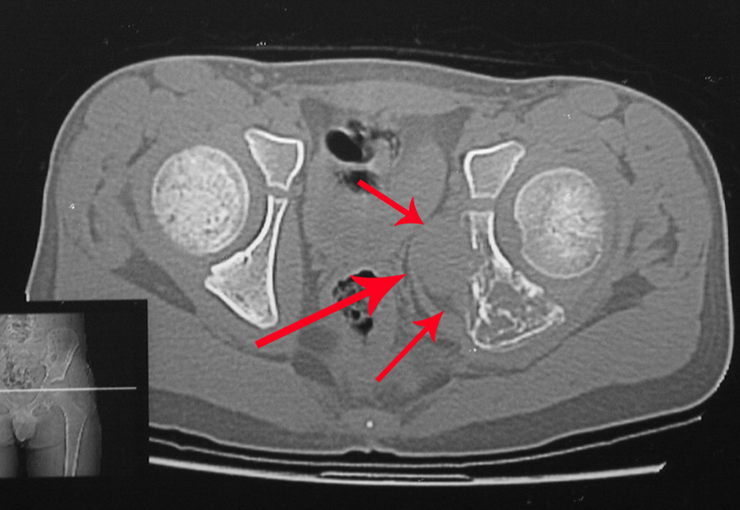
She presented with clinical symptoms of pain and numbness in her left leg. The X-ray revealed an enlarged foramen in the S-3 sacral vertebra. The CT scan showed a well-defined area, and the MRI exhibited a circumscribed lesion, bounded by a thick pseudocapsule. In the sagittal section, we observed the formation of a sacral tumor and its continuity with the nerve root, suggesting it was a schwannoma. An arteriography was performed for further study. The CT reconstruction indicated that the lesion was likely slow-growing, chronic, and benign.

A surgeon performed the laparoscopy, opening the retroperitoneum and isolating the iliac vessels under our guidance, to ligate and cut the left internal iliac artery and vein. Then, the tumor was exposed, and we confirmed its continuity with the S-3 root, resembling a horse’s tail. Next, he placed the tumor inside a surgical glove and aspirated the contents, removing everything through the laparoscopic tube. The patient was able to walk on the second day after surgery.

We discussed the possibility of performing the dissection of sacral tumors by passing the Gigli saws laparoscopically. To acquire the learning curve, we started with a case of Ewing’s sarcoma affecting the sacrum below S-3. In this case, we only needed to pass one saw horizontally. The tumor was successfully resected oncologically.

A Kirschner wire is placed as a guide, and we position two segments of the tube: the first is shorter and will exit through the patient’s back, while the second will push the first and be removed first, as per the diagram. We repeated this operation for the passage of the other saws. This way, we have the three saws safely positioned to make the osteotomies through the posterior approach. Currently, we use laparoscopy to perform all the procedures that were previously done through the anterior approach.

The case of a patient with a large volume chordoma, involving the entire sacrum and with difficulty in defecating and urinating, who had been bedridden for eight months in another hospital, was decisive in the approval of this technique. The patient had pressure ulcers and a posture of hip and knee flexion. She was operated on using the Gigli saw technique, positioned laparoscopically, and then we performed the release and lengthening of the hip and knee flexors, realigning the lower limbs. This chordoma case, despite its severity, was successfully operated on, and the total sacrectomy with laparoscopic assistance was successful. The patient started physiotherapy and rehabilitation of bladder and bowel excretory functions through abdominal maneuvers. She can walk with the aid of a walker and is reintegrated into daily life, independently, despite motor deficits in the ankle dorsiflexor muscles.

Sacrectomy with laparoscopic assistance is advantageous, presenting shorter surgery times, reduced need for blood transfusions, and satisfactory functional outcomes. Currently, we see no need to perform reconstructions.

Total sacrectomy

Total sacrectomy technique using Gigli saws Part I

Total sacrectomy

Check out the video of the lecture

Total sacrectomy technique using Gigli saws.

Sacrectomy is a complex and delicate surgical procedure that involves the total or partial removal of the sacrum bone, which is the lower part of the spine located between the lumbar spine and the coccyx. This bone plays a fundamental role in the body’s support structure and the stability of the pelvic region.

There are several reasons why a sacrectomy may be necessary. For example, in cases of malignant bone tumors or severe traumatic injuries, such as severe sacral fractures, partial or total removal of the sacrum bone may be necessary to avoid further complications and alleviate pain.

Sacrectomy is a highly specialized surgical intervention, requiring a multidisciplinary team of orthopedic surgeons, neurosurgeons, oncologists, among other healthcare professionals. The surgical approach may vary depending on the extent of sacrum bone removal and the underlying condition of the patient.

During the procedure, it is essential to ensure the preservation of nearby nerve structures, such as the sacral nerves, to minimize the risk of neurological complications and preserve the function of the lower limbs and bowel and bladder.

After sacrectomy, rehabilitation is a crucial part of the recovery process. Patients typically undergo physiotherapy and occupational therapy to aid in the recovery of mobility and adaptation to potential changes in musculoskeletal function.

Although sacrectomy is a complex surgery with significant risks and challenges, it can be vital for improving quality of life and prolonging survival in cases of severe conditions affecting the sacrum bone. It is essential for patients to receive close monitoring and specialized care before, during, and after the procedure to optimize outcomes and minimize complications.

Let’s describe the video of the conference presented at Mount Sinai Hospital – Toronto, May 25, 2011.

Summary

Video 1 of the first part of the total sacrectomy technique for treating tumor lesions. This technique uses Gigli saws to perform an oncologically safe resection of sacral neoplasms, whether in bone or soft tissue, in the region.

Watch the video part 1 of the sacrectomy technique using Gigli saws.

We will discuss the topic of sacral resection and reconstruction, with a focus on the anterior approach using video laparoscopy.

Total sacrectomy refers to the complete removal of this segment. This procedure typically results in deformities, functional loss, and compromised control of the bladder and anal sphincters.

What are the functions and quality of life for a patient following a total sacrectomy without reconstruction?

We will first demonstrate improvements in the surgical approach for sacral resection, utilizing a Gigli saw as previously presented in this case of a recurrent giant cell tumor (GCT) of the sacrum after the third surgery involving intralesional curettage.

We plan to perform a total sacrectomy using both anterior and posterior approaches to achieve an oncological resection.

 To perform this procedure, two osteotomies will be made on the iliac bone, and a third will be made below the L5 vertebra.

 To minimize bleeding during these pelvic osteotomies in spongy bone, we perform an anterior intra-abdominal approach and ligate the internal iliac vessels (arteries and veins) before positioning the three Gigli saws. Two are placed through the sciatic notch and the iliac crest, and the third below the level of the L5 vertebra.

 To facilitate the placement of the Gigli saws from the inside of the pelvic cavity to the dorsal region of the patient, a cushion should be placed under the pelvis and sacrum. This cushion will be removed during the surgery, allowing the surgeon to easily pull out the saws afterwards.”       

The patient is positioned supine in a gynecological position for either a transperitoneal or retroperitoneal anterior approach. In this case, we isolate the vessels and nerves and use a puncture through the sciatic notch to place the saws.

Gigli saws should be sheathed in a plastic tube, such as a catheter, which is then passed through this metal tube and exits through the back. This diagram illustrates the passage of the saws at the level of L5-S1, through the posterior portion of the right and left iliac bones. An X-ray is then performed to verify the correct positioning of the saws.

Once the saw is positioned, the anterior approach is closed, and the patient is repositioned prone. We can then see the saws on their back. The posterior approach is commenced, and all osteotomies are performed as shown in this diagram. This results in an en bloc resection.

Here we are performing the osteotomy using the saws through the posterior approach. Hemostasis has been achieved, and the resection of the sacrum is performed according to this schema. We performed an “en bloc resection.”

Postoperative X-rays confirm the total resection of the sacrum.

The patient has started bed-based physiotherapy and will remain on a tilt table for eight weeks to train for orthostatic tolerance.

After this training, the patient will begin exercises on parallel bars and walking with Canadian crutches. After about three months, the patient is able to walk with the aid of crutches.

This technique has been enhanced with the use of video laparoscopy.

Video of the second part of sacrectomy, with the assistance of videolaparoscopy, coming soon on this channel! 

Tumor de Células Gigantes

Giant Cell Tumor

Giant Cell Tumor

Characteristics, Diagnosis and Treatment

Giant cell tumor, also known as TGC, is a mesenchymal neoplasm that is notable for the proliferation of large multinucleated cells, called gigantocytes. These cells resemble osteoclasts and are found within a stroma of mononucleated cells. Due to its peculiar histological morphology, accurate diagnosis often requires a thorough analysis of the clinical and radiographic picture in order to avoid confusion with other pathological processes.

The main manifestation of a giant cell tumor is intermittent local pain, often accompanied by an increase in volume in the affected region and restriction of adjacent joint movements. The evolution period varies from 6 to 12 months, depending on the affected bone, with reports of trauma as the initial trigger of symptoms being common.

This type of tumor tends to affect a single bone, mainly long bones such as the femur, tibia, humerus and radius. However, in rarer cases, it can occur in bones of the axial skeleton, with a predilection for the sacrum. The incidence is more common between the third and fourth decades of life, affecting both sexes equally.

Radiographically, TGC appears as an epiphyseal lesion characterized by bone rarefaction, initially eccentric and later compromising the cortex. Diagnostic confirmation is obtained through histological analysis, which reveals the presence of multinucleated giant cells and spindle cell stroma.

The treatment of giant cell tumors is well established and aims at segmental resection of the lesion, whenever possible, ensuring safety margins for both bone and soft tissues. In cases where segmental resection is not feasible, such as in the cervical spine, endocavitary curettage followed by adjuvant therapy is indicated. Among the adjuvant therapeutic options are the CO2 laser, phenol diluted in 4% alcohol, liquid nitrogen and electrothermia.

The electrothermal technique has been shown to be effective in complementing curettage, providing a more complete cleaning of the tumor cavity. After electrothermia, milling of the cavity using appropriate instruments such as the slow drill is performed to ensure complete removal of the remaining tumor cells.

Filling the treated cavity can be done with different materials, such as autologous bone graft, bone substitutes or methylmethacrylate, each with its advantages and disadvantages. Post-treatment follow-up is essential to monitor disease recurrence and ensure the effectiveness of the treatment performed.

In summary, giant cell tumor is a complex condition that requires a multidisciplinary approach to ensure the best therapeutic outcome and the patient’s quality of life. Advances in diagnostic and treatment techniques have contributed significantly to improving the prognosis of these patients, offering increasingly effective and safe therapeutic options.

Author: Prof. Dr. Pedro Péricles Ribeiro Baptista

 Orthopedic Oncosurgery at the Dr. Arnaldo Vieira de Carvalho Cancer Institute

Sarcomas em Tecidos Moles

Sarcomas in Soft Tissues

Sarcomas in Soft Tissues

Introduction:

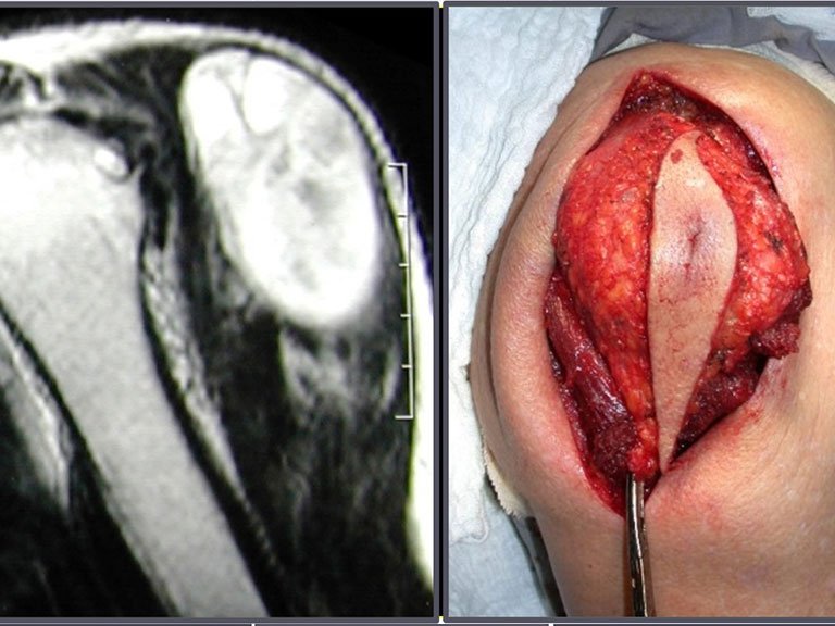
Orthopedic oncological surgery encompasses the treatment of musculoskeletal lesions, including benign and malignant bone neoplasms, pseudo-tumor lesions and benign and malignant soft tissue neoplasms. Soft tissue sarcoma is a malignant neoplasm derived from the mesoderm, occurring in soft tissues, such as muscles, fascia, tendons, etc., differentiating itself from carcinomas, which have embryonic origin in the ectoderm.

Etiology:

Most soft tissue sarcomas do not have a defined cause, but some risk factors are well described, such as previous radiotherapy, lymphedema, Li-Fraumeni syndrome, type I neurofibromatosis, individual genetic propensity and HIV infection.

Incidence:

It is a rare tumor, representing approximately 12% of pediatric neoplasms and only 1% of all malignant tumors in adults. In the USA, there are an estimated 12,000 new cases per year, resulting in around 4,700 deaths. Around 60% of soft tissue sarcomas arise in the limbs, predominantly in the thigh, followed by the chest wall and retroperitoneum.

Classification:

The classification of soft tissue sarcoma is based on histological subtype such as liposarcoma, synovial sarcoma, rhabdomyosarcoma etc. Histological grade is also used, being divided into Grade 1 (well differentiated), Grade 2 (moderately differentiated) and Grade 3 (poorly differentiated).

Clinical condition:

The initial clinical picture is characterized by a palpable tumor bulge, often painless, with progressive growth, mainly in the thigh. Some patients may experience pain and paresthesia due to the compressive effect of the tumor. Fever or weight loss are exceptional symptoms.

Staging:

At diagnosis, soft tissue sarcoma rarely metastasizes, occurring more frequently as large-volume, deep-to-muscular, high-grade tumors. The pattern of dissemination is mainly hematogenous, with predominant lung metastases.

Imaging exams:

Exams include radiography, magnetic resonance imaging, tomography, PET-CT and scintigraphy. Biopsy is indicated for histological diagnosis, and can be performed in several ways, including percutaneous needle biopsy, incisional (surgical), ultrasound or tomography-guided biopsy.

Pathology:

The pathologist plays a crucial role in the diagnosis, carrying out examinations such as freezing, to ensure the representativeness of the sample, and subsequently microscopic study in paraffin, for histopathological diagnosis as well as determining the histological grade of the tumor. Immunohistochemistry is an important resource to complement the study of the sample.

Author: Prof. Dr. Pedro Péricles Ribeiro Baptista

 Orthopedic Oncosurgery at the Dr. Arnaldo Vieira de Carvalho Cancer Institute

Sarcoma de Ewing Diagnóstico e Tratamento

Ewing’s sarcoma

Ewing's sarcoma

Diagnosis and Treatment

Ewing sarcoma diagnosis and treatment. Ewing sarcoma is a malignant tumor composed of small, round, undifferentiated cells that represents a significant clinical challenge due to its aggressive nature and rapid spread. This type of neoplasia has a maximum incidence in the first and second decades of life, being less common after the third decade, and exhibiting a 2:1 ratio between male and female sexes.

Although the exact origin of Ewing sarcoma cells has been the subject of controversy, recent studies suggest a neuroectodermal origin. These tumors frequently arise in the meta-diaphyseal region of the long tubular bones and in the pelvis, and their macroscopic characteristics include a bone lesion with a whitish-gray color and soft consistency. A distinctive feature is the formation of a thin lamellar reaction, resulting in an “onion skin” radiographic appearance and a large amount of extracortical lesion.

On histological examination, Ewing’s sarcoma presents uniformly distributed, small, round cells, similar to lymphocytes, but larger in size. The argent impregnation technique reveals a shortage of reticulin fibers, commonly found only around blood vessels and in lymphoma. Clinical manifestations include pain, swelling, local hypersensitivity and increased erythrocyte sedimentation rate, which may initially resemble osteomyelitis.

The differential diagnosis involves distinction with osteosarcoma, eosinophilic granuloma, rhabdomyosarcoma and osteomyelitis. The treatment currently adopted consists of preoperative multidrug therapy followed by surgery to resect the lesion, complemented by postoperative multidrug therapy. Reconstruction of the segment can be performed after resection, using endoprosthesis or biological solutions with autologous bone grafts. In children, growth plate autotransplantation techniques can be used, translating the fibula plate to the tibia or to other locations such as the shoulder and wrist, in these cases with the help of microsurgery.

Assessment of the response to preoperative chemotherapy is essential, having prognostic value and guiding subsequent treatment. This assessment is classified into grades, depending on the percentage of tumor necrosis observed. With advances in chemotherapy treatment, patients have achieved excellent responses and prospects for a “cure”, which has driven the use of biological solutions in surgical treatment, aiming to avoid complications associated with endoprostheses or bank grafts, whose durability may be limited.

Therefore, a comprehensive understanding of Ewing sarcoma, from diagnosis to therapeutic options, is essential to providing the best possible care to patients affected by this challenging malignancy.

Author: Prof. Dr. Pedro Péricles Ribeiro Baptista

 Orthopedic Oncosurgery at the Dr. Arnaldo Vieira de Carvalho Cancer Institute

Osteossarcoma de Superfície

Surface Osteosarcoma

Surface Osteosarcoma

Clinical Aspects, Diagnosis and Treatment

Surface osteosarcoma, also known as paraosteal or juxta-cortical, is a specific form of this type of bone tumor that originates on the external surface of the bone, without involvement of the bone marrow. In this type of lesion, the tumor growth zone is visible on the bone surface, indicating a more immature region of the bone. Surface osteosarcoma can present two distinct histological representations:

– Low Grade: Characterized by the almost complete absence of atypical mitoses and morphologically similar to mature bone. The diagnosis is usually confirmed through clinical evaluation and imaging tests.

– High Grade: It has similar clinical characteristics, but exhibits atypia and a polymorphic pattern of immature neoplasia.

Clinically, surface osteosarcoma generally presents a slow evolution and tends to occur in individuals in the third and fourth decade of life. The most common locations include the posterior and distal metaphyseal region of the femur, popliteal cavus, proximal humerus, and distal radius.

Radiologically, this form of osteosarcoma is characterized by a dense lesion of tumoral bone neoformation, with its base in continuity with the cortex of the affected bone. The surface of the lesion may have a cartilaginous layer, representing the most immature area of ​​the tumor.

One of the main differential diagnoses is myositis ossificans, which exhibits an immature central area in the lesion, while the periphery is more mature and calcified.

Treatment of low-grade surface osteosarcoma generally involves oncological resection of the lesion, which may be partial, parietal or segmental, followed by reconstruction with an autologous bone graft or endoprosthesis. High-grade osteosarcoma is treated in a similar way to central osteosarcoma, with protocols that include chemotherapy, surgery and then adjuvant chemotherapy.

In summary, understanding the clinical, diagnostic and therapeutic aspects of surface osteosarcoma is crucial to ensure an effective and personalized approach for each patient, always aiming to achieve the best clinical results and good quality of life.

Author: Prof. Dr. Pedro Péricles Ribeiro Baptista

 Orthopedic Oncosurgery at the Dr. Arnaldo Vieira de Carvalho Cancer Institute

Osteocondroma

Osteochondroma

Osteochondroma

Diagnosis and Treatment

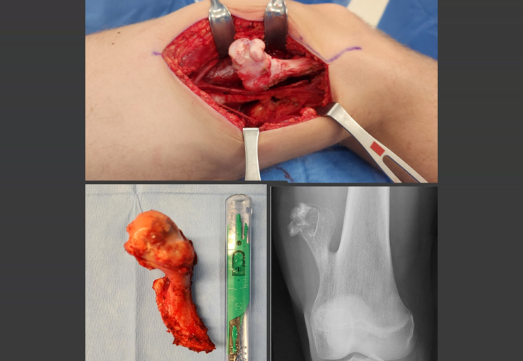
Osteochondroma, also known as osteocartilaginous exostosis, represents the most common benign bone lesion, although its incidence may be even higher than that reported in the literature due to many patients presenting asymptomatic osteochondromas.

This condition generally develops in the first and second decades of life, being located in the metaphyseal region of long bones. Radiographically, it is characterized by presenting a tumor composed of cartilage and bone. A distinctive feature is that the central cancellous bone of the exostosis continues with the medullary of the affected bone, while the dense cortical layer of the tumor continues with the normal cortical bone. On the surface of the lesion, there is a band of cartilage, through which the growth of the lesion occurs, hence the name “osteochondroma” – tumor forming cartilage and bone.

Osteochondromas can present as sessile bases (with an enlarged base) or pedunculated bases. They can be single or multiple, characterizing hereditary multiple osteochondromatosis.

The standard treatment for osteochondromas is surgery, generally involving resection, especially when the lesion compromises aesthetics, compresses vascular or nervous structures or limits function. It is important to note that these tumors usually continue to grow while the patient is in the growth phase.

When an osteochondroma increases in size after skeletal maturity, it may indicate post-traumatic bursitis or, more worryingly, malignancy to chondrosarcoma, which requires resection with oncological margins.

The risk of malignancy is low, with solitary osteochondromas having a malignancy rate of about 1%, while multiple osteochondromatosis can be as high as 10%.

In summary, a comprehensive understanding of the characteristics, diagnosis and treatment of osteochondroma is essential to ensure an effective and appropriate therapeutic approach, always aiming for the patient’s well-being and quality of life.

Author: Prof. Dr. Pedro Péricles Ribeiro Baptista

 Orthopedic Oncosurgery at the Dr. Arnaldo Vieira de Carvalho Cancer Institute

Osteoblastoma

Osteoblastoma

Osteoblastoma

Treatment and Differentiation of Bone Lesions

Osteoblastoma is a rare bone neoplasm, radiographically characterized by areas of bone rarefaction, with denser foci of ossification. This tumor has two distinct clinical forms: genuine osteoblastoma, which is more common and is generally located in the pedicle of the spine or, less commonly, in the metaphysis of long bones; and aggressive osteoblastoma, also known as “malignant”.

Mainly occurring in the first and second decades of life, the clinical picture of osteoblastoma is marked by intense pain, which can lead to fractures and functional and neurological deficits, especially when it occurs in the spine, and can even cause antalgic scoliosis in some cases.

The differential diagnosis of osteoblastoma includes other bone lesions such as osteoid osteoma, aneurysmal bone cyst and osteosarcoma, and a careful evaluation is essential to determine the most appropriate therapeutic course.

Treatment of osteoblastoma generally involves en bloc resection of the lesion, with bone grafting when necessary. In regions such as the spine, where en bloc resection may be challenging, judicious curettage is performed. Furthermore, the use of local adjuvants, such as phenol and electrothermia, has been increasingly used in order to minimize the risk of recurrence.

In short, osteoblastoma is a pathology that demands specialized attention and a multidisciplinary therapeutic approach to guarantee the best clinical result and avoid future complications. Detailed knowledge about this condition is essential for an accurate diagnosis and an effective treatment plan, always aiming for the patient’s quality of life and well-being. Understanding the diagnostic nuances and available therapeutic options is critical to successful management of this condition. Therefore, it is crucial that healthcare professionals stay up to date on the latest advances in the field of osteoblastoma diagnosis and treatment.

Author: Prof. Dr. Pedro Péricles Ribeiro Baptista

 Orthopedic Oncosurgery at the Dr. Arnaldo Vieira de Carvalho Cancer Institute

Metástases Ósseas

Bone Metastases

Bone Metastases

Bone metastases appear as one of the most common complications in cancer patients, representing an important medical issue. This is in part due to bone’s ability to act as a filter for circulating tumor cells. Like the lung and liver, skeletal tissue has the potential to harbor metastatic cells, especially due to its slower sinusoidal circulation.

With the advancement of cancer diagnosis and treatment methods, we have seen an increase in patient survival. However, this also means that more individuals end up developing bone metastases, with tumors such as breast, prostate, lung, kidney and thyroid being the main responsible for these spreads to the skeleton.

The clinical manifestations of bone metastases often involve pain and, in some cases, can lead to pathological fractures, significantly compromising the patient’s quality of life. In these situations, orthopedic surgical intervention becomes essential to alleviate pain, restore function and avoid serious complications.

The most common sites for these injuries include the femur, humerus, vertebrae, pelvis, scapula, and tibia. The therapeutic approach generally involves resection and reconstruction surgeries, using techniques such as osteosynthesis with cement or endoprosthetic implants. In some cases, radiotherapy may be used as a palliative measure to relieve pain in patients who are not candidates for surgery.

In the context of multiple myeloma, one of the most common primary bone tumors, treatment usually involves chemotherapy and, in certain cases, radiotherapy. However, when bone lesions become significant or present a risk of fracture, the orthopedic surgical approach follows the same principles adopted for bone metastases from other types of cancer.

In summary, the management of bone metastases requires a multidisciplinary approach, involving oncologists, orthopedic surgeons and radiation oncologists, with the aim of providing pain relief, preserving function and improving the quality of life of patients affected by this complication of cancer.

Author: Prof. Dr. Pedro Péricles Ribeiro Baptista

 Orthopedic Oncosurgery at the Dr. Arnaldo Vieira de Carvalho Cancer Institute

Granuloma Eosinófilo

Eosinophilic Granuloma

Eosinophilic granuloma

History and Evolution of Understanding:

The history of Eosinophilic Granuloma dates back to 1938, when Schaerer diagnosed a lesion on the skull of a child, initially considered to be eosinophilic myeloma or eosinophilic osteomyelitis. It was later described as a new clinical entity by Otani and Ehrlich in 1940, called Solitary Granuloma of Bone. Farber and Green, in 1942, identified its possible relationship with Hand-Schuller-Christian disease and Letter-Siwe disease. In 1944, Jaffe and Lichtenstein introduced the term Eosinophilic Granuloma of Bone, consolidating its association with systemic forms of the disease, now called Langerhans Cell Histiocytosis.

Introduction and Epidemiology:

Eosinophilic Granuloma of Bone, which is the most common form of Langerhans Cell Histiocytosis, represents between 60% and 80% of cases. Despite being a rare benign bone lesion, it occurs mainly in children and adolescents, with a predominance of males. The majority of cases occur in individuals under 21 years of age, with the most affected age group being between five and 15 years. Axial involvement of the skeleton is predominant, with a variety of bones affected, such as the skull, pelvis, ribs and vertebrae, with the spine being responsible for around 10% of cases in children.

Clinical Manifestations and Diagnosis:

The most common symptom of Eosinophilic Granuloma is localized pain, often confused with other causes of headache when it affects the skull. Other forms of Langerhans Cell Histiocytosis can present systemic symptoms, such as fever and diabetes insipidus. The diagnosis is made based on imaging tests, which reveal a bone rarefaction lesion with cortical erosion, in addition to laboratory changes, such as increased ESR and CRP.

Treatment and Prognosis:

The treatment of Eosinophilic Granuloma varies according to the extent of the disease. In isolated cases, an expectant approach or percutaneous biopsy followed by intralesional corticosteroid infusion may be effective. Spontaneous resolution is possible, especially in children, due to the potential for bone remodeling. In more extensive cases or with multiple lesions, systemic treatment with corticosteroids and Vinblastine may be indicated. The prognosis is generally good, with a favorable resolution rate in 97% of cases of solitary lesions.


Conclusion :

Eosinophilic Granuloma of Bone, as the most common form of Langerhans Cell Histiocytosis, represents a diagnostic and therapeutic challenge. However, with a multidisciplinary approach and appropriate therapeutic options, most patients can have a favorable outcome. A thorough understanding of this clinical entity is essential for effective management and an optimistic prognosis.

Author: Prof. Dr. Pedro Péricles Ribeiro Baptista

 Orthopedic Oncosurgery at the Dr. Arnaldo Vieira de Carvalho Cancer Institute

1 2 3 8
Hello! How can we assist you?