This digital library houses the book on Oncology and Orthopedic Oncosurgery.

It includes academic lectures, presentations from national and international congresses, published papers, case discussions, performed surgical procedures, and proprietary techniques developed.

The digital format was chosen because the web allows the inclusion of texts with numerous visual resources, such as images and videos, which would not be possible in a printed book.

The content is intended for students, healthcare professionals, and the general public interested in the field.

Osteoid Osteoma

7651301cabf91a1be8e3cf0b72e8734f 3
[auto_translate_button]
Osteoid Osteoma Osteoid osteoma is a benign osteoblastic lesion, smaller than one centimeter, with precise limits and with reactive bone sclerosis around osteoid tissue, with highly vascularized stroma and histologically mature bone.

Osteoid osteoma

It is a lesion that is preferably located in the cortex of long bones or in the pedicle of the spinal column (compact bones). It can occur in three different locations in the bone:

  • Cortical  : the vast majority, figures 1 and 2a, 2b and 2c.
Figura 1: Quadro clínico de tumefação devido a osteoma osteóide da cortical medial da tíbia .
Figure 1: Clinical picture of swelling due to osteoid osteoma of the medial cortical bone of the tibia.
Figura 2: Aumento de densidade da cortical medial, devido à esclerose óssea (a); corte tomográfico mostrando a lesão de rarefação na cortical, circundada por halo de esclerose (b) e nicho de osteoma osteóide com a esclerose, já ressecado (c).
Figure 2: Increased density of the medial cortex, due to bone sclerosis (a); tomographic section showing the rarefaction lesion in the cortex, surrounded by a halo of sclerosis (b) and an osteoid osteoma niche with sclerosis, already resected (c).
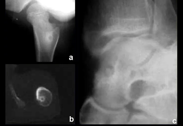
Figura 3: Osteoma osteóide na medular do fêmur (a); tomografia confirmando a localização central (b); osteoma osteóide medular do tálus (c).
Figure 3: Osteoid osteoma in the medullary bone of the femur (a); tomography confirming central location (b); Medullary osteoid osteoma of the talus (c).
  • Medullary : or spongy (endostal), figure 3a, 3b and 3c.
  • Figura 4: Radiografia do antebraço mostrando lesão subperiosteal na face ulnar do rádio, com nicho central do osteoma osteóide e halo de esclerose óssea (a), e tumor ressecado (b).
    Figure 4: Radiograph of the forearm showing a subperiosteal lesion on the ulnar surface of the radius, with a central niche of osteoid osteoma and a halo of bone sclerosis (a), and resected tumor (b).
    Figura 5: Osteoma osteóide subperiosteal na face medial do fêmur.
    Figure 5: Subperiosteal osteoid osteoma on the medial surface of the femur.
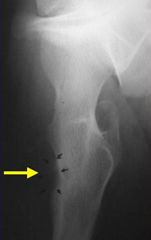
    Figura 6: Nicho de osteoma osteóide na superfície do fêmur, periosteal.
    Figure 6: Osteoid osteoma niche on the surface of the femur, periosteal.
    • Sub-periosteal,  figures 4, 5 and 6.

    It occurs in the first and second decade of life, occurring twice as often in males.

    The clinical picture is nighttime pain (mild pain that the child does not manifest while playing), which improves with the administration of salicylates. Local swelling (fig. 4) and arthralgia may occur.

    Makes differential diagnosis with steoma, osteoblastoma and Brodie’s abscess  (sequestration of chronic osteomyelitis).

    Treatment consists of resection of the central niche (fig. 5). It is not necessary to remove all the surrounding sclerosis. When the bone failure caused by resection poses a risk for fracture (as in the femoral neck (fig. 6), a bone graft must be placed.

    Author: Prof. Dr. Pedro Péricles Ribeiro Baptista

     Orthopedic Oncosurgery at the Dr. Arnaldo Vieira de Carvalho Cancer Institute

    Leave a Reply

    Hello! How can we assist you?