This digital library houses the book on Oncology and Orthopedic Oncosurgery.

It includes academic lectures, presentations from national and international congresses, published papers, case discussions, performed surgical procedures, and proprietary techniques developed.

The digital format was chosen because the web allows the inclusion of texts with numerous visual resources, such as images and videos, which would not be possible in a printed book.

The content is intended for students, healthcare professionals, and the general public interested in the field.

Giant Cell Tumor

36d838a7f08b7ac8295662b7b104440b 3
[auto_translate_button]
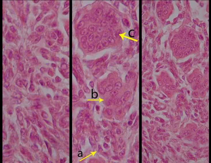
Giant cell tumor is a neoplasm of mesenchymal nature, characterized by the proliferation of multinucleated giant cells (gigantocytes) that resemble osteoclasts, in a stroma of mononucleated cells (fig. 1a). It is also known as osteoclastoma and gigantocellular tumor, and the acronyms TCG or TGC are commonly used. It was first described by Sir Astley Cooper(1) in 1818. Later, Paget (1853)(2) called it “brown or myeloid tumor”. Nelaton (1860)(3) described its clinical and histological characteristics, highlighting its local aggressiveness and giving it the name “myeloplaxis tumor”. Gross (1879)(4) insisted on its benignity and highlighted the difficulties of differential diagnosis with “the aneurysmal variant of medullary sarcoma”. With the advent of radiology, the differential diagnosis of this lesion was determined and Bloodgood (1923)(5) proposed the name “benign giant cell tumor”.

Giant cell tumor

In recent decades, much has been discussed about the nature of gigantocellular tumors. For Geschikter and Copeland (1949)(6) and Willis (1949)(7), the gigantocellular tumor would be a neoplasm of osteoclasts in the mesenchymal stroma, given the similarity between the gigantocyte and the normal osteoclast.

Jaffe et al (1940)(8) described its origin as being derived from stromal cells. Sherman (1965)(9) stated that the bone disappeared at the site of tumor growth and the gigantocytes resulted from the fusion of stromal mesenchymal cells, taking into account the similarity between optical microscopy of stromal nuclei and giant cells. The histochemistry and tissue culture studies carried out by Schajowicz (1961)(10) did not demonstrate significant differences between tumor gigantocytes and normal osteoclasts. On the other hand, studies using electron microscopy(11) confirmed that giant cells are syncytia made up of stromal cells. Thus, the undifferentiated mesenchymal cells from the bone marrow would give rise to the tumor stroma, whose cells, in turn, when differentiating, would form clusters with the characteristics of gigantocytes. The numerous giant cells that resemble osteoclasts, in a stroma of spindle cells, are the most important elements of this tumor. The histological aspect of GCT presents characteristics common to several tumor and pseudotumor lesions(12,13), requiring joint analysis with clinical and imaging characteristics to confirm the diagnosis(14,15).

Figura 1
The main differential diagnoses, both from a clinical, radiographic and anatomopathological point of view, are: aneurysmal bone cyst, teleangectatic osteosarcoma and chondroblastoma(16,17). TGC generally affects a single bone. When a lesion suggestive of this tumor is found in several bones, the possibility of it being a “brown tumor of hyperparathyroidism” should be checked, which presents a similar radiographic appearance, but with multiple lesions and suggestive changes in serum calcium and phosphorus(18 ). GCT occurs in the third and fourth decades of life, affecting both sexes equally(19-22). The main manifestation is intermittent local pain, accompanied or not by an increase in volume in the affected region. The length of history is variable and depends on the bone and the affected region(23-26).
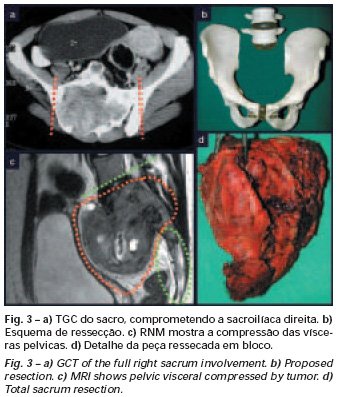
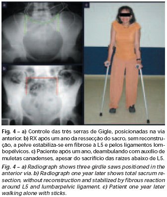
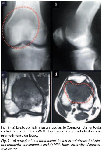
Some patients seek treatment due to pain, others because of the perception of the tumor or a pathological fracture(27,28). Generally, they relate the beginning of the clinical history to some trauma(29,30). As the tumor is epiphyseal, joint involvement with limited movement is frequent, with progressive functional changes, and intra-articular effusion may occur (fig. 1c), simulating the clinical picture of meniscal processes or arthritis(31,32). TGC is most frequent in the distal epiphysis of the femur (28.2%) (figs. 1c and 1d) and proximal tibia(19,23,31,33) (fig. 1b), followed by the proximal and distal regions of the humerus of the radio. It is rare in the axial skeleton and, when it occurs, it predominates in the sacrum. When located in the ilium (fig. 2) or sacrum (figs. 3 and 4), it generally presents a large volume, intense pain, and can cause neurological manifestations(34,35). On the radiograph, an epiphyseal bone rarefaction lesion was observed, initially eccentric and respecting the limits of the cortex. With progression, cortical rupture and joint involvement may occur (Figs. 1c and 1d). Computed tomography can help assess the degree of joint involvement and cortical erosion, facilitating the choice of the best surgical reconstruction technique.
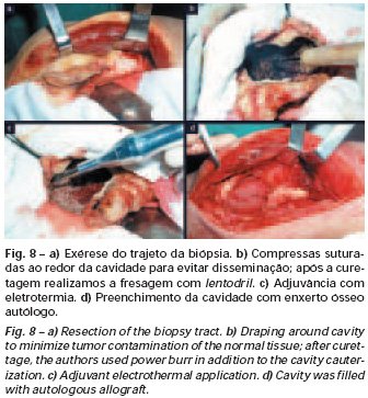
Bone mapping is characterized by an area of ​​uniform hyperuptake in the affected epiphysis. More recently, we can also use magnetic resonance imaging to evaluate the limits of the tumor and its characteristics of a homogeneous solid lesion, which may present areas of liquid content, resulting from tumor necrosis or association with areas of aneurysmal bone cyst. The treatment of giant cell tumor is currently well established. Whenever possible, segmental resection of the lesion should be chosen, with an oncological safety margin in both the bone and soft tissues (figs. 2, 3 and 4). This surgery provides a greater opportunity for cure, with a lower risk of recurrence(36-39). However, in regions where segmental resection is not feasible, endocavitary curettage must be performed (fig. 8), carefully, complemented with adjuvant therapy; laser, CO2, 4% diluted phenol, liquid nitrogen or electrothermia (fig. 8b). Methylmethacrylate has a low adjuvant effect. When used to fill the cavity, it must be preceded by one of the adjuvant therapies mentioned(37,40).
In the past, curettage had high recurrence rates because the bone was not opened to allow effective cleaning and local adjuvants were not used. Currently, when endocavitary curettage is indicated, it is recommended to create a large bone window, providing a broad view of the lesion. At the SCMSP DOT, we complement curettage with cavity milling; For this purpose, we used the Lentodrill with a spherical dental milling cutter (fig. 8c)(33,37).
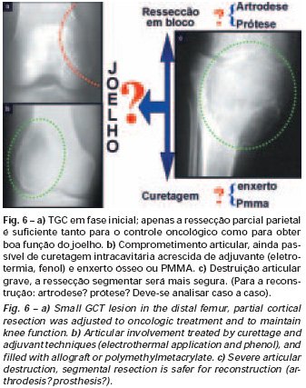
We used electrothermia(33,37,41) as a local adjuvant, using an electric scalpel for this purpose. This electrothermal technique is effective, as with the curved tip of the scalpel we can reach areas that are more difficult to access. Electrothermy, in addition to cauterization, also complements curettage, as the tumor cells, remaining in the small “cavities” of the bone wall, are destroyed and become detached, facilitating their removal. Electrothermy must precede milling, avoiding possible cell dissemination due to the rotation of the Lentodrill. In the knee region (fig. 5), we frequently recommend endocavitary curettage, followed by electrothermia and milling with Lentodrill. This is because segmental resection of this region would imply arthrodesis or replacement with an endoprosthesis or homologous osteoarticular graft.
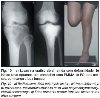
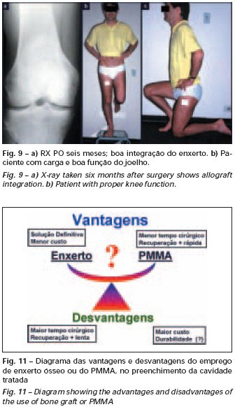
Arthrodesis of the knee joint creates great limitations for the patient, which restricts its indication. Prosthetic replacements in young patients can result in problems in the near future and their indication must be judicious. The osteoarticular homologous graft presents numerous restrictions. Therefore, for the knee region, we initially recommend the most conservative therapy: curettage followed by local adjuvant, milling and filling with autologous bone graft. For advanced cases, with significant destruction of the bone structure, in which both joint function and local control of the disease may be compromised with the curettage technique, we recommend segmental resection and reconstruction with an endoprosthesis and, exceptionally, arthrodesamis(37 ). A brief comment remains regarding filling the treated cavity. This can be done with an autologous bone graft, with a homologous graft or with methylmethacrylate (fig. 10). Each of these techniques has its advantages and disadvantages (fig. 11)(33,37,39,42,43).
Figura 10
Figura
Methylmethacrylate makes it possible to easily visualize possible recurrences, is easy to use and allows early loading; however, it is not a biological solution and fractures may occur in the region(39).
Bone grafting is a biological and definitive solution; however, it makes it difficult to visualize possible early recurrence, which can be confused with the physiological reabsorption of the graft integration process, in addition to requiring around six months, on average, for full load. The non-autologous homologous graft has a longer integration period and is not always available, but, on the other hand, it shortens surgical time. The autologous graft has the advantage of immunocompatibility and faster integration, but it prolongs surgical time. Due to the risk of malignant transformation, radiotherapy can only be considered as a treatment option for giant cell tumors located in structures that are difficult to access surgically. Therefore, especially for the knee region, the orthopedist familiar with the treatment of oncological lesions must evaluate the clinical and radiographic aspects, the degree of joint destruction, the patient’s profession, in short, all pertinent factors, in order to make the best indication. therapy(37). Complications inherent to this tumor are recurrences, sinking of the articular surface, leading to varus, valgus, antecurvatum or retrocurvatum deviations. Exceptionally, lung metastases or malignancy may occur(43-46).REFERENCES1. Cooper A., ​​Travers B.: Surgical Essays. London, Cox and Son, 1818. 2. Paget J.: Lectures on Surgical Pathology. London, Longmans, 1853. 3. Nelaton E.: D’une Nouvelle Espece de Tumeurs à Mieloplaxes. Paris, Adrien Delahaye, 1860. 4. Gross SW: Sarcoma of the long bones: based upon a study of one hundred and sixty five cases. Am J Med Sci 78: 17-19, 1879. 5. Bloodgood JC: Benign giant cell tumor of bone: its diagnosis and conservative treatment. Am J Surg 37:105-106, 1923. 6. Geschikter DF, Copeland NM: Tumors of Bone. Philadelphia, JB Lippincott, 1949. 7. Willis GE: The pathology of osteoclastoma or giant cell tumors of bone. J Bone Joint Surg [Br] 31: 236-238, 1949. 8. Jaffe HL, Lichtenstein L., Portis R., RB: Giant cell tumor of bone. Pathological appearance, grading, supposed variants and treatment. Arch Pathol 30: 993-995, 1940. 9. Sherman M.: “Giant cell tumor of bone” in Tumors of bone and soft tissue. Chicago, Year Book Medical Publishers, 1965. 10. Schajowics F.: Giant cell tumors of bone (osteoclastoma): a pathological and histochemical study. J Bone Joint Surg [Am] 43: 1-3, 1961. 11. Hanaoka H., Friedman NB, Mack RP: Ultrastructure and histogenesis of giant cell tumor of bone. Cancer 25: 1408-1423, 1970. 12. Wilson Jr. PD: A clinical study of the biochemical behavioral defects. Clin Orthop 87: 81-109, 1972. 13. Ackerman M., Berg NO, Person BM: Fine needle aspiration biopsy in the evaluation of tumor-like lesions of bone. Acta Orthop Scand 47: 129-136, 1976. 14. Eideken J., Hodes PJ: Roentgen Diagnosis of Diseases of Bone. Baltimoremore, Williams and Wilkins, pp 6588-6593, 1967. 15. Chaix C., Trifaud A.:Evolution and treatment of giant cell tumors of bone. Rev Chir Orthop 61: 429-434, 1975. 16. Derqui JC: Diagnosis of Giganto-cellular Osteopathies in Childhood. Buenos Ayres, Macchi HMS, 1962. 17. Lichtenstein L: Current status of problems in diagnosis and treatment. J Bone Joint Surg [Am] 33: 143-152, 1951. 18. Tornberg DN, Dick HM, Johnston AD: Multicentric giant cell tumor in the long bones. A case report. J Bone Joint Surg [Am] 57: 420-422, 1975. 19. Aegerter E., Kirkpatrick JA: Orthopedic Diseases. Philadelphia, WB Saunders, 1975. 20. Dahlin DC, Cupps RE, Johnson Jr. EW: Giant cell tumor: a study of 195 cases. Cancer 25: 1061-1070, 1970. 21. Goldenberg RR, Campbell CJ, Bonfiglio M.: Giant cell tumor of bone. An analysis of 218 cases. J Bone Joint Surg [Am] 52: 619-664, 1970. 22. Larsson SE, Lorentzon R., Boquist L.: Giant cell tumor of bone. A demographic, clinical and histopathological study recorded in the Swedish Cancer Registry. J Bone Joint Surg [Am] 57: 167-173, 1975. 23. Lichtenstein L.: Bone Tumors. St. Louis, CV Mosby, 1972. 24. McGrath PJ: Giant cell tumor of bone. An analysis of fifty two cases. J Bone Joint Surg [Br] 54: 216-227, 1972. 25. Marcove RC, Weiss LD, Vaghaiwalla MR: Cryo surgery in the treatment of giant cell tumors of bone. A report of 52 consecutive cases. Cancer 41: 957-964, 1978. 26. Rodrigues JS: Giant cell tumors of bone [Thesis]. Recife, Pernambuco, Faculty of Medicine of the Federal University of Pernambuco, 1972. 27. Sissons HA: “Bone tumors” in Wright P., Symmers W. St. C.: Systemic pathology. London, Longmans, Green and Co., 1966. 28. Huvos AG: Bone Tumors Diagnosis, Treatment and Prognosis. Philadelphia, WB Saunders, vol. 17, pp 429-467, 1991. 29. Hutter RVP, Worcester Jr. JN, Francis KC: Benign and malignant giant cell tumors of bone. A clinicopathological analysis of the natural history of the disease. Cancer 15: 663-672, 1962. 30. Levine HA, Eurile F.: Giant cell tumor of patellar tendon coincident with Paget’s disease. J Bone Joint Surg [Am] 53: 335-341, 1971. 31. Jaffe HL: Tumors and tumorous conditions of the bones and joints. Philadelphia, Lea & Febiger, 1958. 32. Cameron GW: Giant cell tumor of the patella. J Bone Joint Surg [Am] 37: 184-187, 1955. 33. Baptista PPR: Treatment of giant cell tumors by curettage, electrothermal cauterization, drill regularization and autologous bone graft. Rev Bras Ortop 30: 819-827, 1995. 34. Ottolenghi CE: Massive osteo and osteoarticular bone graft. Technique and results of 62 cases. Clin Orthop 87: 156-164, 1972. 35. Kambin P.: Giant cell tumor of thoracic spine with pathological fracture paraparesis: a method of stabilization. J Bone Joint Surg [Am] 48: 774-779, 1966. 36. Parrish FF: Treatment of bone tumors by total excision and replacement with massive autologous and homologous grafts. J Bone Joint Surg [Am] 48: 968-990, 1966. 37. Baptista PPR:Treatment of giant cell tumor of the distal end of the femur and proximal end of the tibia – Curettage, electrothermal cauterization and autologous bone graft [Thesis]. São Paulo: Faculdade de Ciências Médicas da Santa Casa de São Paulo, 1994. 38. Murphy WR, Ackerman L.: Benign and malignant giant cell tumor of bone. Cancer 9: 317-324, 1956. 39. Camargo FP: Segmental resection in bone tumors and surgical reconstitution of the skeleton [Thesis]. São Paulo: Faculty of Medicine of the University of São Paulo, 1968. 40. Forrest M.: The pathology of giant cell tumors of bone. Rev Chir Ortho 61: 359-366, 1975. 41. Barnes R.: Giant cell tumor of bone. J Bone Joint Surg [Br] 54: 213-215, 1972. 42. Campbell CJ, Akbarnia BA: Giant cell tumor of the radius treated by massive resection and tibial bone graft. J Bone Joint Surg [Am] 57: 982- 986, 1975. 43. Frangakis EK: Soft tissue spread of giant cell tumor. J Bone Joint Surg [Am] 52: 994-998, 1971. 44. Caballos RL: The mechanism of metastasis in the so-called “benign giant cell tumor of bone”. Hum Pathol 12: 762-768, 1981. 45. Schajowics F.: About the degeneration and malignant variety of giant cell tumors. Rev Orthop Traum 10: 349-355, 1941. 46. Riley LD, Hartmann W.: Soft tissue recurrence of giant cell tumor of bone after irradiation and excision. J Bone Joint Surg [Am] 49: 365-368, 1967.
 

Author: Prof. Dr. Pedro Péricles Ribeiro Baptista

 Orthopedic Oncosurgery at the Dr. Arnaldo Vieira de Carvalho Cancer Institute

Leave a Reply

Hello! How can we assist you?