This digital library houses the book on Oncology and Orthopedic Oncosurgery.

It includes academic lectures, presentations from national and international congresses, published papers, case discussions, performed surgical procedures, and proprietary techniques developed.

The digital format was chosen because the web allows the inclusion of texts with numerous visual resources, such as images and videos, which would not be possible in a printed book.

The content is intended for students, healthcare professionals, and the general public interested in the field.

Reconstruction of Femoral Neck Injury with Fracture

83d3d4b6c9579515e1679aca8cbc8033 3
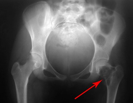
Reconstruction of Femoral Neck Injury with Fracture. A 9-year-old patient, reporting pain in the left lower limb, evaluated in January 2001, underwent an x-ray of the pelvis, figure 1.

Reconstruction of femoral neck injury with fracture; Access route and special plate with autologous graft; Femoral neck bone cyst with fracture in a child

Figura 1: Radiografia da bacia, de 20/01/2001, com lesão de rarefação óssea no colo femoral esquerdo, seta amarela. Paciente iniciando a puberdade, com potencial de crescimento bem evidente analisando-se a linha de Risser, seta azul.
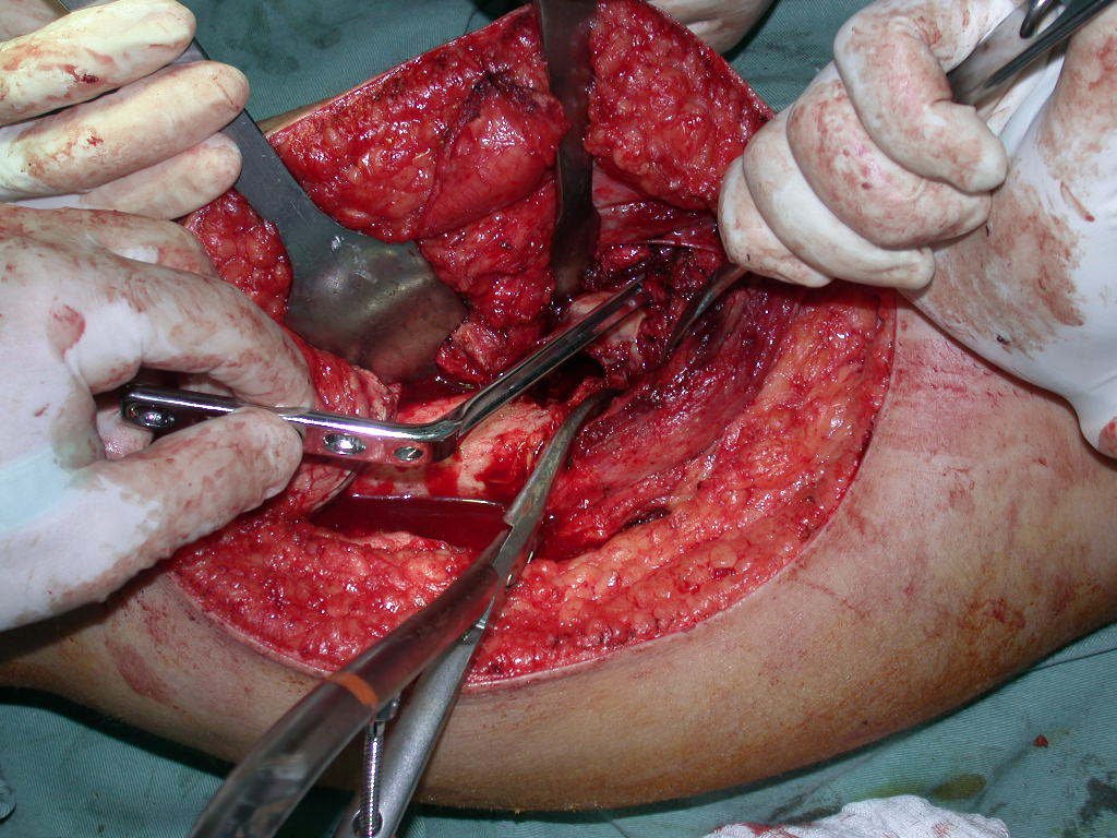
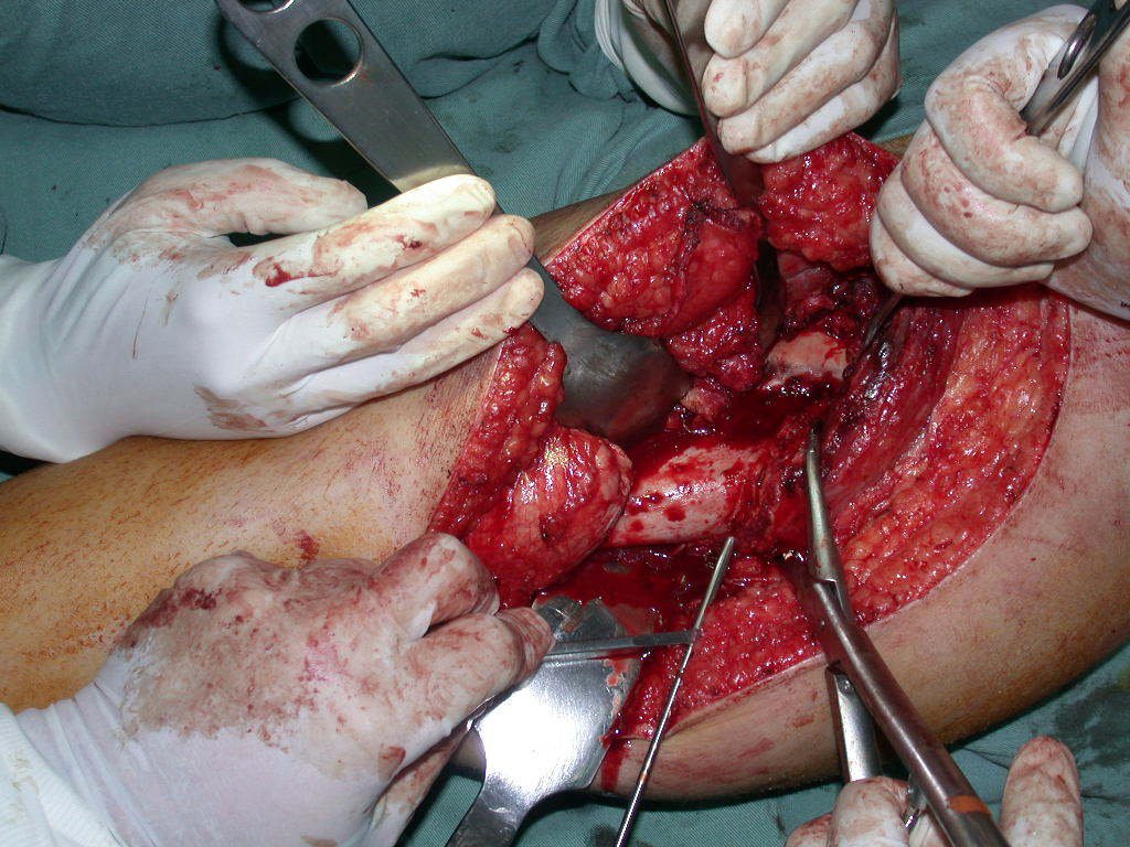
Figure 1: X-ray of the pelvis, from 01/20/2001, with bone rarefaction lesion in the left femoral neck, yellow arrow. Patient starting puberty, with growth potential clearly evident when analyzing the Risser line, blue arrow.
On the X-ray on that occasion, we could see a bone rarefaction lesion in the femoral neck, which was not noticed. The complaint was interpreted as growing pains, and the patient was followed up for three years. In mid-2004, the doctor accompanying her asked another colleague to evaluate the patient’s X-ray, in a meeting in the hospital corridor. Both do not see the injury and believe it could be a “pelvis tilt”, due to the probable discrepancy of the limbs and choose to request a scanogram of the lower limbs, figures 2 to 4.
Figura 2: Radiografia da bacia, frente, de maio de 2014, apresentando lesão lítica no colo femoral esquerdo, com a cortical medial ainda íntegra (analise a imagem com o recurso da ampliação).
Figure 2: X-ray of the pelvis, front, from May 2014, showing a lytic lesion in the left femoral neck, with the medial cortex still intact (analyze the image using magnification).
Figura 3: Escanometria de membros inferiores.
Figure 3: Scanometry of lower limbs.
Figura 4: Radiografia em posição de Lowenstein, focalizada no quadril esquerdo.
Figure 4: Radiograph in Lowenstein position, focused on the left hip.
Figura 5: Radiografia da bacia de 14/10/2004, frente, com aumento da lesão e fratura do colo femoral esquerdo.
Figure 5: X-ray of the pelvis from 10/14/2004, front, with enlargement of the lesion and fracture of the left femoral neck.
Figura 6: Radiografia da bacia em posição de Lowenstein, documenta a fratura do quadril esquerdo.
Figure 6: X-ray of the pelvis in Lowenstein position, documenting the fracture of the left hip.
Figura 7: Radiografia da bacia em alar, de outubro de 2004, mostrando o desvio posterior da cabeça femoral.
Figure 7: X-ray of the alar pelvis, from October 2004, showing the posterior deviation of the femoral head.
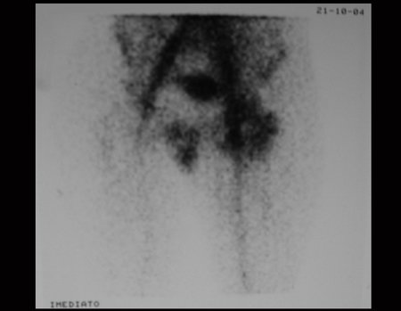
Figura 8: Cintilografia óssea na fase de fluxo, com aumento de captação no quadril esquerdo.
Figure 8: Bone scintigraphy in the flow phase, with increased uptake in the left hip.
Figura 9: Cintilografia óssea, fase tardia, frente, inclinação pélvica e captação no quadril esquerdo devido à fratura.
Figure 9: Bone scintigraphy, late phase, front, pelvic tilt and uptake in the left hip due to the fracture.
Figura 10: Cintilografia óssea, fase tardia, incidência posterior, inclinação pélvica e captação no quadril esquerdo devido à fratura.
Figure 10: Bone scintigraphy, late phase, posterior view, pelvic tilt and uptake in the left hip due to the fracture.
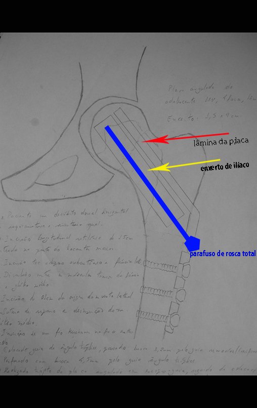
After these x-rays, the diagnosis of simple bone cyst with fracture of the femoral neck was made. The patient was referred for our evaluation and we proposed surgery to reconstruct the bone defect, curettage of the lesion and reconstruction with an autologous bone graft taken from the iliac crest, on the same side, with correction of the angular deformity. For this surgery, we planned and created a special plate that allowed the placement of a full-thread screw, internally fixing the graft block, which would serve as a column in the reconstruction, figures 11 to 19.
Figura 11: Planejamento para a reconstrução, frente.
Figure 11: Planning for reconstruction, front.
Figura 12: Planejamento para a reconstrução, perfil.
Figure 12: Planning for reconstruction, profile.
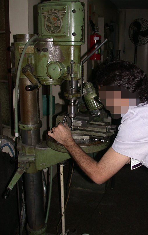
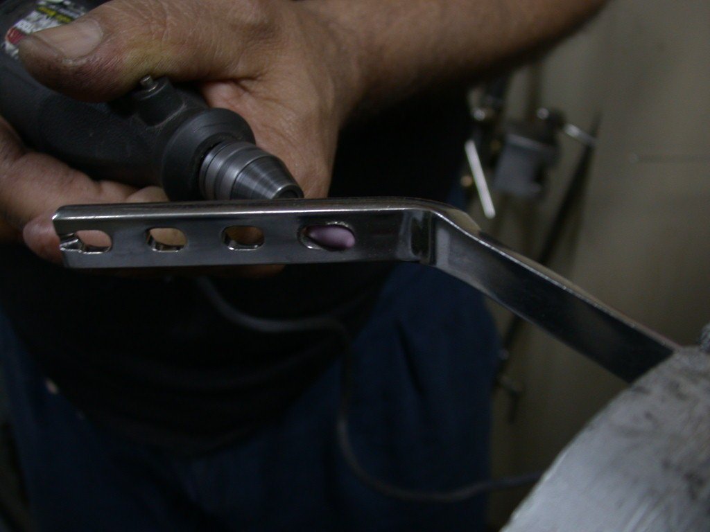
Figura 13: Preparação de placa especial para a reconstrução. Alargamento do primeiro orifício da placa para entrada do parafuso especial de rosca total.
Figure 13: Preparation of a special plate for reconstruction. Enlargement of the first hole on the plate to allow the special full-thread screw to enter.
Figura 14: Parafuso de rosca total, feito especialmente para esta situação.
Figure 14: Full thread screw, made especially for this situation.
Figura 15: Correção do ângulo de inclinação do parafuso.
Figure 15: Correction of the screw inclination angle.
Figura 16: Escarificação e ajustes da largura do orifício.
Figure 16: Scarification and hole width adjustments.
Figura 17: Fresagem e lixamento das bordas no novo orifício.
Figure 17: Milling and sanding the edges of the new hole.
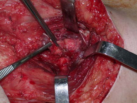
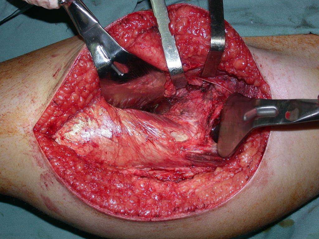
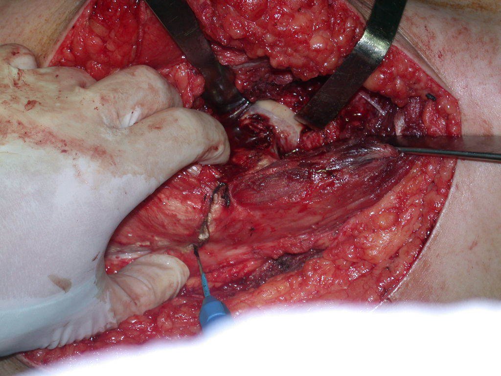
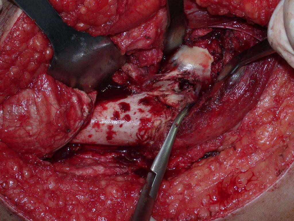
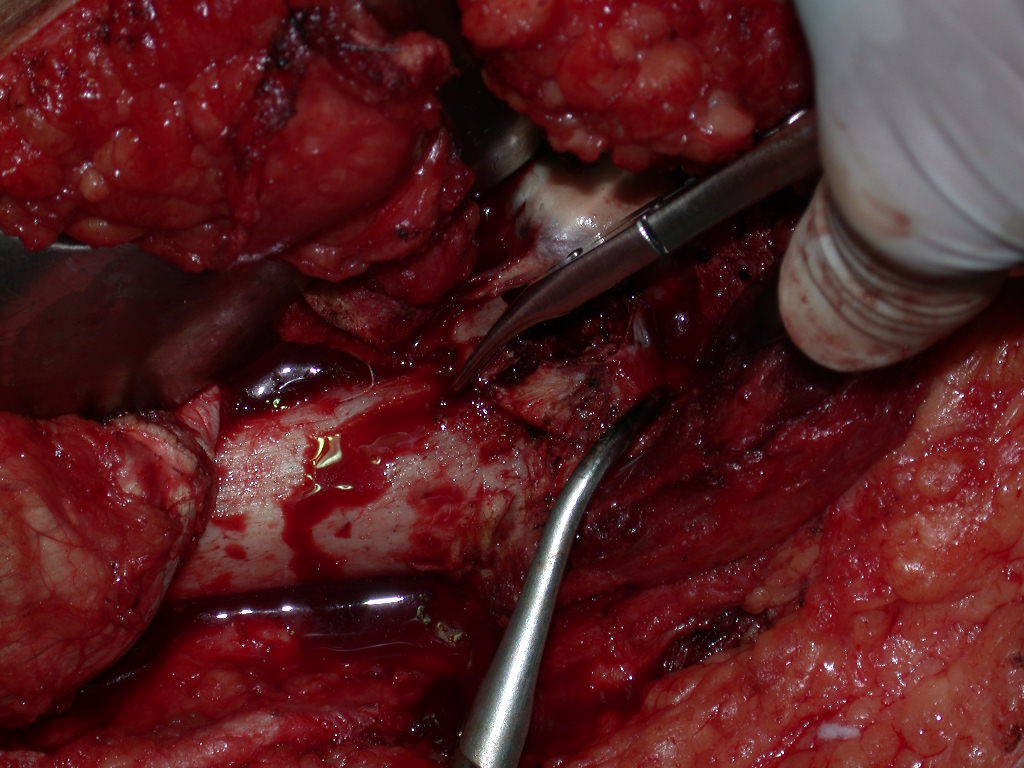
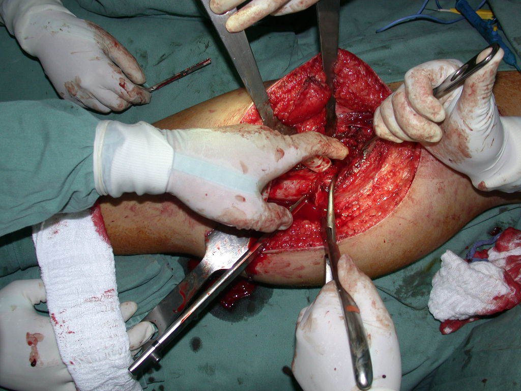
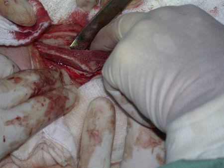
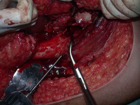
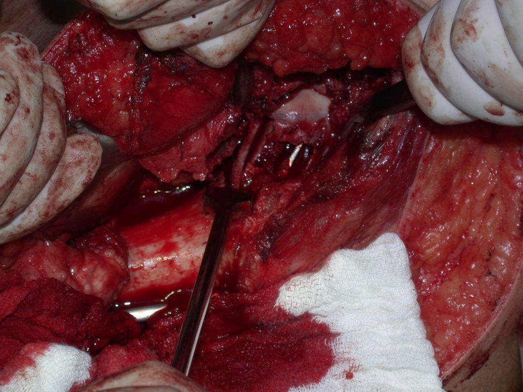
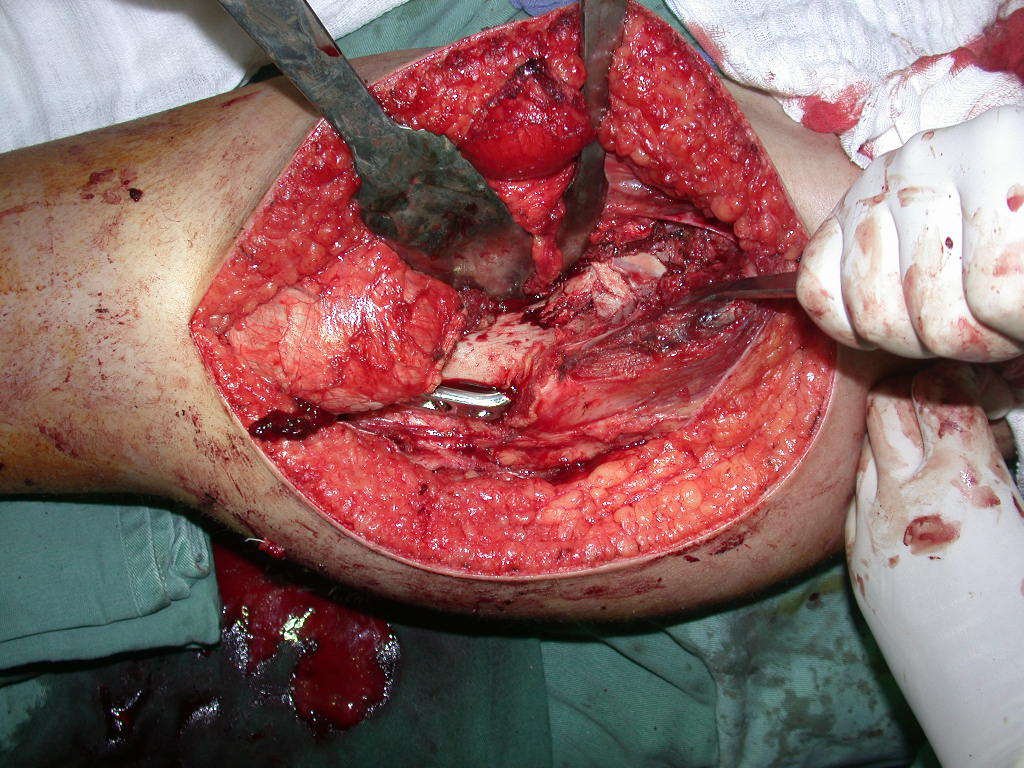
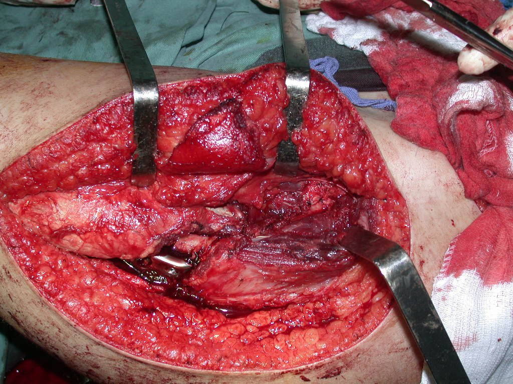
To perform this technique, an adequate surgical access is necessary, which allows for a broad approach and for this access to have good exposure, so as not to hinder reconstruction, reduction or osteosynthesis. Surgical access and reconstruction are shown in figures 20 to 81.
Figura 20: Incisão póstero-lateral na coxa, curvando-se bem posterior ao grande trocânter.
Figure 20: Posterolateral incision on the thigh, curving well posterior to the greater trochanter.
Figura 21: A fáscia lata deve ser seccionada conforme demonstra a figura. A seta azul salienta a secção do contorno que cobre o grande trocânter.
Figure 21: The fascia lata must be sectioned as shown in the figure. The blue arrow highlights the section of the outline that covers the greater trochanter.
Figura 22: Rebatimento da fáscia lata, seta azul. As setas amarelas salientam o contorno da borda posterior da fáscia lata.
Figure 22: Reflection of the fascia lata, blue arrow. The yellow arrows highlight the contour of the posterior edge of the fasciae latae.
Figura 23: Dissecção do tendão do músculo reto anterior da coxa.
Figure 23: Dissection of the tendon of the anterior rectus muscle of the thigh.
This incision allows access that leaves the lateral aspect of the thigh completely exposed, without the need to remove the posterior part of the fasciae latae. This situation is essential to facilitate reduction and fixation with the angled plate.
Figura 24: Identificação do tendão em Y do músculo reto anterior.
Figure 24: Identification of the Y-shaped tendon of the anterior rectus muscle.
Figura 25: Secção da porção circunflexa do tendão do músculo reto anterior da coxa.
Figure 25: Section of the circumflex portion of the tendon of the anterior rectus muscle of the thigh.
Figura 26: Dissecção da porção medial ao colo femoral.
Figure 26: Dissection of the medial portion of the femoral neck.
Figura 27: Colocação de afastador de Bennet medialmente ao colo femoral, dissecção da porção lateral ao colo e colocação do segundo afastador de Bennet.
Figure 27: Placement of the Bennet retractor medially to the femoral neck, dissection of the portion lateral to the neck and placement of the second Bennet retractor.
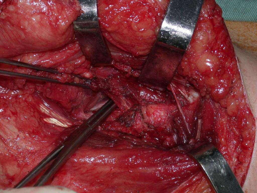
Figura 28: Abertura em T da cápsula articular.
Figure 28: T-shaped opening of the joint capsule.
Figura 29: Exposição da cabeça femoral.
Figure 29: Exposure of the femoral head.
Figura 30: Desinserção do vasto lateral, superiormente.
Figure 30: Disinsertion of the vastus lateralis, superiorly.
Figura 31: Desinserção do vasto lateral, retro vasto.
Figure 31: Disinsertion of the vastus lateralis, retro vastus.
Figura 32: Descolamento do músculo vasto anterior e lateral, desperiostizando a porção antero-lateral da região metafisária proximal do fêmur.
Figure 32: Detachment of the vastus anterior and lateral muscle, deperiostizing the anterolateral portion of the proximal metaphyseal region of the femur.
Figura 33: Desimpactação da fratura do colo femoral.
Figure 33: Disimpaction of the femoral neck fracture.
Figura 34: Tração e redução da cabeça femoral.
Figure 34: Traction and reduction of the femoral head.
Figura 35: Curetagem da lesão e retirada da membrana do cisto ósseo unicameral.
Figure 35: Curettage of the lesion and removal of the unicameral bone cyst membrane.
Figura 36: Alinhamento da redução e correção da deformidade em varo.
Figure 36: Alignment of reduction and correction of varus deformity.
Figura 37: Estudo do posicionamento da placa e conferência da correção da deformidade angular e rotacional.
Figure 37: Study of the positioning of the plate and checking the correction of angular and rotational deformity.
Figura 38: Cabeça, colo e metáfise do fêmur expostos e alinhados. Cavidade do cisto curetada, limpa e preparada para reconstrução.
Figure 38: Head, neck and metaphysis of the femur exposed and aligned. Cyst cavity cured, cleaned and prepared for reconstruction.
Figura 39: Passagem de fio de Kirschner para servir de guia para o parafuso de fixação do enxerto ósseo autólogo em bloco.
Figure 39: Passage of Kirschner wire to serve as a guide for the fixation screw of the autologous bone graft en bloc.
Figura 40: Posicionamento da placa angulada sobre o colo, para orientação do formão que fará o trajeto guia da lâmina.
Figure 40: Positioning the angled plate on the neck, to guide the chisel that will guide the blade.
Figura 41: Introdução do formão guia da lâmina da placa.
Figure 41: Introduction of the plate blade guide chisel.
Figura 42: Finalização do aprofundamento do formão guia.
Figure 42: Completion of the deepening of the guide chisel.
Figura 43: Conferência do posicionamento da lâmina na cabeça femoral.
Figure 43: Checking the positioning of the blade in the femoral head.
Figura 44: Incisão sobre a crista ilíaca esquerda, para a retirada de enxerto ósseo autólogo.
Figure 44: Incision over the left iliac crest, to remove an autologous bone graft.
Figura 45: Incisão na borda de inserção do músculo oblíquo do abdome, feita com eletrocautério.
Figure 45: Incision at the insertion edge of the abdominal oblique muscle, made with electrocautery.
Figura 46: Descolamento do periósteo da tábua interna do ilíaco.
Figure 46: Detachment of the periosteum from the internal table of the iliac bone.
Figura 47: Descolamento do periósteo das tábuas do osso ilíaco, desinserindo a musculatura do músculo oblíquo e glúteos.
Figure 47: Detachment of the periosteum from the iliac bone plates, detaching the muscles of the oblique muscle and glutes.
Figura 48: Realização de orifício no bloco do enxerto que será utilizado na reconstrução e confecção do passo de rosca.
Figure 48: Making a hole in the graft block that will be used in the reconstruction and creation of the thread pitch.
Figura 49: Passagem de fio de Kirschner no orifício, para servir de guia à osteotomia de retirada do enxerto ósseo.
Figure 49: Passing a Kirschner wire through the hole, to serve as a guide for the osteotomy to remove the bone graft.
Figura 50: Enxerto ressecado em um grande fragmento.
Figure 50: Graft resected into a large fragment.
Figura 51: Bloco de enxerto que será encavilhado dentro do colo femoral e fixado com parafuso de rosca total.
Figure 51: Graft block that will be nailed into the femoral neck and fixed with a full-thread screw.
Figura 52: Bloco de enxerto com o fio guia.
Figure 52: Graft block with guide wire.
Figura 53: Passagem prévia do parafuso rosqueado para teste final e moldagem do enxerto.
Figure 53: Previous passage of the threaded screw for final testing and molding of the graft.
Figura 54: Alargamento de anteriorização do orifício de entrada da lâmina, para correção da rotação.
Figure 54: Anterior enlargement of the blade entry hole, to correct rotation.
Figura 55: Regularização do canal de entrada da lâmina e conferência da medida do seu comprimento.
Figure 55: Regularization of the blade entry channel and checking its length measurement.
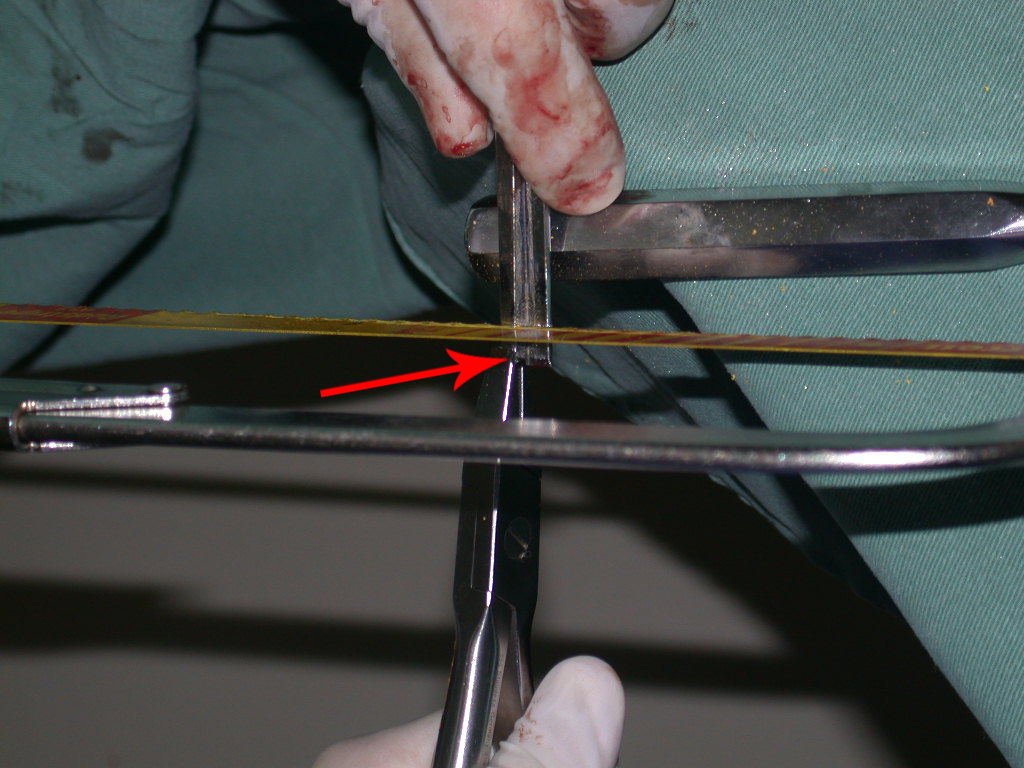
Figura 56: Corte de 0.8 cm da lâmina, por segurança, para evitar que ocorra perfuração da cabeça femoral.
Figure 56: Cut 0.8 cm from the blade, for safety reasons, to prevent perforation of the femoral head.
Figura 57: Complementação do corte de 0,8 cm do comprimento da lâmina da placa.
Figure 57: Complementary cut of 0.8 cm in the length of the plate blade.
Figura 58: Posicionamento da placa e perfuração da cortical.
Figure 58: Positioning of the plate and perforation of the cortex.
Figura 59: Realização do canal de rosca na cortical do fêmur.
Figure 59: Creation of the thread channel in the femoral cortex.
Note that the fascia lata does not appear in the operative field. If we did not use this type of access, we would have a rope over the lateral surface of the thigh, making it difficult to position the plate and reduce the fragments.
Figura 60: Checagem da redução.
Figure 60: Checking the reduction.
Figura 61: Correção do posicionamento do enxerto.
Figure 61: Correction of graft positioning.
Figura 62: Passagem do fio de Kirschner dentro do orifício do bloco de enxerto que foi encavilhado dentro do colo femoral.
Figure 62: Passage of the Kirschner wire inside the orifice of the graft block that was embedded inside the femoral neck.
Figura 63: Checagem final da redução e do posicionamento do enxerto.
Figure 63: Final check of reduction and graft positioning.
Figura 64: Passagem final da fresa na cortical e no enxerto em bloco para colocação do parafuso.
Figure 64: Final passage of the drill in the cortex and in the block graft for screw placement.
Figura 65: Colocação do parafuso e fixação do bloco de enxerto ósseo.
Figure 65: Placement of the screw and fixation of the bone graft block.
Figura 66: Retirada de enxerto em tiras para complementação do preenchimento da lesão do colo femoral.
Figure 66: Removal of graft in strips to complement the filling of the femoral neck lesion.
Figura 67: O enxerto é retirado da tábua interna, mantendo-se a tábua externa intacta para preservar o contorno da anatomia do ilíaco.
Figure 67: The graft is removed from the inner table, keeping the outer table intact to preserve the contour of the iliac anatomy.
Figura 68: Impactação final da placa, e colocação dos parafusos diafisários.
Figure 68: Final impaction of the plate and placement of diaphyseal screws.
Figura 69: Impactação das tiras de enxerto ao redor da coluna de enxerto em bloco, colocada no centro do colo e transfixada com parafuso.
Figure 69: Impaction of the graft strips around the block graft column, placed in the center of the neck and transfixed with a screw.
Figura 70: Toda a lesão cavitaria ocasionada pelo cisto ósseo deve ser preenchida com enxerto para permitir uma reparação completa da anatomia do colo femoral.
Figure 70: Any cavitary lesion caused by the bone cyst must be filled with a graft to allow complete repair of the femoral neck anatomy.
Figura 71: Aspecto da tábua externa do ilíaco após a retirada de enxerto do lado interno.
Figure 71: Aspect of the external table of the iliac bone after graft removal from the internal side.
Figura 72: A impactação do enxerto é importante para uma boa reconstrução.
Figure 72: Graft impaction is important for good reconstruction.
Figura 73: Colocação de lasca de enxerto para cobertura da janela óssea.
Figure 73: Placement of graft sliver to cover the bone window.
Figura 74: Impactação da lasca de cobertura da janela.
Figure 74: Impaction of the window covering chip.
Figura 75: Colocação de cunha óssea para contenção do enxerto.
Figure 75: Placement of a bone wedge to contain the graft.
8beca3817318ec14b010b1e4a39b4380 3
Figure 76: Neck and metaphysis reduced, cavity filled with autologous graft, osteosynthesis completed.
Ecfb953d9d17e80aa226d13ff9dd04a8 3
Figure 77: Reconstruction completed.
Figura 78: Fechamento da cápsula articular do colo femoral.
Figure 78: Closure of the femoral neck joint capsule.
Figura 79: Aproximação da porção reflexa do tendão do reto anterior.
Figure 79: Approximation of the reflex portion of the anterior rectus tendon.
Figura 80: Sutura do tendão da porção reflexa do reto anterior.
Figure 80: Suture of the tendon of the reflex portion of the anterior rectus.
Figura 81: Tendão reconstruído.
Figure 81: Reconstructed tendon.
After surgery, control radiographs were taken, figures 82 and 83.
Figura 82: Radiografia do pós-operatório imediato, frente.
Figure 82: Radiograph of the immediate postoperative period, front.
Figura 83: Radiografia do pós-operatório imediato, perfil.
Figure 83: Radiograph of the immediate postoperative period, profile.
Figura 84: Função da paciente em 15/04/2005, após sete meses da cirurgia.
Figure 84: Patient function on 04/15/2005, seven months after surgery.
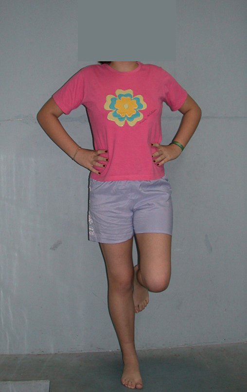
Figura 85: Carga monopodal após sete meses da cirurgia de reconstrução do colo femoral, com enxerto autólogo e placa especial, frente.
Figure 85: Single-leg loading after seven months of femoral neck reconstruction surgery, with autologous graft and special plate, front.
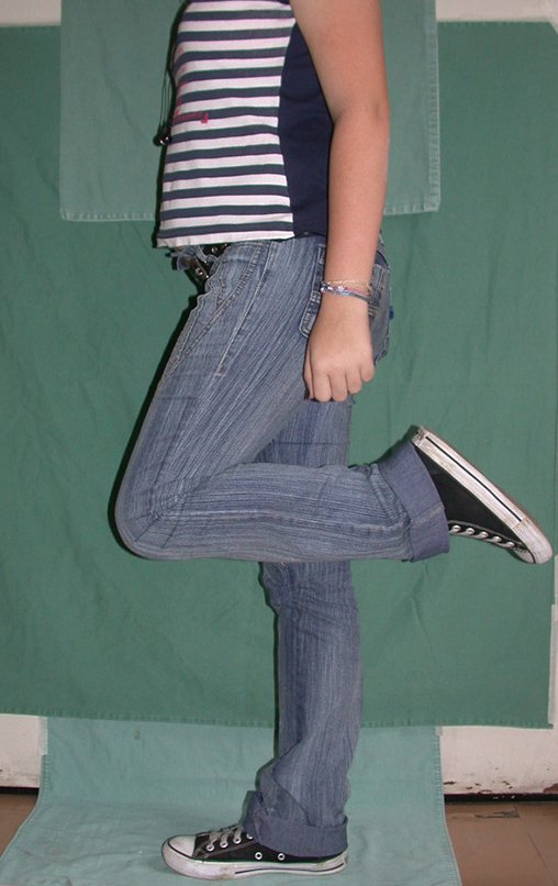
Figura 86: Flexão do quadril e do joelho, sem carga, após sete meses da cirurgia.
Figure 86: Flexion of the hip and knee, without weight, seven months after surgery.
Figura 87: Carga total monopodal, após sete meses da cirurgia de reconstrução do colo femoral com enxerto autólogo e placa especial, perfil.
Figure 87: Total single-leg load, seven months after femoral neck reconstruction surgery with autologous graft and special plate, profile.
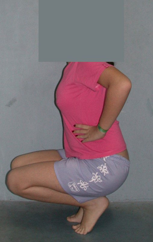
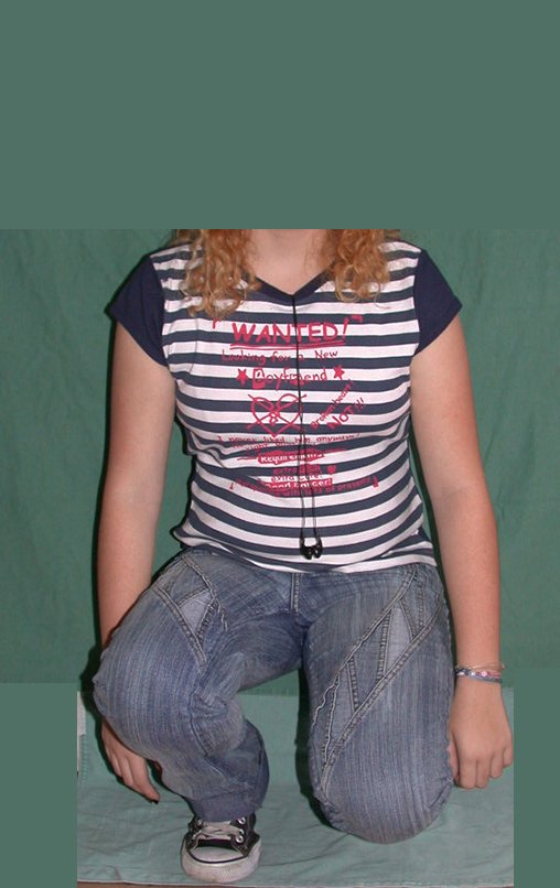
Figura 88: Flexão dos quadris e joelhos simétricos, com carga total, após sete meses da cirurgia.
Figure 88: Symmetrical flexion of the hips and knees, with full load, seven months after surgery.
Figura 89: Radiografias de controle de 12/04/2005, perfil, após sete meses da cirurgia, reconstituição anatômica do colo femoral.
Figure 89: Control radiographs from 04/12/2005, profile, seven months after surgery, anatomical reconstruction of the femoral neck.
Figura 90: Radiografias de controle de 12/04/2005, frente, após sete meses da cirurgia, reconstituição anatômica do colo femoral.
Figure 90: Control radiographs from 04/12/2005, front, seven months after surgery, anatomical reconstruction of the femoral neck.
Figura 91: Flexão ativa do joelho e do quadril operado, sem carga, após um ano da cirurgia.
Figure 91: Active flexion of the operated knee and hip, without load, one year after surgery.
Figura 92: Carga total, monopodal, sobre o quadril operado, um ano da cirurgia.
Figure 92: Total load, single leg, on the operated hip, one year after surgery.
Figura 93: Flexão com carga total, perfil, após um ano da cirurgia.
Figure 93: Flexion with full load, profile, one year after surgery.
Figura 94: Flexão com carga total, frente, após um ano da cirurgia.
Figure 94: Flexion with full load, front, one year after surgery.
Figura 95: Radiografia da bacia frente, com os quadris simétricos e com a integração total do enxerto autólogo, reconstituindo o colo femoral.
Figure 95: X-ray of the front pelvis, with symmetrical hips and with full integration of the autologous graft, reconstituting the femoral neck.
Figura 96: Radiografia do quadril operado, integração total do enxerto autólogo, reconstituição anatômica do colo femoral.
Figure 96: Radiograph of the operated hip, full integration of the autologous graft, anatomical reconstruction of the femoral neck.
Figura 97: Paciente com dez anos de idade, após um ano de pós-operatório, iniciando a puberdade, com um bom prognóstico.
Figure 97: Ten-year-old patient, one year after surgery, starting puberty, with a good prognosis.
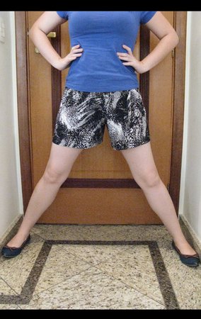
The autologous graft provides earlier and better bone integration. The patient is doing well, with normal movement of the operated hip. On March 19, 2016, we reevaluated the patient clinically and with imaging studies, figures 98 to 108 and video 1.
Figura 98: Radiografia da bacia de março de 2016, após doze anos da reconstrução. Detalhe do ilíaco esquerdo, área doadora do enxerto.
Figure 98: X-ray of the basin in March 2016, twelve years after reconstruction. Detail of the left iliac bone, graft donor area.
Figura 99: Radiografia de quadril operado, após doze anos. Boa congruência articular.
Figure 99: Radiograph of the operated hip, after twelve years. Good joint congruence.
Figura 100: Radiografia em perfil, após doze anos, bom alinhamento do colo femoral.
Figure 100: Profile x-ray, after twelve years, good alignment of the femoral neck.
Figura 101: Tomografia da congruência do quadril esquerdo após doze anos.
Figure 101: Tomography of left hip congruence after twelve years.
Figura 102: Tomografia da cabeça femoral, após doze anos, observamos pequenas áreas císticas remanescentes.
Figure 102: Tomography of the femoral head, after twelve years, we observed small remaining cystic areas.
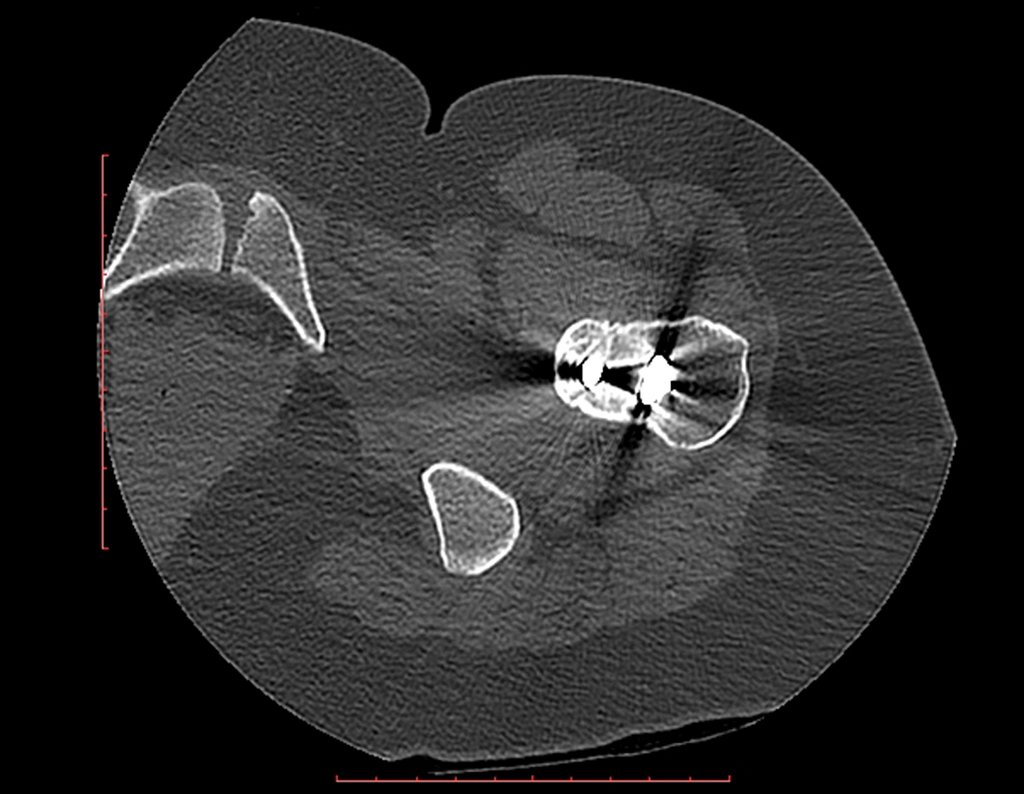
Figura 103: Tomografia do colo femoral operado, após doze anos, imagem com artefato da lâmina e do parafuso.
Figure 103: Tomography of the operated femoral neck, after twelve years, image with blade and screw artifact.
Figura 104: Tomografia do final, há haste da placa, na diáfise femoral, em março de 2016.
Figure 104: Tomography of the end, there is a plate stem, in the femoral diaphysis, in March 2016.
Figura 105: Paciente com 24 anos de idade, após doze anos da reconstrução do quadril, bem sem queixa.
Figure 105: 24-year-old patient, twelve years after hip reconstruction, with no complaints.
Figura 106: Carga total, monopodal, após doze anos da reconstrução biológica do quadril.
Figure 106: Full weight, single leg, after twelve years of biological hip reconstruction.
Figura 107: Flexão com carga total, após doze anos da reconstrução biológica do quadril. Boa função
Figure 107: Flexion with full load, twelve years after biological hip reconstruction. Good function
Figura 108: Abdução normal dos quadris, após doze anos da reconstrução biológica do quadril. Boa função
Figure 108: Normal hip abduction, twelve years after biological hip reconstruction. Good function
Video 1: Patient on 03/19/2016, twelve years after surgery. Good hip function.
The patient, now married, is well and satisfied with her role. No complaints.

Author: Prof. Dr. Pedro Péricles Ribeiro Baptista

 Orthopedic Oncosurgery at the Dr. Arnaldo Vieira de Carvalho Cancer Institute

Leave a Reply

Hello! How can we assist you?