This digital library houses the book on Oncology and Orthopedic Oncosurgery.

It includes academic lectures, presentations from national and international congresses, published papers, case discussions, performed surgical procedures, and proprietary techniques developed.

The digital format was chosen because the web allows the inclusion of texts with numerous visual resources, such as images and videos, which would not be possible in a printed book.

The content is intended for students, healthcare professionals, and the general public interested in the field.

Soft tissue sarcoma of the shoulder

032abcd424b4312e7087f434ef1c0094 2

Soft Tissue Sarcoma of the Shoulder

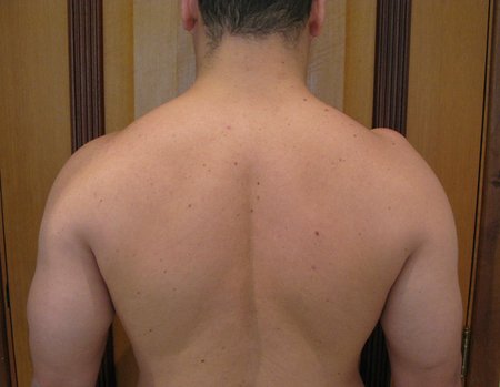
Soft tissue sarcoma of the shoulder. A 29-year-old male patient reports the appearance of a nodule on his right shoulder eight months ago.
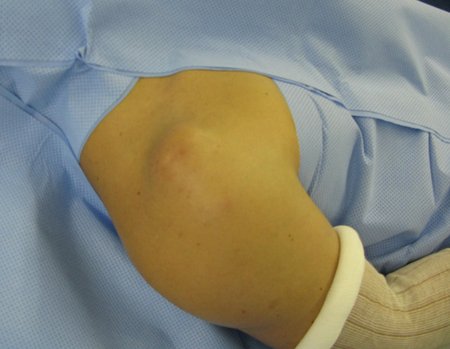
Figura 1: Abaulamento no ombro direito, sobre o acrômio e extremidade distal da clavícula.
Figure 1: Bulging in the right shoulder, over the acromion and distal end of the clavicle.
Figura 2: Aspecto posterior da lesão nodular.
Figure 2: Posterior aspect of the nodular lesion.
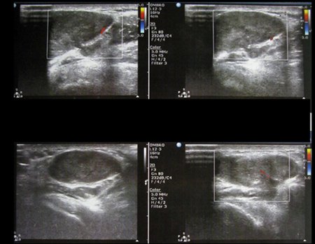
In August 2014, an ultrasound was performed, figures 3 and 4.
Figura 3: Ultrassonografia realizada três meses antes da nossa consulta.
Figure 3: Ultrasound performed three months before our consultation.
Figura 4: Laudo: lesão sólida, heterogênea e expansiva medindo 5,5 x 3,4 x 1,7 cm. Sugere a realização de Ressonância magnética.
Figure 4: Report: solid, heterogeneous and expansive lesion measuring 5.5 x 3.4 x 1.7 cm. Suggests magnetic resonance imaging.
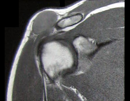
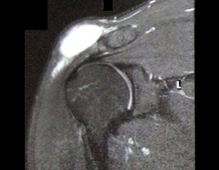
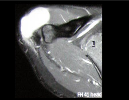
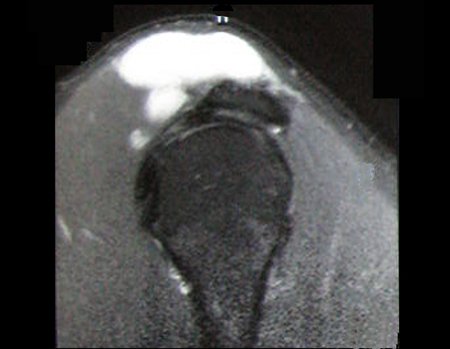
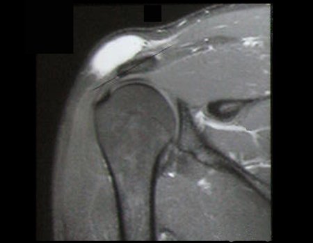
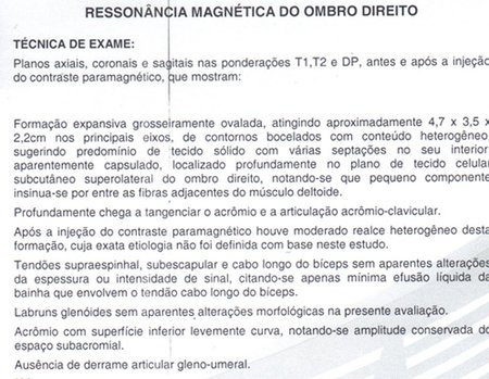
This exam is operator dependent, and we are responsible for limiting the report carried out. There is a need for magnetic resonance imaging, which is the best imaging test for soft tissue tumors. An MRI was then performed, the main images of which are shown in figures 5 to 10.
Figura 5: Rm cor, com lesão sólida superficial, com baixo sinal em T1, localizada acima do acrômio e extremidade distal da clavícula direita.
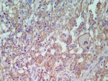
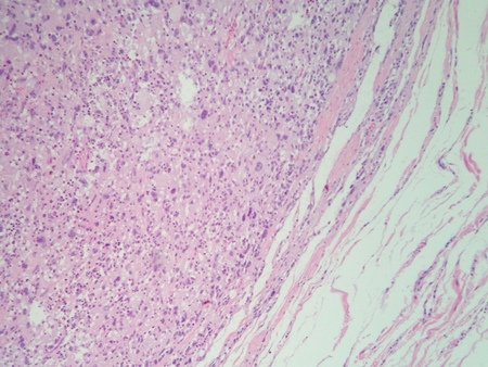
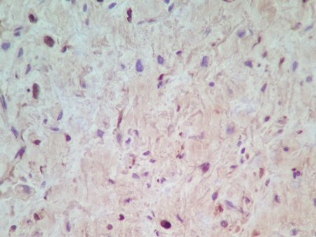
Figure 5: Color MRI, with superficial solid lesion, with low signal on T1, located above the acromion and distal end of the right clavicle.
Figura 6: Rm cor com alto sinal em T1 com supressão de gordura.
Figure 6: Color MRI with high signal on T1 with fat suppression.
Figura 7: Rm axial com alto sinal em T1 com supressão de gordura.
Figure 7: Axial MRI with high signal on T1 with fat suppression.
Figura 8: Rm sagital com alto sinal em T1 com supressão de gordura.
Figure 8: Sagittal MRI with high signal on T1 with fat suppression.
Figura 9: Rm coronal com captação de contraste, em T1 com supressão de gordura e gadolínio.
Figure 9: Coronal MRI with contrast uptake, on T1 with fat and gadolinium suppression.
Figura 10: Laudo do exame de ressonância magnética.
Figure 10: Magnetic resonance imaging report.
In October 2014, a needle biopsy was performed, guided by ultrasound, the pathology reports and the requested review are shown in figures 11 and 12, respectively.
Figura 11: Laudo do anatomopatológico da biópsia.
Figure 11: Biopsy anatomopathological report.
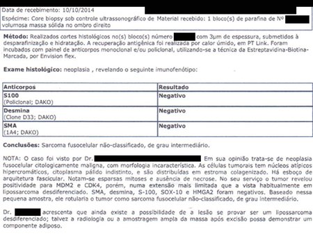
Figura 12: Laudo da revisão de lâminas, blocos e imuno-histoquímica solicitada.
Figure 12: Report of the review of slides, blocks and immunohistochemistry requested.
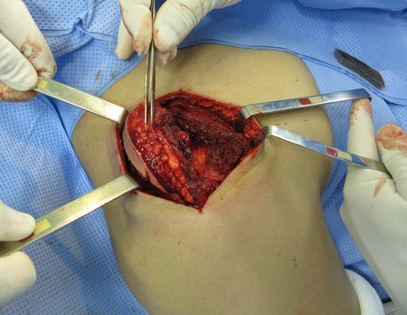
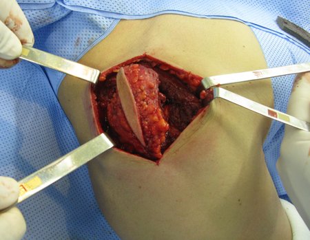
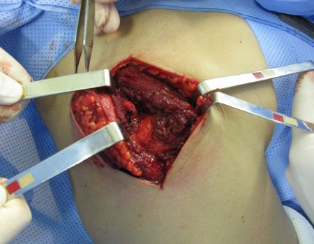
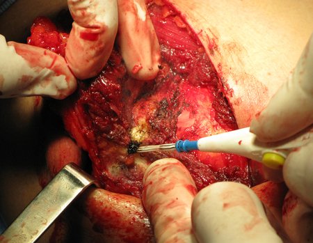
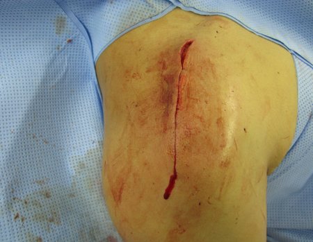
With these data, the patient was referred for our evaluation and management. We performed surgical resection of the lesion, en bloc, with a wide margin, resecting the skin and the biopsy path and having as a deep margin the periosteum of the acromion and the distal end of the right clavicle, figures 13 to 25.
Figura 13: Paciente posicionado em decúbito dorsal semissentado.
Figure 13: Patient positioned in semi-sitting supine position.
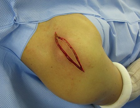
Figura 14: Incisão sobre a articulação acromioclavicular, estendendo-se do terço externo da clavícula até a articulação glenoumeral, contornando todo o trajeto da biópsia.
Figure 14: Incision over the acromioclavicular joint, extending from the outer third of the clavicle to the glenohumeral joint, contouring the entire biopsy path.
Figura 15: Dissecção do subcutâneo, contornando o trajeto da biópsia no plano profundo e realização cuidadosa da hemostasia.
Figure 15: Subcutaneous dissection, contouring the biopsy path in the deep plane and carefully performing hemostasis.
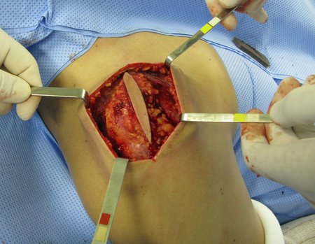
Figura 16: Secção de fibras do músculo deltoide e exposição da borda anterior do acrômio.
Figure 16: Section of deltoid muscle fibers and exposure of the anterior edge of the acromion.
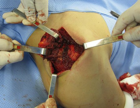
Figura 17: Dissecção nos planos profundos com margem circunferencial da lesão e hemostasia.
Figure 17: Dissection in deep planes with circumferential margin of the lesion and hemostasis.
Figura 18: Exposição da borda anterior do acrômio e início da desperiostização do mesmo, utilizando o termo cautério.
Figure 18: Exposure of the anterior edge of the acromion and beginning of its deperiostization, using the term cautery.
Figura 19: Com o emprego do bisturi elétrico vamos "ruginando" o periósteo do acrômio e do terço distal da clavícula, removendo-o juntamente com a neoplasia.
Figure 19: Using the electric scalpel, we "roar" the periosteum of the acromion and the distal third of the clavicle, removing it together with the neoplasm.
Figura 20: Cuidadosamente o tumor vai sendo ressecado, com margem ampla em toda a sua circunferência.
Figure 20: The tumor is carefully resected, with a wide margin around its entire circumference.
Figura 21: O tumor é totalmente removido.
Figure 21: The tumor is completely removed.
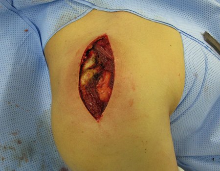
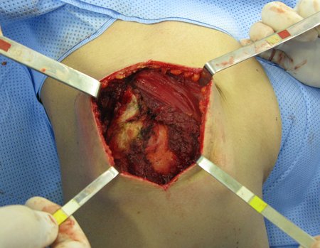
Figura 22: Podemos verificar o leito operatório, com parte do deltoide ressecado e a superfície exposta do tecido ósseo das extremidades.
Figure 22: We can see the operating bed, with part of the deltoid resected and the exposed surface of the bone tissue of the extremities.
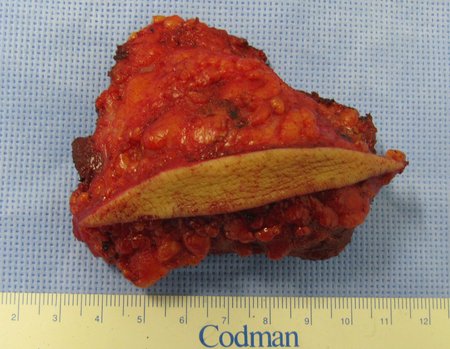
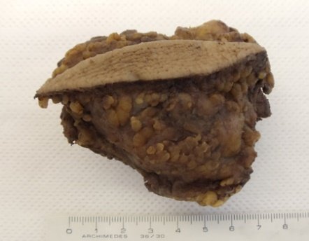
Figura 23: Peça ressecada com a pele e o trajeto da biópsia, observando-se tecido adiposo sadio em toda a superfície da lesão, nesta face.
Figure 23: Resected piece with the skin and the biopsy path, observing healthy adipose tissue on the entire surface of the lesion, on this face.
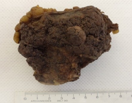
Figura 24: Peça ressecada com fibras do deltoide e periósteo, como margem no plano profundo.
Figure 24: Resected piece with fibers from the deltoid and periosteum, as a margin in the deep plane.
Figura 25: O fechamento é realizado deslizando-se parte das fibras do deltoide por sobre o tecido ósseo exposto, recobrindo-o.
Figure 25: Closure is performed by sliding part of the deltoid fibers over the exposed bone tissue, covering it.
Figura 27: Peça cirúrgica, ressecada com o periósteo e fibras do músculo deltoide.
Figure 26: Surgical specimen, resected with the skin and the biopsy path. Figure 27: Surgical specimen, resected with the periosteum and deltoid muscle fibers.
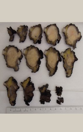
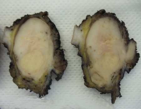
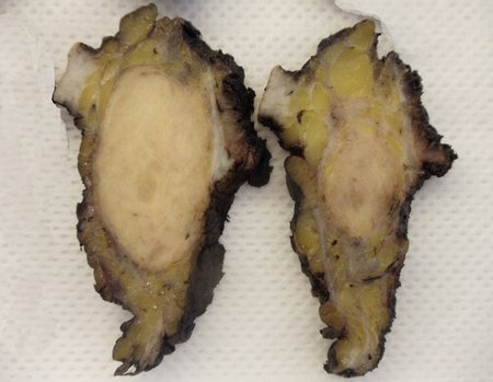
Figura 28: Cortes da peça cirúrgica, cujas bordas foram pintadas com Nankin.
Figure 28: Sections of the surgical piece, whose edges were painted with Nankin.
Figura 29: Corte da peça cirúrgica, para estudo e avaliação das margens oncológicas.
Figure 29: Section of the surgical specimen, for study and evaluation of the oncological margins.
Figura 30: Vários cortes da peça são realizados, para estudo.
Figure 30: Several cuts of the piece are made for study.
Figura 31: teste de edição
Figure 31: Edit test
Figura 32
Figure 32
Figura 33
Figure 33
Figura 34
Figure 34
Figura 35
Figure 35
Figura 36
Figure 36
Figura 37
Figura 37
Figura 38
Figure 38
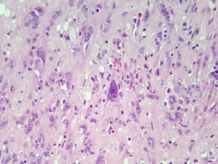
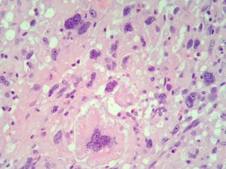
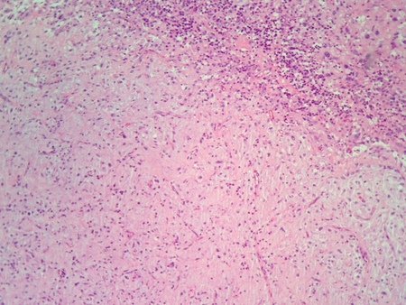
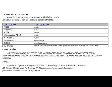
Figura 39: Relatório final da anatomia patológica da neoplasia.
Figure 39: Final report on the pathological anatomy of the neoplasm.
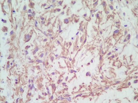
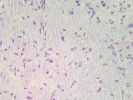
Figura 40: Laudo da patologia e imuno-histoquímica da peça cirúrgica.
Figure 40: Pathology and immunohistochemistry report of the surgical specimen.

Author: Prof. Dr. Pedro Péricles Ribeiro Baptista

 Orthopedic Oncosurgery at the Dr. Arnaldo Vieira de Carvalho Cancer Institute

Hello! How can we assist you?