This digital library houses the book on Oncology and Orthopedic Oncosurgery.

It includes academic lectures, presentations from national and international congresses, published papers, case discussions, performed surgical procedures, and proprietary techniques developed.

The digital format was chosen because the web allows the inclusion of texts with numerous visual resources, such as images and videos, which would not be possible in a printed book.

The content is intended for students, healthcare professionals, and the general public interested in the field.

Bone Metastasis – Breast Cancer

Ac2ef27277eab81da1d8dea067dd80c1 3

Bone Metastasis of Breast Cancer in the Humerus

Bone Metastasis – Breast Cancer
Figura 1: Paciente em decúbito dorsal com o membro superior direito sobre a barriga, preparado para a incisão cirúrgica.
Figure 1: Patient in supine position with the right upper limb on the belly, prepared for the surgical incision.
Figura 2: Incisão cirúrgica no membro superior direito.
Figure 2: Surgical incision in the right upper limb.
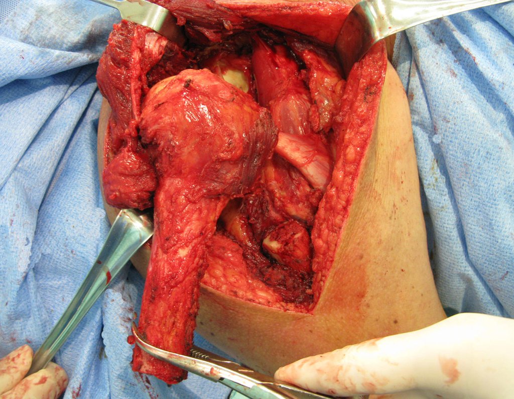
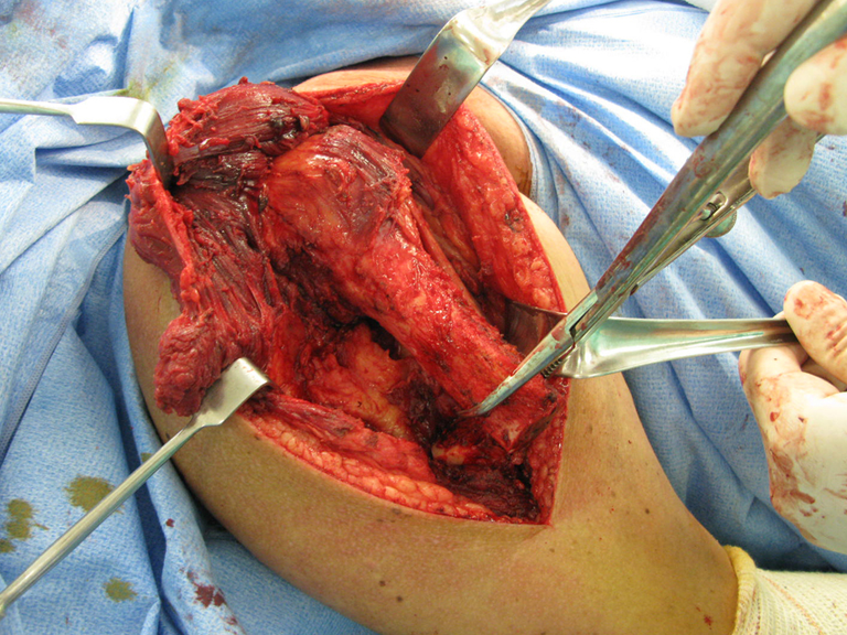
Figura 3: Evidenciamos a musculatura do braço direito.
Figure 3: We highlight the muscles of the right arm.
Figura 4: Neste momento a musculatura está sendo separada do osso.
Figure 4: At this moment the muscles are being separated from the bone.
Figura 5: Evidenciamos a passagem da Serra de Gigle para começar a serra o osso.
Figure 5: We can see the passage of Serra de Gigle to begin sawing the bone.
Figura 6: Neste momento o osso foi serrado.
Figure 6: At this point the bone was sawn.
Figura 7
Figure 7
Figura 8
Figure 8
Figura 9
Figure 9
Figura 10
Figure 10
Figura 11
Figure 11
Figura 12
Figure 12
Figura 13: Evidenciamos a comparação da prótese de prova com o úmero proximal ressecado.
Figure 13: We show the comparison of the trial prosthesis with the resected proximal humerus.
Figura 14: Evidenciamos a comparação da prótese final com o úmero proximal retirado.
Figure 14: We show the comparison of the final prosthesis with the removed proximal humerus.
Figura 15: Evidenciamos a colocação da prótese.
Figure 15: We show the placement of the prosthesis.
Figura 16: Evidenciamos a colocação da prótese.
Figure 16: We show the placement of the prosthesis.
Figura 17: Fechamento da ferida operatória sobre a prótese.
Figure 17: Closing the surgical wound over the prosthesis.
Figura 18: Fechamento da ferida operatóriaso sobre a prótese.
Figure 18: Closing the surgical wound over the prosthesis.
Figura 19: Colocação do dreno de Portovac e fechamento da ferida operatória.
Figure 19: Placement of the Portovac drain and closure of the surgical wound.
Figura 20: Ferida operatória fechada e com curativo.
Figure 20: Surgical wound closed and with dressing.
Figura 21: Radiografia pós operatória imediato do úmero direito frente, evidenciando a parte proximal da prótese.
Figure 21: Immediate post-operative radiograph of the front right humerus, showing the proximal part of the prosthesis.
Figura 22: Radiografia pós operatória imediato evidenciando a parte distal da prótese.
Figure 22: Immediate post-operative radiograph showing the distal part of the prosthesis.
Figura 23: Radiografia pós operatória imediato da prótese do úmero.
Figure 23: Immediate postoperative radiograph of the humerus prosthesis.

Author: Prof. Dr. Pedro Péricles Ribeiro Baptista

 Orthopedic Oncosurgery at the Dr. Arnaldo Vieira de Carvalho Cancer Institute

Hello! How can we assist you?