This digital library houses the book on Oncology and Orthopedic Oncosurgery.

It includes academic lectures, presentations from national and international congresses, published papers, case discussions, performed surgical procedures, and proprietary techniques developed.

The digital format was chosen because the web allows the inclusion of texts with numerous visual resources, such as images and videos, which would not be possible in a printed book.

The content is intended for students, healthcare professionals, and the general public interested in the field.

Ankle Metastasis

801a089759389ea9fa5f77ecc339f4be 2

Bone metastasis from squamous cell cancer in the ankle
Ankle Metastasis. Female patient, 61 years old, with a history of uterine cancer, underwent total hysterectomy in May 2014. She presented with a new primary tumor, now breast cancer, and underwent surgery in July 2014. She underwent adjuvant chemotherapy and radiotherapy. The patient developed recurrence of the uterine tumor in the vagina and underwent reoperation twice, the last time being vaginal amputation. Until now, she had no signs of tumor recurrence. She started experiencing pain in the posterior region of her right ankle, which had worsened after walking for 3 months. The gynecologist requested x-rays of the ankle shown below.
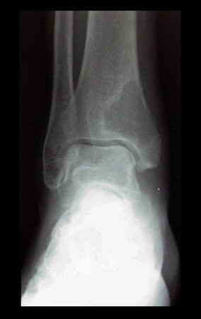
Figura 1: Rx de frente do tornozelo direito, onde observamos lesão radio transparente bem delimitada na região meta epifisária distal da tíbia, envolvendo a região medial e o maléolo. Existe halo de esclerose contornando a lesão na região lateral e na face medial, parece afilar a cortical, porém não a rompe e não vemos reação periosteal.
Figure 1: X-ray of the front of the right ankle, where we observed a well-defined radio-transparent lesion in the distal meta-epiphyseal region of the tibia, involving the medial region and the malleolus. There is a halo of sclerosis surrounding the lesion on the lateral and medial sides. It appears to thin the cortex, but does not rupture it and we do not see a periosteal reaction.
Figura 2: Rx de perfil do tornozelo direito. Observamos que a lesão está na porção posterior do tornozelo e, apesar do afilamento cortical não existe ruptura aparente e não vemos reação periosteal posterior.
Figure 2: Profile x-ray of the right ankle. We observed that the lesion is in the posterior portion of the ankle and, despite the cortical thinning, there is no apparent rupture and we do not see a posterior periosteal reaction.
Figura 3: Rx de perfil do pé direito. Vemos com clareza que a cortical posterior da tíbia está preservada e não sofreu destruição pelo tumor.
Figure 3: Profile x-ray of the right foot. We clearly see that the posterior cortex of the tibia is preserved and has not been destroyed by the tumor.

When we analyzed the images, we observed a well-defined lesion in the distal region of the right tibia, with sclerotic edges on the lateral surface, without destruction of the medial cortex and without periosteal reaction. We can imagine that this area represents a lesion of low aggressiveness and likely benign origin. However, we cannot forget that we are not facing an examination finding, that is, the injury is causing symptoms of pain, which led the patient to complain to her doctor. Furthermore, his history of two neoplasms creates the need to rule out or confirm the metastatic etiology of this lesion.

The next step in managing this case is to perform a complete tumor staging. We request tomography scans of the thorax, abdomen and pelvis to search for other neoplasms in parenchymal viscera. We did not find other tumor sites. We request a total body bone scan to check for other skeletal injuries. This exam, which is very sensitive and not very specific, allows the investigation of other skeletal sites with possible neoplastic involvement, which did not reveal other affected sites other than the right ankle. Serum protein electrophoresis is mandatory to investigate multiple myeloma, another neoplasm compatible with the patient’s age that often presents bone lesions not visualized on scintigraphy. This patient did not present changes in electrophoresis. Magnetic resonance imaging of the ankle is necessary for local tumor staging and surgical planning.

Figura 4: Rm sag T1. observamos grande lesão tumoral na região meta epifisária posterior da tíbia, de limites não precisos, rompendo totalmente a cortical posterior, crescendo em direção aos tecidos moles, com aparente preservação da superfície articular com o tálus.
Figure 4: Rm sag T1. we observed a large tumor lesion in the posterior metaepiphyseal region of the tibia, with unclear limits, completely disrupting the posterior cortex, growing towards the soft tissues, with apparent preservation of the articular surface with the talus.
Figura 5: Rm sag T2 Spir. Lesão de alto sinal, com grande extensão para os tecidos moles adjacentes
Figure 5: Rm sag T2 Spir. High signal lesion, with great extension to adjacent soft tissues
Figura 6: Rm cor T2 Spir. Observamos que apesar da agressividade da lesão, a superfície articular com o tálus mantem-se preservada. Isso é relevante para o planejamento cirúrgico.
Figure 6: Rm color T2 Spir. We observed that despite the aggressiveness of the injury, the articular surface with the talus remains preserved. This is relevant for surgical planning.
Figura 7: Rm axial T2 Spir. nesta imagem podemos situar a relação do tumor com as estruturas nobres da região. O feixe vascular tibial posterior parece estar livre da neoplasia, da mesma forma o tendão calcâneo e a articulação fíbulo talar distal.
Figure 7: Axial Rm T2 Spir. In this image we can see the relationship between the tumor and the noble structures of the region. The posterior tibial vascular bundle appears to be free of neoplasia, as do the Achilles tendon and the distal fibulotalar joint.
Figura 8: Laudo da RM.
Figure 8: MRI report.
Figura 9: Laudo da RM do tornozelo direito.
Figure 9: MRI report of the right ankle.
Fifteen days passed between the initial x-ray and the MRI and we observed more aggressive features than those previously seen. It is clear that we have an aggressive lesion in the posteromedial region of the ankle, thinning and destroying the posterior cortex of the tibia, and growing towards the soft tissues. Although we did not find other sites affected by the disease, the neoplastic history and age of this patient favor the hypothesis of a bone metastasis originating from another neoplasm. Possibly originating from one of the patient’s previous tumors, or less likely a third neoplasm.
Figura 10: Cintilografia. Nesta paciente não observamos lesões em outros locais do esqueleto, além do tornozelo.
Figure 10: Scintigraphy. In this patient, we did not observe lesions in other locations of the skeleton, besides the ankle.

The possibility of it being a primary bone tumor cannot yet be completely ruled out. Academically, the best approach would be a biopsy of the ankle with the aim of differentiating a sarcoma, which would represent a primary tumor, from a carcinoma that would confirm the metastatic origin of this lesion. We initially recommended percutaneous biopsy for this patient. However, between the first evaluation and the return, this patient presented a major destructive evolution of the ankle seen in this x-ray below, taken one month after the first. If we performed the biopsy and waited for the results to then plan a resection, this joint would probably be doomed by rapid tumor progression. At this moment, the patient is at high risk of a pathological bone fracture in the ankle, with a risk of collapse of the remaining bone architecture and severe local functional impairment. It has 11 points out of 12 according to the Mirrels classification, which predicts the risk of fracture in metastatic lesions.
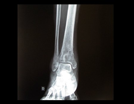
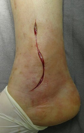
Figura 11: Rx de frente do tornozelo direito realizada um mês após o primeiro exame. Lesão nitidamente mais agressiva quando comparada a radiografia inicial. Apresenta destruição da parede medial da tíbia e reação periosteal. Os limites da lesão já não estão nítidos como anteriormente e a reação esclerótica não delimita mais o tumor.
Figure 11: X-ray of the front of the right ankle performed one month after the first examination. Clearly more aggressive injury when compared to the initial x-ray. It presents destruction of the medial wall of the tibia and periosteal reaction. The limits of the lesion are no longer as clear as before and the sclerotic reaction no longer delimits the tumor.
Figura 12: Rx de perfil do tornozelo direito no pré-operatório. Observamos aqui a fragmentação da parede posterior da região distal da tíbia, com grande falha do trabeculado ósseo metafisário e limites imprecisos da lesão.
Figure 12: Profile x-ray of the right ankle preoperatively. Here we observed the fragmentation of the posterior wall of the distal region of the tibia, with a large failure of the metaphyseal bone trabeculation and imprecise limits of the lesion.
We chose to perform an open biopsy with intralesional resection of the tumor and reconstruction with bone cement. Once the articular surface is preserved, we can provide mechanical support for the bone defect produced by the tumor using polymethylmethacrylate (PMM) or bone cement. In this way we can interrupt the local growth of the tumor, and its consequent neoplastic destruction that was in progress. The collected material is sent for anatomical pathological examination and we will have diagnostic confirmation.
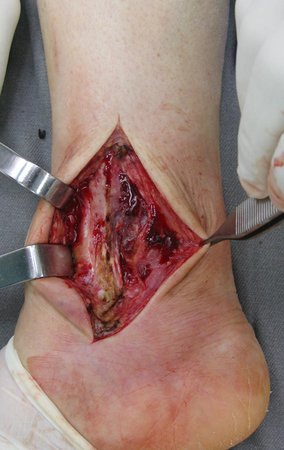
Figura 14: Incisão. A dissecção em planos profundos no tornozelo se restringe a fáscia e ao início do ventre muscular do tibial posterior. Logo nos deparamos com a tíbia e a lesão tumoral na sua região posterior. Note que é difícil definir o plano de clivagem entre o tumor com os tecidos adjacentes.
Figure 14: Incision. Dissection in deep planes of the ankle is restricted to the fascia and the beginning of the posterior tibialis muscle belly. We soon came across the tibia and the tumor lesion in its posterior region. Note that it is difficult to define the cleavage plane between the tumor and adjacent tissues.
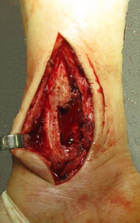
Figura 15: fragmento ósseo retirado da face lateral da tíbia para acessar o tumor subjacente.
Figure 15: bone fragment removed from the lateral aspect of the tibia to access the underlying tumor.
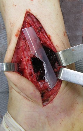
Figura 16: Realizamos uma janela óssea na face medial da tíbia para a cessar a lesão abaixo. Note que quando expomos e manipulamos o tumor surge um grande sangramento no campo cirúrgico. Isso é típico, e ocorre porque as lesões tumorais costumam ser muito vascularizadas. Devemos ter especial cuidado ao abordarmos alguns tumores pelo risco de sangramento excessivo como metástases de carcinoma renal.
Figure 16: We create a bone window on the medial aspect of the tibia to stop the injury below. Note that when we expose and manipulate the tumor, a large amount of bleeding appears in the surgical field. This is typical, and occurs because tumor lesions tend to be very vascular. We must be especially careful when approaching some tumors due to the risk of excessive bleeding, such as metastases from renal carcinoma.
Figura 17: Ao curetarmos completamente a lesão o sangramento se interrompe. Podemos ter uma nítida visão da grande destruição óssea provocada pelo crescimento tumoral. Esta falha óssea precisa ser preenchida.
Figure 17: When we completely curet the lesion, the bleeding stops. We can have a clear vision of the great bone destruction caused by tumor growth. This bone gap needs to be filled.
Figura 18: Quando iniciarmos o preenchimento da falha óssea com o cimento, a tendencia é que ele se espalhe além dos limites desejados e crie saliências que podem prejudicar o deslizamento de tendões, causar dor e pressionar a cicatriz da incisão. Para evitar que isso ocorra criamos uma canaleta com uma seringa de 60ml para ser usada como apoio posterior no momento da cimentação.
Figure 18: When we start filling the bone gap with cement, the tendency is for it to spread beyond the desired limits and create protrusions that can impair tendon sliding, cause pain and put pressure on the incision scar. To prevent this from happening, we created a channel with a 60ml syringe to be used as subsequent support at the time of cementation.
Figura 19: Visualizamos o aspecto final da cimentação. Note que graças ao apoio da canaleta utilizada, o cimento respeita o limite posterior da tíbia. Desta forma não irá interferir no deslizamento do tendão Calcâneo ou impactar outras estruturas.
Figure 19: We visualize the final appearance of the cementation. Note that thanks to the support of the channel used, the cement respects the posterior limit of the tibia. This way it will not interfere with the sliding of the Achilles tendon or impact other structures.
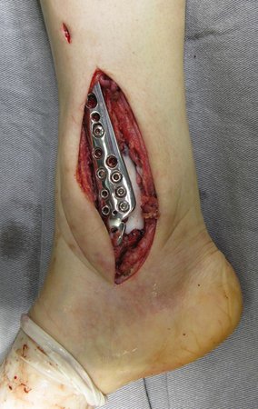
Figura 20: Para garantir a estabilidade optamos por aplicar uma placa de grandes fragmentos de baixo perfil, bloqueada na face medial da tíbia. Note que os parafusos são fixados diretamente no cimento ósseo. Isso aumenta a estabilidade e impede o deslocamento do cimento. A passagem dos parafusos deve ser feita quando o cimento estiver em estado sólido e duro, dessa forma cria-se uma "rosca" no trajeto e caso seja necessário a retirada de algum parafuso no futuro, isso será possível.
Figure 20: To guarantee stability, we chose to apply a low-profile large-fragment plate, locked on the medial surface of the tibia. Note that the screws are fixed directly into the bone cement. This increases stability and prevents the cement from shifting. The passage of the screws must be done when the cement is in a solid and hard state, this way a "thread" is created in the path and if it is necessary to remove any screws in the future, this will be possible.
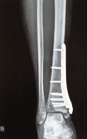
We performed intralesional resection of the tumor and reconstructed it with bone cement. We use a locked plate of large medial support fragments to stabilize the medial column and prevent the cement from shifting in relation to the tibia. Postoperatively, we kept the patient immobilized for the first few days for analgesic purposes and then released her for active mobilization. Partial weight-bearing protection was maintained for two weeks and then allowed for full weight-bearing with the aid of a contralateral crutch. In the immediate postoperative radiograph, below, we observed the almost complete filling of the bone cavity by the cement. The high viscosity of the cement and the hardening time of this material influence the complete filling of bone cavities.
 
Figura 21: Início do fechamento da incisão. O tornozelo é uma articulação muito superficial e com poucos tecidos para cobertura. É importante o correto fechamento dos tecidos moles com cobertura do implante pela fáscia muscular para minimizar complicações de ferida operatória.
Figure 21: Beginning of incision closure. The ankle is a very superficial joint with few tissues for coverage. It is important to correctly close the soft tissues with coverage of the implant by the muscular fascia to minimize surgical wound complications.
Figura 22: Aspecto final após a sutura da incisão. Vemos uma pequena incisão proximal a sutura principal. Esta pequena via foi utilizada para a colocação do parafuso mais proximal da placa de maneira percutânea, dessa forma reduzindo o tamanho final da incisão e a agressão cirúrgica.
Figure 22: Final appearance after suturing the incision. We see a small incision proximal to the main suture. This small route was used to place the most proximal screw of the plate percutaneously, thus reducing the final size of the incision and surgical aggression.
Figura 23: Rx de frente do tornozelo direito no pós-operatório imediato. Vemos a falha óssea produzida pela retirada do tumor quase totalmente preenchida pelo cimento. A coluna medial da tíbia foi refeita e estabilizada pelo implante colocado.
Figure 23: X-ray of the front of the right ankle in the immediate postoperative period. We see the bone defect produced by the removal of the tumor almost completely filled by cement. The medial column of the tibia was reconstructed and stabilized by the implant placed.
Figura 24: Rx de perfil do tornozelo direito no pós-operatório imediato. Vemos a margem posterior do cimento ósseo lisa e alinhada com a parede posterior da tíbia.
Figure 24: Lateral X-ray of the right ankle in the immediate postoperative period. We see the posterior margin of the bone cement smooth and aligned with the posterior wall of the tibia.
Figura 25: Anátomo Patológico sugestivo de neoplasia maligna indiferenciada de grandes células.
Figure 25: Pathological anatomy suggestive of undifferentiated large cell malignant neoplasm.
Figura 26: Laudo de Imunohistoquímica com diagnóstico de Carcinoma espinocelular metastático
Figure 26: Immunohistochemistry report with diagnosis of metastatic squamous cell carcinoma
Figura 27: Tomografia de tórax.
Figure 27: Chest tomography.
Figura 28: Tomografia de tórax.
Figure 28: Chest tomography.
Figura 29: Imagem de frente do tornozelo direito no pós-operatório de 1 mês e 5 dias, mostrando o local onde será realizado a radioterapia.
Figure 29: Image of the front of the right ankle 1 month and 5 days postoperatively, showing the location where radiotherapy will be performed.
Figura 30: Imagem de perfil do tornozelo direito no pós-operatório de 1 mês e 5 dias, mostrando a cicatriz do local da incisão cirúrgica.
Figure 30: Profile image of the right ankle 1 month and 5 days postoperatively, showing the scar at the site of the surgical incision.
Figura 31: Paciente em pé, e deambulando no pós-operatório de 1 mês e 5 dias.
Figure 31: Patient standing and walking 1 month and 5 days postoperatively.

Author: Prof. Dr. Pedro Péricles Ribeiro Baptista

 Orthopedic Oncosurgery at the Dr. Arnaldo Vieira de Carvalho Cancer Institute

Hello! How can we assist you?