This digital library houses the book on Oncology and Orthopedic Oncosurgery.

It includes academic lectures, presentations from national and international congresses, published papers, case discussions, performed surgical procedures, and proprietary techniques developed.

The digital format was chosen because the web allows the inclusion of texts with numerous visual resources, such as images and videos, which would not be possible in a printed book.

The content is intended for students, healthcare professionals, and the general public interested in the field.

Desmoid Fibroma of the Foot

9bc99c590be3511b8d53741684ef574c 2

Desmoid fibroma of the foot Reconstruction of the fifth metatarsal

Desmoid Fibroma of the Foot. A 43-year-old patient with a “callus” on the fifth metatarsal of his left foot for twenty years, which developed deformity, increased volume and pain in the last year.
Figura 1: lesão na cabeça do quinto metatarso esquerdo com deformidade, erosão óssea e insuflação da cortical
Figure 1: injury to the head of the left fifth metatarsal with deformity, bone erosion and cortical inflation
Figura 2: Tomografia com lesão agressiva do quinto dedo do pé esquerdo.
Figure 2: Tomography with aggressive lesion of the fifth toe of the left foot.
Figura 3: tomografia em perfil, evidenciando erosão da cortical e agressividade local.
Figure 3: Tomography in profile, showing cortical erosion and local aggressiveness.
Figura 4: Rm axial T1 lesão de baixo sinal.
Figure 4: Axial MRI T1 low signal lesion.
Figura 5: Rm axial com captação de contraste.
Figure 5: Axial MRI with contrast capture.
Figura 6: Rm axial T1, com saturação de gordura.
Figure 6: Axial T1 MRI, with fat saturation.
Figura 7: Rm axial com contraste.
Figure 7: Axial MRI with contrast.
Figura 8: Rm sagital contraste.
Figure 8: Sagittal MRI contrast.
Figura 9: Biópsia da lesão, sob controle de radioscopia.
Figure 9: Biopsy of the lesion, under radioscopy control.
Figura 10: Incisão anterior, sobre o quinto metacarpeano, com exerese do trajeto da biópsia.
Figure 10: Anterior incision, over the fifth metacarpeal, with excision of the biopsy path.
Figura 11: Ressecção de 2/3 terços distais do quinto metacarpeano.
Figure 11: Resection of the distal 2/3 thirds of the fifth metacarpal bone.
Figura 12: Retirada de enxerto em bloco do ilíaco esquerdo, mesmo lado da lesão.
Figure 12: Removal of a block graft from the left iliac, same side of the lesion.
Figura 13: Bloco de enxerto estruturado que será modelado para substituir o segmento ressecado.
Figure 13: Structured graft block that will be modeled to replace the resected segment.
Figura 14: Enxerto sendo esculpido, para substituir os 2/3 distais do quinto metatarsiano.
Figure 14: Graft being carved to replace the distal 2/3 of the fifth metatarsal.
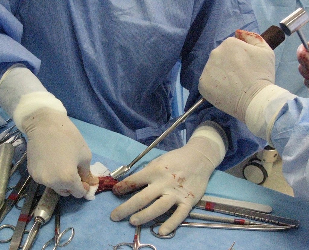
Figura 15: Enxerto sendo aparado com saca bocado, no formato do quinto metatarsiano.
Figure 15: Graft being trimmed with a bit puller, in the shape of the fifth metatarsal.
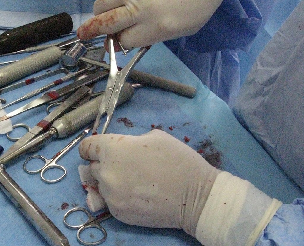
Figura 16: bloco do enxerto sendo cortado no tamanho adequado para a reconstrução.
Figure 16: graft block being cut to the appropriate size for reconstruction.
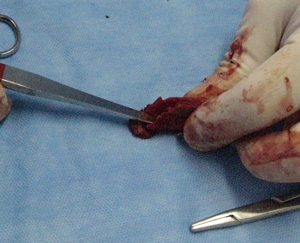
Figura 17: Realização de um sulco no enxerto para encaixar o segmento remanescente do metatarseano, visando melhor acomodação e integração.
Figure 17: Creating a groove in the graft to fit the remaining metatarsal segment, aiming for better accommodation and integration.
Figura 18: Bloco de enxerto já preparado para a reconstrução.
Figure 18: Graft block already prepared for reconstruction.
Figura 19: Enxerto encaixado no segmento remanescente do quinto metatarseano.
Figure 19: Graft fitted into the remaining segment of the fifth metatarsal.
figura 20: Passagem de fios de Kirshnner 2.0, retrogadamente, pelo canal medular do segmento remanescente do quinto metatarseano, para a fixação do enxerto
figure 20: Passage of Kirshnner 2.0 wires, retrogradely, through the medullary canal of the remaining segment of the fifth metatarsal bone, to fix the graft
Figura 21: posicionamento do enxerto e fixação com o fio.
Figure 21: positioning of the graft and fixation with the thread.
Figura 22: Detalhe do reposicionamento do enxerto.
Figure 22: Detail of the graft repositioning.
Figura 23: Reposicionamento final da reconstrução e corte do fio, na base do quinto metatarso.
Figure 23: Final repositioning of the reconstruction and cutting of the wire, at the base of the fifth metatarsal.
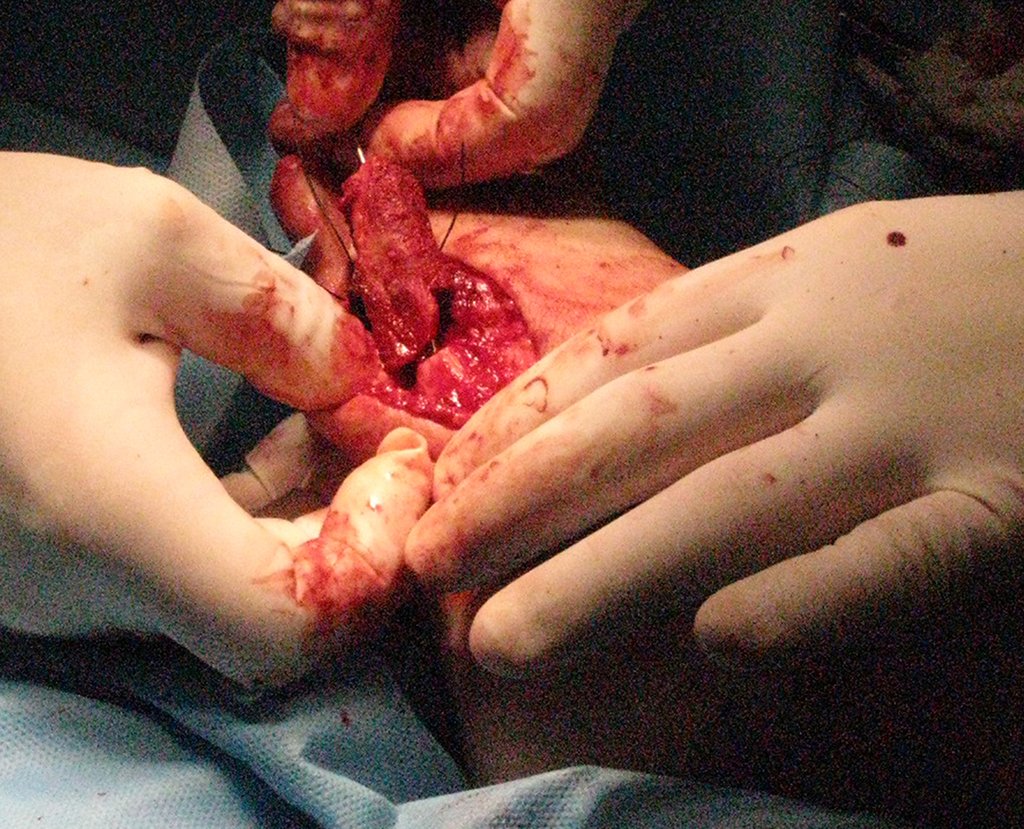
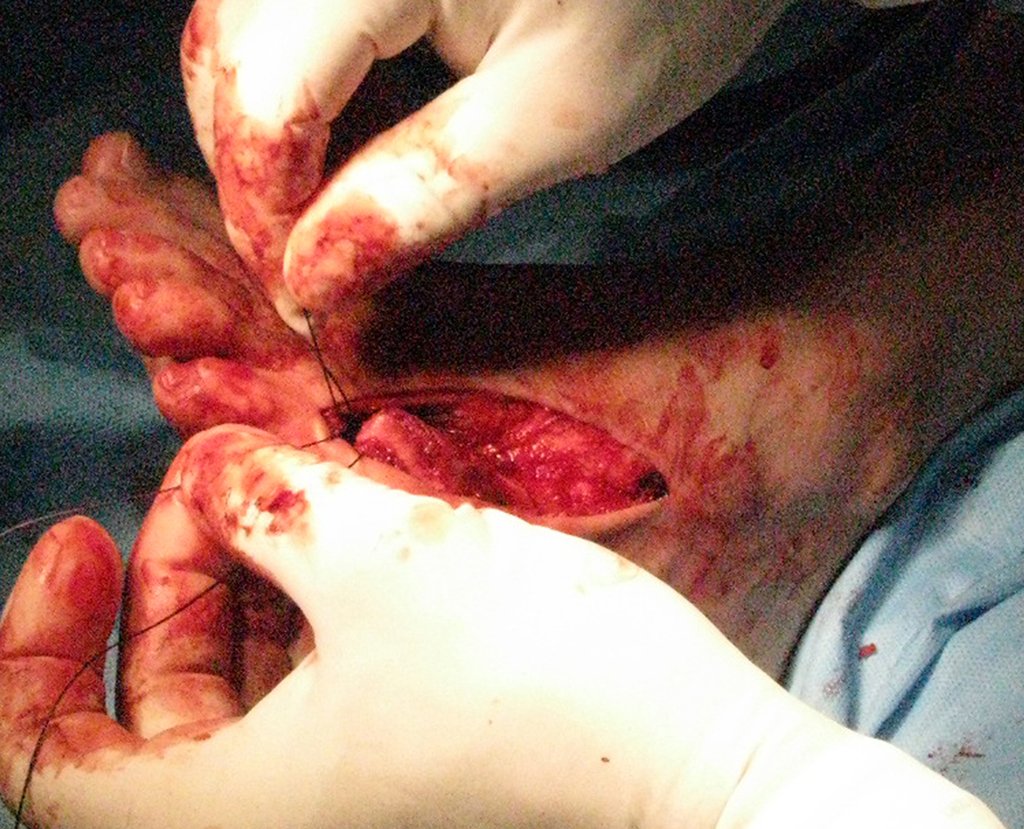
Figura 24: Reparo da capsuloplastia para sutura e fixação no enxerto, mantendo-o alinhado.
Figure 24: Capsuloplasty repair for suturing and fixing the graft, keeping it aligned.
Figura 25: Reposicionamento final da reconstrução e corte do fio, na base do quinto metatarso.
Figure 25: Final repositioning of the reconstruction and cutting of the wire, at the base of the fifth metatarsal.
Figura 26: Realização de orifícios no enxerto para a ancoragem dos fios de sutura.
Figure 26: Making holes in the graft to anchor the sutures.
Figura 27: Enxerto fixado e posicionado com bom alinhamento.
Figure 27: Graft fixed and positioned with good alignment.
Figura 28: Peça ressecada, face dorsal, com pele e trajeto da biópsia.
Figure 28: Resected piece, dorsal side, with skin and biopsy path.
Figura 29: Peça ressecada, face plantar.
Figure 29: Dried piece, plantar surface.
Figura 30: Radiografia intra-operatória documentando a reconstrução, frente.
Figure 30: Intraoperative radiograph documenting the reconstruction, front.
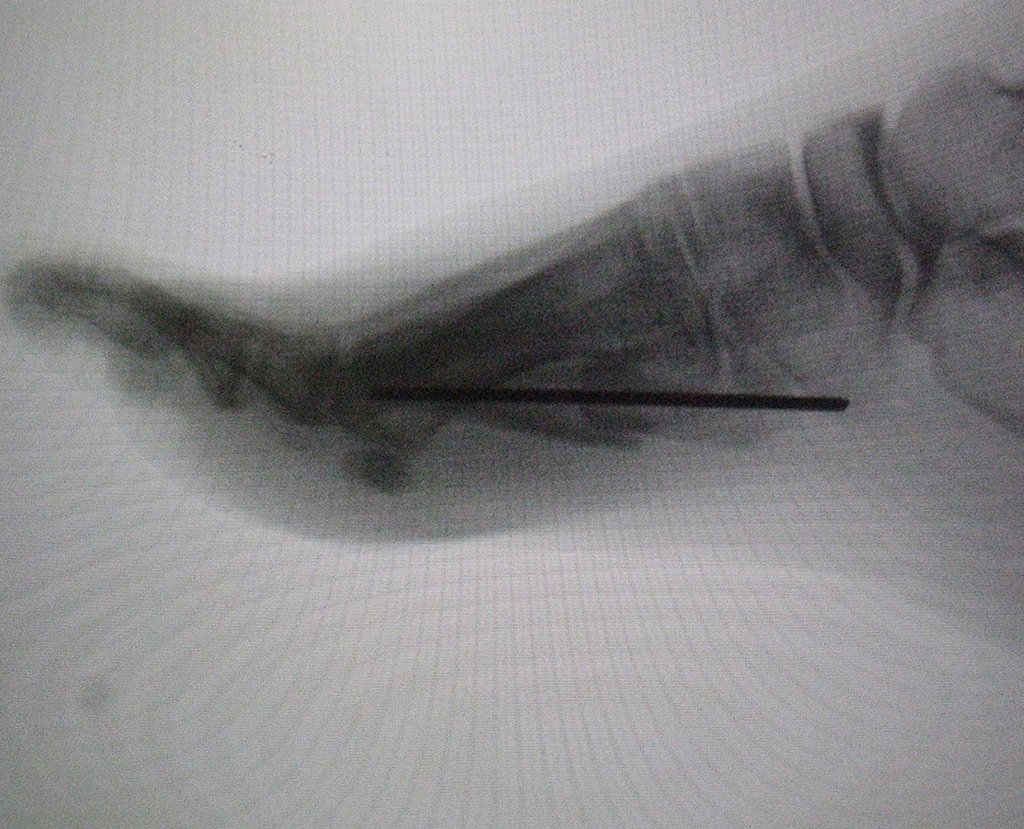
Figura 31: Radiografia intra-operatória documentando a reconstrução, perfil.
Figure 31: Intraoperative radiograph documenting the reconstruction, profile.
Figura 32: Reconstrução em 3D da lesão.
Figure 32: 3D reconstruction of the lesion.
Figura 33: Planejamento pré-operatório.
Figure 33: Pre-operative planning.
Figura 34: Pós operatório 12 dias. Boa cicatrização.
Figure 34: 12 days post-operative. Good healing.

Video 1: Function 12 days after surgery. Walking with Robofoot.

Figura 35: Radiogtrafia da reconstrução, frente, pós operatório 3 meses, em 13/12/2016.
Figure 35: X-ray of the reconstruction, front, 3 months postoperatively, on 12/13/2016.
Figura 36: Radiogtrafia da reconstrução, perfil, pós operatório 3 meses, em 13/12/2016.
Figure 36: X-ray of the reconstruction, profile, 3 months postoperatively, on 12/13/2016.
Figura 37: Aspecto clínico e função, pós operatório 3 meses, em 13/12/2016.
Figure 37: Clinical appearance and function, 3 months after surgery, on 12/13/2016.
Figura 38: Cicatriz da incisão da crista ilíaca, para retirada de enxerto autólogo. Bom aspecto.
Figure 38: Scar from the iliac crest incision, for autologous graft removal. Good looking.
Figura 39: Aspecto cosmético do pé, pós operatório 3 meses, em 13/12/2016.
Figure 39: Cosmetic appearance of the foot, 3 months after surgery, on 12/13/2016.

Video 1: Function 12 days after surgery. Walking with Robofoot.

Author: Prof. Dr. Pedro Péricles Ribeiro Baptista

 Orthopedic Oncosurgery at the Dr. Arnaldo Vieira de Carvalho Cancer Institute

Hello! How can we assist you?