This digital library houses the book on Oncology and Orthopedic Oncosurgery.

It includes academic lectures, presentations from national and international congresses, published papers, case discussions, performed surgical procedures, and proprietary techniques developed.

The digital format was chosen because the web allows the inclusion of texts with numerous visual resources, such as images and videos, which would not be possible in a printed book.

The content is intended for students, healthcare professionals, and the general public interested in the field.

Diagnosis between Callus and Nodule

2109366d68770c5528aa56705516f364 3

Differential Diagnosis Lung Nodule and Fracture Callus

Diagnosis between Callus and Nodule. An 85-year-old patient, a smoker since the age of eighteen, reports “shortness of breath” on exertion. Look for a Basic Health Unit where a chest x-ray is performed, figures 1 and 2.
Figura 1: Radiografia de tórax AP, campos pulmonares, com círculo em vermelho, destacando imagem condensante na projeção do lobo inferior do pulmão esquerdo.
Figure 1: AP chest x-ray, lung fields, with a red circle, highlighting a condensing image in the projection of the lower lobe of the left lung.
Figura 2: Radiografia de tórax em oblíqua (os corpos vertebrais encontran-se sobrepostos aos arcos costais), com seta em vermelho, sinalizando aumento de densidade na projeção do lobo inferior do pulmão esquerdo.
Figure 2: Oblique chest x-ray (the vertebral bodies are superimposed on the costal arches), with a red arrow, signaling increased density in the projection of the lower lobe of the left lung.

This finding was interpreted as a pulmonary nodule and the patient was referred to an oncologist with suspected primary lung cancer.  Lung nodule?

During the diagnostic investigation, tests were requested to stage the lesion and bronchoscopy for biopsy. Among the exams, a chest tomography was performed, figures 3 and 4.

Figura 3: Tomografia axial de tórax, corte superior, evidenciando um pulmão de fumante crônico, com áreas de teleangiectasia e congestão vascular.
Figure 3: Axial tomography of the chest, upper section, showing a chronic smoker's lung, with areas of teleangiectasia and vascular congestion.
Figura 4: Tomografia axial de tórax, corte inferior, evidenciando um pulmão de fumante crônico, com enfisema pulmonar.
Figure 4: Axial tomography of the chest, lower section, showing a lung from a chronic smoker, with pulmonary emphysema.

In the careful analysis of the images from this tomography, the condensing area observed in the radiographs, which suggested the presence of a primary nodule in the pulmonary parenchyma of the lower lobe of the left lung, was not found!?. 

However, changes were found in the 90 and  100 left  posterior costal arch, figures 5 and 6.

Figura 5: Tomografia axial de tórax, estudo do mediastino inferior, sem evidência de nódulo pulmonar. O círculo em amarelo destaca alteração do nono arco costal posterior do lado esquerdo.
Figure 5: Axial tomography of the chest, study of the lower mediastinum, with no evidence of a pulmonary nodule. The yellow circle highlights changes in the ninth posterior costal arch on the left side.
Figura 6: Tomografia axial de tórax, no estudo do parênquima pulmonar inferior, também não se encontra evidência de nódulo pulmonar. A seta em amarelo destaca um calo de fratura no nono arco costal posterior do lado esquerdo !!!
Figure 6: Axial tomography of the chest, in the study of the lower lung parenchyma, there is also no evidence of a pulmonary nodule. The yellow arrow highlights a fracture callus in the ninth posterior costal arch on the left side!!!
With this finding, a new x-ray was performed, taking care to obtain an absolute profile of the chest, figures 7 and 8.
Figura 7: Radiografia de tórax AP, repetida após a tomografia, corroborando a presença da imagem condensante, na projeção do lobo inferior do pulmão esquerdo.
Figure 7: AP chest x-ray, repeated after the tomography, corroborating the presence of the condensing image, in the projection of the lower lobe of the left lung.
Figura 8: Radiografia de tórax, em perfil absoluto, não aparece a imagem condensante, que sugeria a presença de nódulo no parênquima pulmonar.
Figure 8: Chest X-ray, in absolute profile, the condensing image does not appear, which suggested the presence of a nodule in the lung parenchyma.

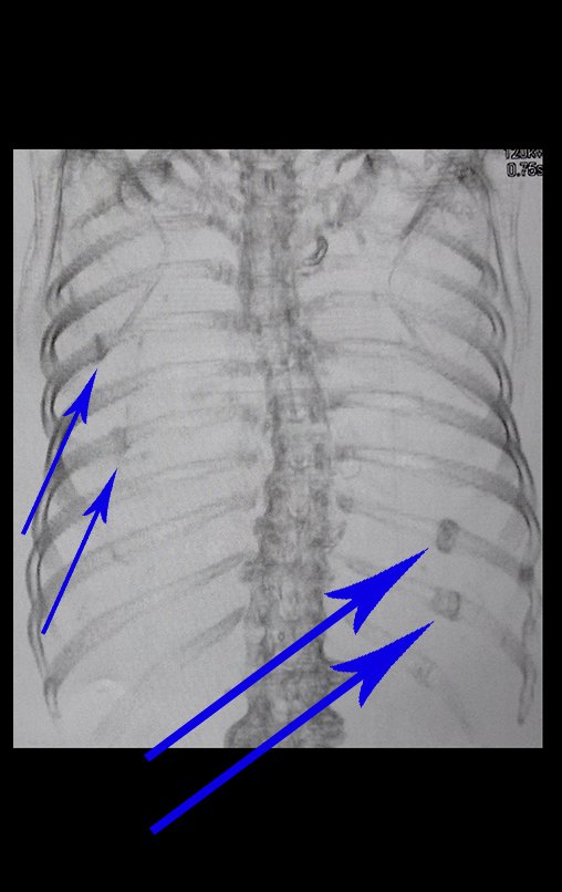
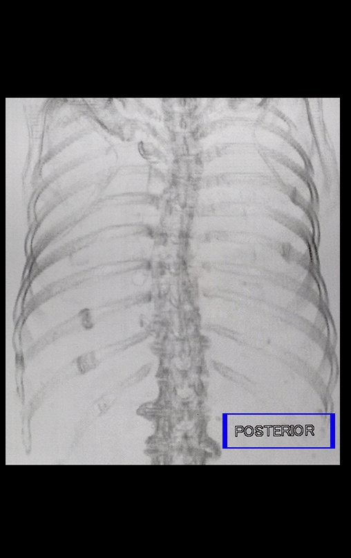
Figures 9, 10 and 11 clarify the false interpretation of fracture callus X pulmonary nodule .

Figura 9: Radiografia de tórax AP, repetida após a tomografia, focalizada, esclarecendo que a condensação corresponde ao calo de fratura do 90 arco costal posterior.
Figure 9: AP chest x-ray, repeated after the tomography, in focus, clarifying that the condensation corresponds to the fracture callus of the posterior costal arch.
Figura 10: Tomografia de tórax AP, destacando calo de fraura no 90 e 100 arcos costais posteriores à esquerda e no 50 e 70 arcos costais porteriores à direita.
Figure 10: AP chest tomography, highlighting fracture callus in the 90th and 100th posterior costal arches on the left and in the 50th and 70th posterior costal arches on the right.
Figura 11: Tomografia de tórax. tomada póstero-anterior, destacando calo de fraura no 90 e 100 arcos costais posteriores à esquerda e no 50 e 70 arcos costais porteriores à direita.
Figure 11: Chest tomography. posteroanterior view, highlighting fracture callus in the 90th and 100th posterior costal arches on the left and in the 50th and 70th posterior costal arches on the right.

In the history, the patient reported falling down stairs two years ago. He had woken up in the early hours of the morning to go to the bathroom and fell, rolling down the stairs and suffering a broken right wrist and several ribs. 

Accurate clinical history + adequate imaging tests + careful analysis  are essential for the correct diagnosis.

Author: Prof. Dr. Pedro Péricles Ribeiro Baptista

 Orthopedic Oncosurgery at the Dr. Arnaldo Vieira de Carvalho Cancer Institute

Hello! How can we assist you?